

Optical coherence tomography images of bell-shaped appearance in late sirolimus-eluting stent restenosis with extension of previous incomplete stent apposition
- PMID: 30532883
- PMCID: PMC6265391
- DOI: 10.1016/j.jccase.2011.08.001
Optical coherence tomography images of bell-shaped appearance in late sirolimus-eluting stent restenosis with extension of previous incomplete stent apposition
Abstract
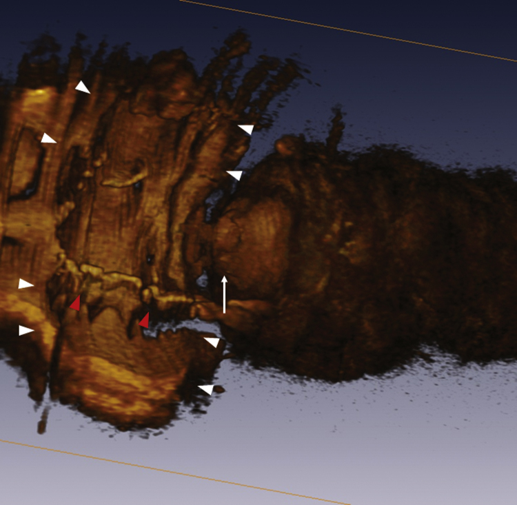
Late adverse events including late stent thrombosis and late catch-up phenomenon after percutaneous coronary intervention have been a serious clinical problem in the drug-eluting stent era. Recently, peri-stent contrast staining, namely extension of incomplete stent apposition was reported following drug-eluting stent implantation. Here, we report a case of late incomplete stent apposition with late stent restenosis 3 years after sirolimus-eluting stent implantation. We evaluated this restenotic site by intravascular ultrasound (IVUS) and optical coherence tomography (OCT). Though IVUS demonstrated irregular structure within stent at the stenotic site, OCT detected unusual bell-shaped image of late stent restenosis with extension of incomplete stent apposition.
Keywords: Drug-eluting stent; In-stent restenosis; Incomplete stent apposition; Optical coherence tomography.
Figures


References
-
- Babapulle M.N., Joseph L., Belisle P., Brophy J.M., Eisenberg M.J. A hierarchical Bayesian meta-analysis of randomised clinical trials of drug-eluting stents. Lancet. 2004;364:583–591. - PubMed
-
- Alfonso F., Perez-Vizcayno M.J., Ruiz M., Suarez A., Cazares M., Hernandez R., Escaned J., Banuelos C., Jimenez-Quevedo P., Macaya C. Coronary aneurysms after drug-eluting stent implantation: clinical, angiographic, and intravascular ultrasound findings. J Am Coll Cardiol. 2009;53:2053–2060. - PubMed
-
- Herrero-Garibi J., Cruz-Gonzalez I., Parejo-Diaz P., Jang I.K. Optical coherence tomography: its value in intravascular diagnosis today. Rev Esp Cardiol. 2010;63:951–962. - PubMed
-
- Kataiwa H., Tanaka A., Kitabata H., Matsumoto H., Kashiwagi M., Kuroi A., Ikejima H., Tsujioka H., Okochi K., Tanimoto T., Yamano T., Takarada S., Nakamura N., Kubo T., Mizukoshi M. Head to head comparison between the conventional balloon occlusion method and the non-occlusion method for optical coherence tomography. Int J Cardiol. 2011;146:186–190. - PubMed
-
- Wilson G.J., Nakazawa G., Schwartz R.S., Huibregtse B., Poff B., Herbst T.J., Baim D.S., Virmani R. Comparison of inflammatory response after implantation of sirolimus- and paclitaxel-eluting stents in porcine coronary arteries. Circulation. 2009;120:141–149. 1–2. - PubMed
LinkOut - more resources
Full Text Sources
