

Three-dimensional visualization of scoring mechanism of 'AngioSculpt' balloon for calcified coronary lesions using optical coherence tomography
- PMID: 30532893
- PMCID: PMC6265381
- DOI: 10.1016/j.jccase.2011.10.008
Three-dimensional visualization of scoring mechanism of 'AngioSculpt' balloon for calcified coronary lesions using optical coherence tomography
Abstract
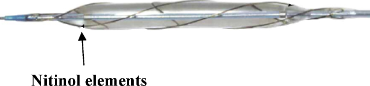
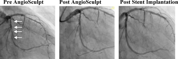
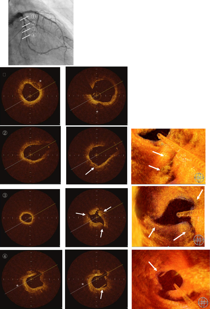
Despite developments in coronary interventional cardiology, plaque calcification is a critical issue of stent expansion. AngioSculpt Scoring Balloon Catheter® (AngioSculpt; AngioScore Inc., Fremont, CA, USA) can produce more 'scoring' marks, which leads to prevention of 'plaque shift' and 'balloon slippage'; moreover, the 'scoring' produces some cutting effect, leading to successful stent implantation even on severe calcified lesions. We have applied AngioSculpt on severe calcified lesions to achieve its adequate expansion, and report the mechanism of the 'scoring' and its efficacy evaluated by three-dimensional stereoscopic reconstruction (3-D) of optical coherence tomography (OCT; LightLab Imaging, Inc., Westford, MA, USA). The patient is a 64-year-old male, who had diffuse stenosis in the left circumflex coronary artery (LCX) with severe calcifications, and was treated using AngioSculpt. AngioSculpt predilatation with a high pressure led to successful stent implantation. The radial scores were clearly imaged by 3-D OCT, demonstrating that radial nitinol wires made spiral indents from the relative weak points at the surface adjacent to calcification, which resulted in a less traumatic and safe dilatation although the scoring mark was not recognized clearly in intravascular ultrasound. This report suggests AngioSculpt might become one of the options for a severe calcified lesion.
Keywords: 3-D OCT; AngioSculpt; Calcified lesion; Optical coherence tomography (OCT); Scoring.
Figures
Similar articles
-
Optical coherence tomography after new scoring balloon angioplasty for in-stent restenosis and de novo coronary lesions.Int J Cardiol. 2010 Jun 11;141(3):e51-3. doi: 10.1016/j.ijcard.2008.11.154. Epub 2009 Jan 6. Int J Cardiol. 2010. PMID: 19128844
-
Intravascular ultrasound assessment of the novel AngioSculpt scoring balloon catheter for the treatment of complex coronary lesions.J Invasive Cardiol. 2008 Jan;20(1):21-7. J Invasive Cardiol. 2008. PMID: 18174614
-
A study of balloon type on calcified coronary lesion predilation: A finite element analysis.Proc Inst Mech Eng H. 2023 Apr;237(4):443-450. doi: 10.1177/09544119231157853. Epub 2023 Mar 16. Proc Inst Mech Eng H. 2023. PMID: 36927166 Free PMC article.
-
Major Complications and Failure Modes of the Angiosculpt Scoring Balloon Catheter: Analysis of the MAUDE Database.Curr Probl Cardiol. 2023 Apr;48(4):101557. doi: 10.1016/j.cpcardiol.2022.101557. Epub 2022 Dec 14. Curr Probl Cardiol. 2023. PMID: 36528205 Review.
-
Current clinical applications of coronary optical coherence tomography.Cardiovasc Interv Ther. 2018 Jan;33(1):1-10. doi: 10.1007/s12928-017-0483-8. Epub 2017 Jul 14. Cardiovasc Interv Ther. 2018. PMID: 28710605 Free PMC article. Review.
Cited by
-
[[New scoring balloon to treat moderate-to-severe calcified coronary lesions. The first-in-man Naviscore study]].REC Interv Cardiol. 2025 Feb 3;7(2):91-98. doi: 10.24875/RECIC.M24000487. eCollection 2025 Apr-Jun. REC Interv Cardiol. 2025. PMID: 40438639 Free PMC article. Spanish.
-
Contemporary Approach to Heavily Calcified Coronary Lesions.Interv Cardiol. 2019 Nov 18;14(3):154-163. doi: 10.15420/icr.2019.19.R1. eCollection 2019 Nov. Interv Cardiol. 2019. PMID: 31867062 Free PMC article. Review.
-
Cracking the Left Main Coronary Artery Nodular Calcium With a Scoring Balloon.Cureus. 2023 Aug 25;15(8):e44123. doi: 10.7759/cureus.44123. eCollection 2023 Aug. Cureus. 2023. PMID: 37750115 Free PMC article.
-
Coronary calcifications, the Achilles heel in coronary interventions.Postepy Kardiol Interwencyjnej. 2024 Mar;20(1):1-17. doi: 10.5114/aic.2024.136415. Epub 2024 Mar 15. Postepy Kardiol Interwencyjnej. 2024. PMID: 38616941 Free PMC article. Review.
References
-
- Moses J.W., Carlier S., Moussa I. Lesion preparation prior to stenting. Rev Cardiovasc Med. 2004;5(Suppl. 2):S16–S21. - PubMed
-
- Finet G., Weissman N.J., Mintz G.S., Satler L.F., Kent K.M., Laird J.R., Adelmann G.A., Ajani A.E., Castagna M.T., Rioufol G., Pichard A.D. Mechanism of lumen enlargement with direct stenting versus predilatation stenting: influence of remodelling and plaque characteristics assessed by volumetric intracoronary ultrasound. Heart. 2003;89:84–90. - PMC - PubMed
-
- Fitzgerald P.J., Ports T.A., Yock P.G. Contribution of localized calcium deposits to dissection after angioplasty. An observational study using intravascular ultrasound. Circulation. 1992;86:64–70. - PubMed
-
- Takano M., Yamamoto M., Murakami D., Takano H., Asai K., Yasutake M., Seino Y., Mizuno K. Optical coherence tomography after new scoring balloon angioplasty for in-stent restenosis and de novo coronary lesions. Int J Cardiol. 2010;141:e51–e53. - PubMed
Publication types
LinkOut - more resources
Full Text Sources
