

Case Reports
doi: 10.1016/j.jccase.2011.09.003.
eCollection 2012 Feb.
Don't forget the memory: Contribution of the T wave vector in localizing the site of origin of a monomorphic idiopathic ventricular tachycardia
Affiliations
- PMID: 30532896
- PMCID: PMC6265211
- DOI: 10.1016/j.jccase.2011.09.003
Item in Clipboard
Case Reports
Don't forget the memory: Contribution of the T wave vector in localizing the site of origin of a monomorphic idiopathic ventricular tachycardia
J Cardiol Cases.
.
Abstract
We report a case of cardiac memory following recurrent episodes of monomorphic idiopathic ventricular tachycardia and explain how it could be helpful in localizing the site of origin of the arrhythmia.
Keywords: Monomorphic ventricular tachycardia; T wave memory.
Figures
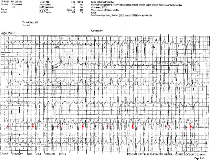
12-Lead electrocardiogram showing a monomorphic ventricular tachycardia with right bundle branch morphology and left axis deviation. Red arrows display P waves in lead II. (For interpretation of the references to color in this figure legend, the reader is referred to the web version of the article.)
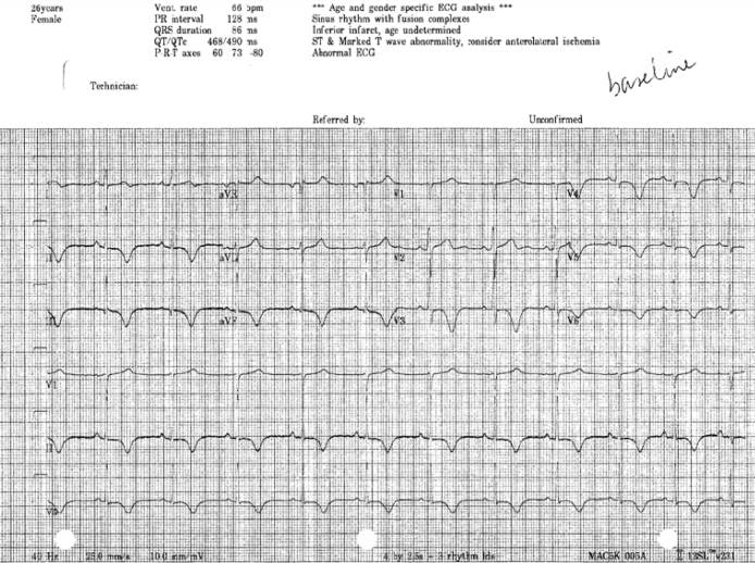
12-Lead baseline electrocardiogram showing T wave memory.
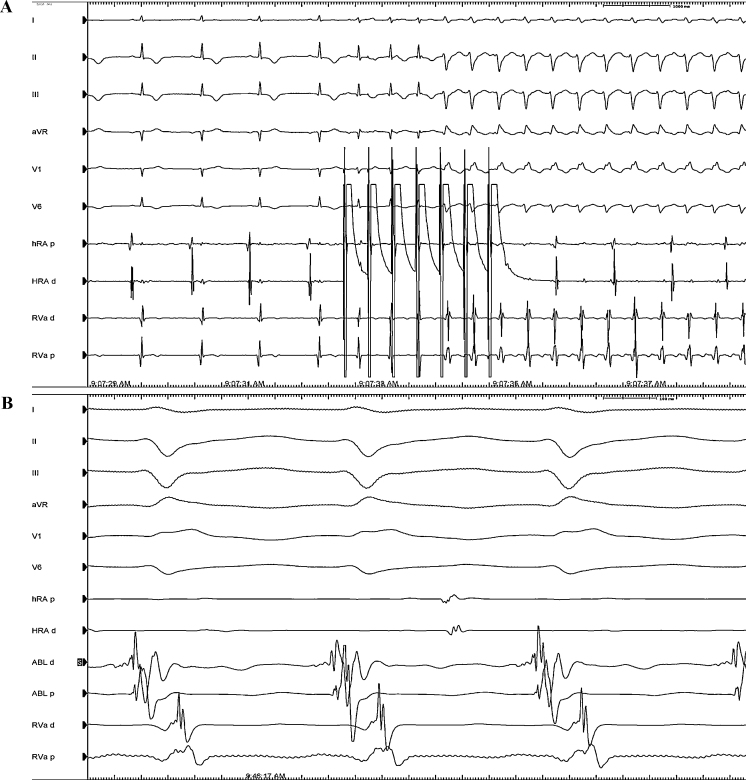
(A) Induction of ventricular tachycardia with atrial bursts from the high right atrium (HRA). From the top to the bottom electrocardiogram (ECG) leads I, II, III, aVR, V1, and V6 are displayed with electrograms from proximal and distal HRA (HRAd and HRAp) and proximal and distal right ventricular apex (RVAd and RVAp). (B) Site of successful ablation. From the top to the bottom ECG leads I, II, III, aVR, V1, and V6 are displayed with electrograms from HRAd and HRAp, proximal and distal ablation catheter (ABLp and ABLd) and RVAd and RVAp.
References
-
- Rosenbaum M.B., Blanco H.H., Elizari M.V., Lázzari J.O., Davidenko J.M. Electrotonic modulation of the T wave and cardiac memory. Am J Cardiol. 1982;50:213–222. - PubMed
-
- Ozgen N., Rosen M.R. Cardiac memory: a work in progress. Heart Rhythm. 2009;6:564–570. - PubMed
-
- Rhinehardt J., Brady W.J., Perron A.D., Mattu A. Electrocardiographic manifestations of Wellens’ syndrome. Am J Emerg Med. 2002;20:638–643. - PubMed
-
- Shvilkin A., Ho K.K.L., Rosen M.R., Josephson M.E. T-vector direction differentiates postpacing from ischemic T-wave inversion in precordial leads. Circulation. 2005;111:969–974. - PubMed
Publication types
LinkOut - more resources
Full Text Sources
