

Acute myocardial infarction as presentation of an infiltrative lung neoplasia
- PMID: 30533123
- PMCID: PMC6275371
- DOI: 10.1016/j.jccase.2012.10.013
Acute myocardial infarction as presentation of an infiltrative lung neoplasia
Abstract
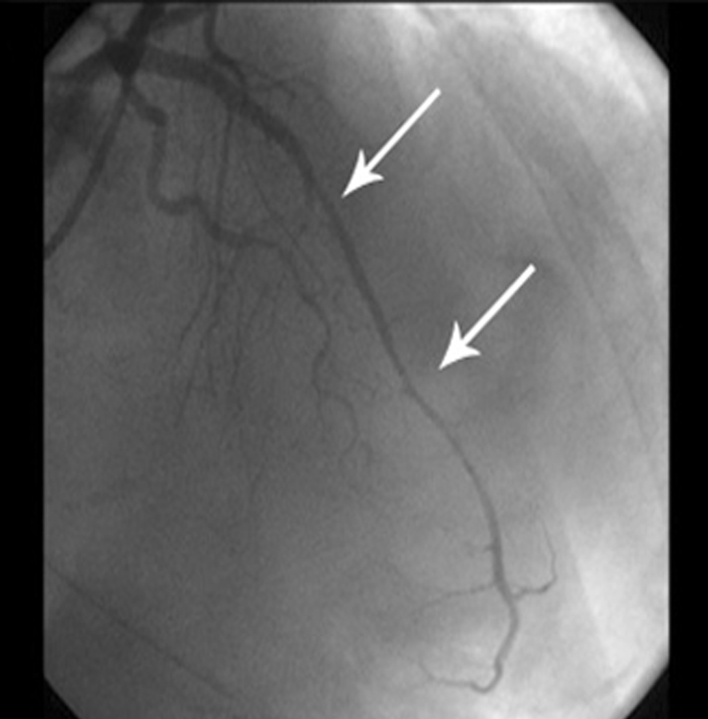
We report the case of a 51-year-old woman who presented with acute myocardial infarction as initial symptom of an infiltrative lung neoplasia. The patient was admitted to our center following an out-of-hospital cardiac arrest due to ventricular fibrillation which was cardioverted. On electrocardiography an anterior wall ST-elevation was found and urgent coronary angiography was performed. Left anterior descending coronary artery was occluded and after thrombus aspiration, an image of diffuse loss of lumen diameter and absence of coronary branches was compatible with an extrinsic compression. Such findings along with a lingula consolidation on chest X-ray examination suggested a thoracic neoplasia. Enhanced-chest computed tomography showed a mass located in the lingula with extensive mediastinal infiltration involving pericardium and myocardium. Anatomopathologic examination confirmed the presence of lung adenocarcinoma. <Learning objective: In patients with neoplasms should be suspected cardiac tumor infiltration in those with acute myocardial infarction. Since differential diagnosis between true AMI electrocardiographic (ECG) changes and pseudo-AMI ECG changes in patients with secondary cardiac tumors assumed to be difficult, such TTE or IVUS findings of extra-cardiac tumor could help physicians to make an accurate diagnosis.>.
Keywords: Cardiac metastasis; Lung adenocarcinoma; Myocardial infarction.
Figures


References
-
- Al-Mamgani A., Baartman L., Baaijens M., de Pree I., Incrocci L., Levendag P.C. Cardiac metastases. Int J Clin Oncol. 2008;3:369–372. - PubMed
-
- Beigelman R.L., Descalzo A.M., Storino R.A., Milei J. Acute myocardial infarction and bronchoalveolar carcinoma. Association or coincidence? Arch Inst Cardiol Mex. 1991;61:231–235. - PubMed
-
- Godwin J.D., Axel L., Adams J.R., Schiller N.B., Simpson P.C., Jr., Gertz E.W. Computed tomography: a new method for diagnosing tumor of the heart. Circulation. 1981;63:448–451. - PubMed
-
- Pérez-David E., García-Lizana M., García-Fernández M.A., Zamorano-Gómez J.L., Ferreirós-Domínguez J., Lafuente-Martínez J. Usefulness of magnetic resonance imaging in the evaluation of cardiac masses and pericardial disease. Rev Esp Cardiol Supl. 2006;6:30E–40E.
LinkOut - more resources
Full Text Sources
