. 2018 Nov 6:2018:3172480.
doi: 10.1155/2018/3172480. eCollection 2018.

Nicotine and Cotinine Inhibit Catalase and Glutathione Reductase Activity Contributing to the Impaired Osteogenesis of SCP-1 Cells Exposed to Cigarette Smoke

Affiliations

Nicotine and Cotinine Inhibit Catalase and Glutathione Reductase Activity Contributing to the Impaired Osteogenesis of SCP-1 Cells Exposed to Cigarette Smoke

Romina H Aspera-Werz et al. Oxid Med Cell Longev. .

Abstract

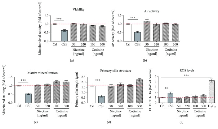
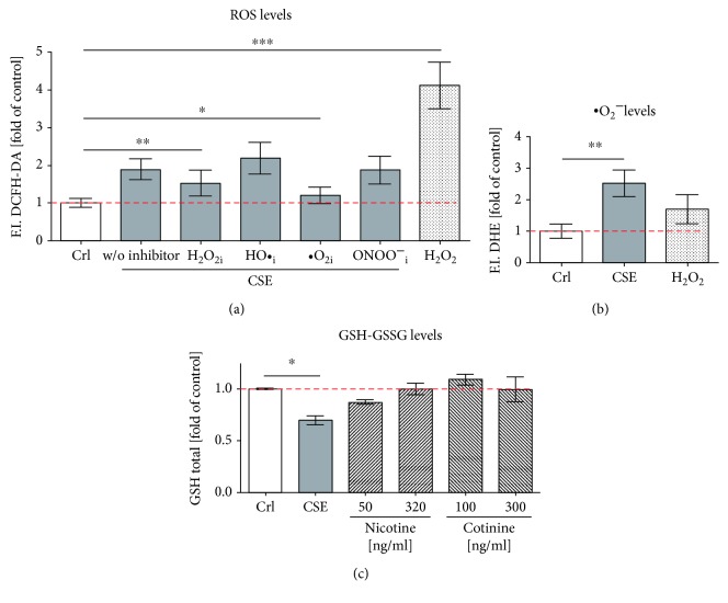
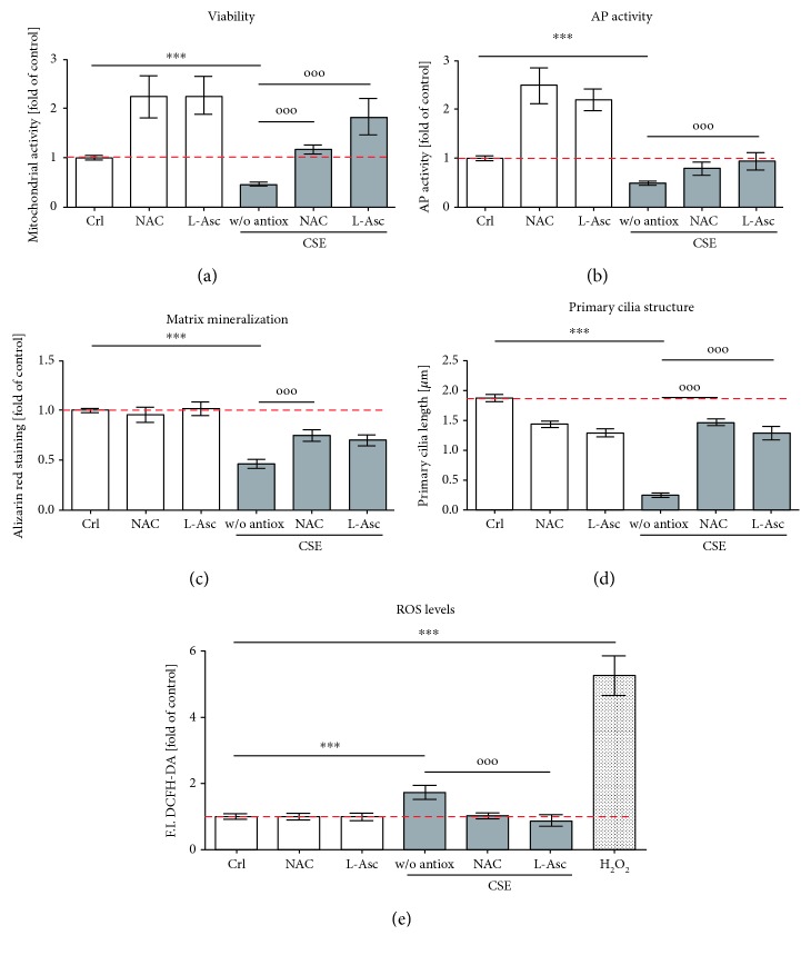
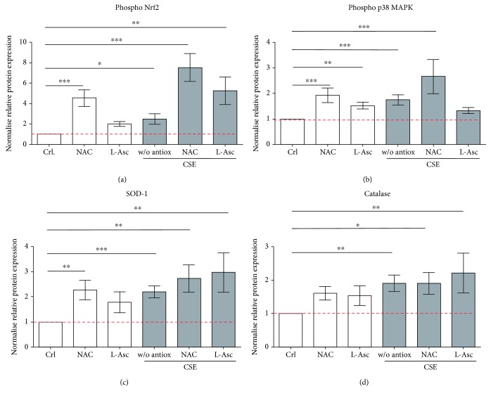
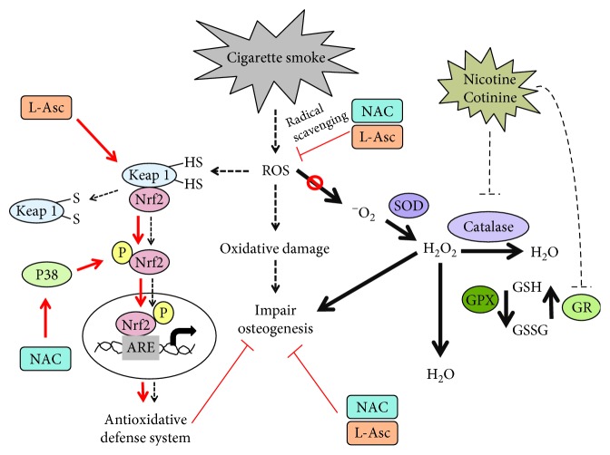
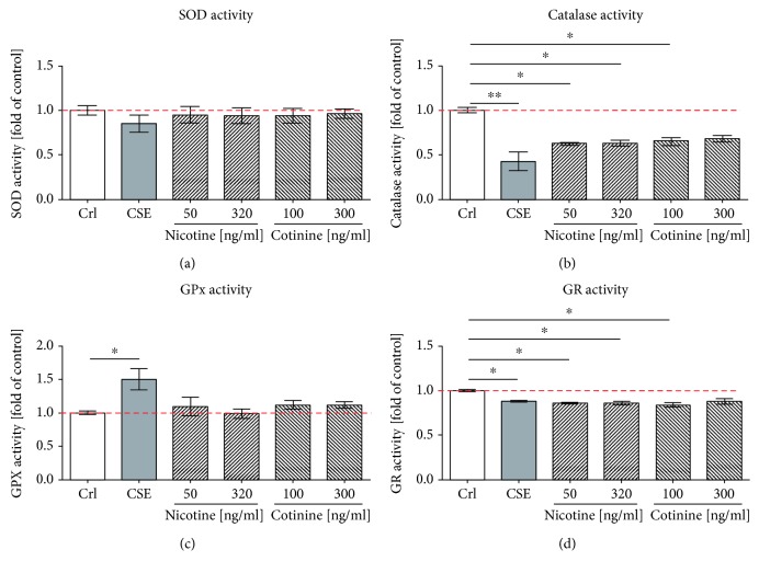
Cigarette smoking has been identified as a major risk factor for osteoporosis decades ago. Several studies have shown a direct relationship between cigarette smoking, decreased bone mineral density, and impaired fracture healing. However, the mechanisms behind impaired fracture healing and cigarette smoking are yet to be elucidated. Migration and osteogenesis of mesenchymal stem/stromal cells (MSCs) into the fracture site play a vital role in the process of fracture healing. In human nicotine, the most pharmacologically active and major addictive component present in tobacco gets rapidly metabolized to the more stable cotinine. This study demonstrates that physiological concentrations of both nicotine and cotinine do not affect the osteogenic differentiation of MSCs. However, cigarette smoke exposure induces oxidative stress by increasing superoxide radicals and reducing intracellular glutathione in MSCs, negatively affecting osteogenic differentiation. Although, not actively producing reactive oxygen species (ROS) nicotine and cotinine inhibit catalase and glutathione reductase activity, contributing to an accumulation of ROS by cigarette smoke exposure. Coincubation with N-acetylcysteine or L-ascorbate improves impaired osteogenesis caused by cigarette smoke exposure by both activation of nuclear factor erythroid 2-related factor 2 (Nrf2) signaling and scavenging of ROS, which thus might represent therapeutic targets to support fracture healing in smokers.


Figures

Figure 1
Figure 1
Nicotine and cotinine do not affect osteogenic differentiation of SCP-1. SCP-1 cells were osteogenically differentiated with nicotine (50 and 320 ng/ml) or its primary metabolite cotinine (100 and 300 ng/ml). Cell viability by Resazurin conversion (a) and AP activity (b) was measured on day 14. (c) Matrix mineralization was evaluated by Alizarin red after 21 days. (d) Primary cilium length was measured on day 21. (e) DCFH-DA assay was used to detect ROS in SCP-1 cells exposed to nicotine and cotinine. 0.01% V/V H2O2 was used as a positive control. Each experiment was conducted at least four times independently with triplicate. The statistical significance was determined by the Kruskal-Wallis H test followed by Dunn's posttest. Data are represented as the mean ± SEM, and the significance was represented as ∗∗∗ p < 0.001 vs the control group.
Figure 2
Figure 2
CSE induces oxidative stress with an increase in ·O2 and GSH reduction in SCP-1. SCP-1 cells were exposed to 5% CSE, and intracellular ROS and GSH levels were measured with different fluorescent probes: (a) DCFH-DA assay was used to detect ·O2 , H2O2, HO·, and ONOO. To trap different ROS, SCP-1 cells were coincubated with 25 μM α-tocopherol (·O2 i), 10 mM sodium-pyruvate (H2O2i), 250 mM DMSO (HO·i), or 100 μM uric acid (ONOO i); (b) DHE assay was used to detect ·O2 , and (c) Ellman assay was used to detect total GSH levels. Results were normalized to control SCP-1 cells (Crl). 0.01% V/V H2O2 was used as positive control (a) or negative control (b) of the assays. Each experiment was conducted at least four times independently with triplicate. The statistical significance was determined by the Kruskal-Wallis H test followed by Dunn's posttest. Data are represented as the mean ± SEM, and the significance was represented as p < 0.05, ∗∗ p < 0.01, and ∗∗∗ p < 0.001 vs the control group.
Figure 3
Figure 3
Antioxidants rescue CSE-impaired osteogenesis in SCP-1. SCP-1 cells were osteogenically differentiated with coincubation of antioxidants NAC 1 mM or L-Asc 200 μM and CSE 5%. After 14 days of treatment, (a) the viability of the cells was measured by Resazurin conversion. The differentiation status of the cells was evaluated by (b) AP activity at day 14 and (c) Alizarin red staining at day 21. (d) Primary cilium length was measured at day 21. (e) DCFH-DA assay was used to detect ROS in SCP-1 cells exposed to 5% CSE and coincubation of antioxidants NAC 1 mM or L-Asc 200 μM. 0.01% V/V H2O2 was used as positive control. Each experiment was conducted at least four times independently with triplicate. The statistical significance was determined by the Kruskal-Wallis H test followed by Dunn's posttest. Data are represented as the mean ± SEM, and the significance was represented as p < 0.05, ∗∗ p < 0.01, and ∗∗∗ p < 0.001 vs control and °p < 0.05, °°p < 0.01, and °°°p < 0.001 vs CSE.
Figure 4
Figure 4
Nrf2-related signaling was activated by NAC and L-ascorbate in SCP-1 during osteogenic differentiation with CSE. SCP-1 cells were osteogenically differentiated with coincubation of antioxidants NAC 1 mM or L-Asc 200 μM and CSE 5%. After 14 days of treatment, phosphorylated Nrf2 (a), p38 MAPKinase (b), SOD-1 (c), and catalase (d) protein expression levels were detected by Western blot. GAPDH was used as internal control. Each experiment was conducted at least three times independently with triplicate. The statistical significance was determined by the Kruskal-Wallis H test followed by Dunn's posttest. Data are represented as the mean ± SEM, and the significance was represented as p < 0.05, ∗∗ p < 0.01, and ∗∗∗ p < 0.001 vs the control group.
Figure 5
Figure 5
NAC and L-ascorbate enhance the osteogenic differentiation in SCP-1 cells exposed to CSE by activation of Nrf-2 signaling and through radical scavenging. Proposed mechanisms for oxidative stress impair osteogenic differentiation under CSE exposure and potential roles of antioxidant. High-level oxidative stress generated by CSE resulted in oxidative damage and impaired SCP-1 cells' osteogenic differentiation. ROS induced through CSE exposure can oxidize the Cys residues on Keap-1, leading to the conformational change and releasing Nrf2. Phospho-Nrf2 can translocate to the nucleus and activates the antioxidant response element (ARE) leading to an activation of antioxidant genes. However, activation of Nrf2 in CSE exposure cells may not be enough to protect the cells from the oxidative stress generated by CSE. NAC activates upstream p38 MAPKinase, which is required to activate Nrf2 and transactivate antioxidant genes that may reduce oxidative stress induced by CSE. L-Asc might act with thiol residues of Keap-1, increasing the levels of Nrf2 available. CSE inhibited catalase activity being not able to process H2O2. GR activity is also affected by CSE to a decrease of total GSH. NAC and L-Asc treatment decreased CSE-induced ROS production by increasing the biosynthesis of GSH via Nrf2 signaling and also by radical scavenging. CSE decreased the enzymatic activity of SOD and catalase, leading to accumulation of ·O2 and H2O2 in the cells. Additionally, CSE decreased total GSH and decreased GR activity causing that there is no GSH availed. Therefore, GPx cannot catalyze the reduction of H2O2 to H2O. Nicotine and cotinine, despite not affecting the osteogenic differentiation of the cells, evidenced negative inhibitory effects on the enzymatic activity of catalase and GR. Nicotine and cotinine imbalance the antioxidative system contributing in part to the negative effects in the osteogenic differentiation of SCP-1 cell exposure to CSE.
Figure 6
Figure 6
CSE generated an imbalance in the antioxidative system. Enzyme activities SOD (a), catalase (b), GPx (c), and GR (d) were determined with and without exposure to 5% CSE, 50 ng/ml or 320 ng/ml nicotine, and 100 ng/ml or 300 ng/ml cotinine. The enzymatic activity was expressed as the fold of control. Each experiment was conducted at least three times independently with triplicate. The statistical significance was determined by the Kruskal-Wallis H test followed by Dunn's posttest. Data are represented as the mean ± SEM, and the significance was represented as p < 0.05, ∗∗ p < 0.01, and ∗∗∗ p < 0.001 vs the control group.

References

    1. Abate M., Vanni D., Pantalone A., Salini V. Cigarette smoking and musculoskeletal disorders. Muscle, Ligaments and Tendons Journal. 2013;3(2):63–69. doi: 10.11138/mltj/2013.3.2.063. - DOI - PMC - PubMed
    1. Kanis J. A., Johnell O., Oden A., et al. Smoking and fracture risk: a meta-analysis. Osteoporosis International. 2005;16(2):155–162. doi: 10.1007/s00198-004-1640-3. - DOI - PubMed
    1. Rothem D. E., Rothem L., Soudry M., Dahan A., Eliakim R. Nicotine modulates bone metabolism-associated gene expression in osteoblast cells. Journal of Bone and Mineral Metabolism. 2009;27(5):555–561. doi: 10.1007/s00774-009-0075-5. - DOI - PubMed
    1. Pappas R. S. Toxic elements in tobacco and in cigarette smoke: inflammation and sensitization. Metallomics. 2011;3(11):1181–1198. doi: 10.1039/c1mt00066g. - DOI - PMC - PubMed
    1. Daffner S. D., Waugh S., Norman T. L., Mukherjee N., France J. C. Nicotine increases osteoblast activity of induced bone marrow stromal cells in a dose-dependent manner: an in vitro cell culture experiment. Global Spine Journal. 2012;2(3):153–158. doi: 10.1055/s-0032-1326946. - DOI - PMC - PubMed

MeSH terms