

When and where to hatch? Red-eyed treefrog embryos use light cues in two contexts
- PMID: 30533307
- PMCID: PMC6283037
- DOI: 10.7717/peerj.6018
When and where to hatch? Red-eyed treefrog embryos use light cues in two contexts
Abstract
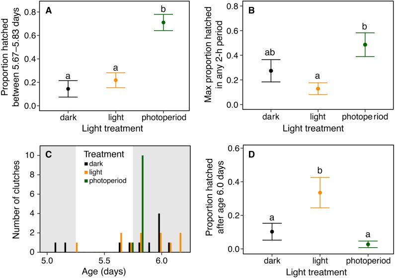
Hatching timing is under strong selection and environmentally cued in many species. Embryos use multiple sensory modalities to inform hatching timing and many have spontaneous hatching patterns adaptively synchronized to natural cycles. Embryos can also adaptively shift their hatching timing in response to environmental cues indicating immediate threats or opportunities. Such cued shifts in hatching are widespread among amphibians; however, we know little about what, if anything, regulates their spontaneous hatching. Moreover, in addition to selection on hatching timing, embryos may experience benefits or suffer costs due to the spatial orientation of hatching. Amphibian eggs generally lack internal constraints on hatching direction but embryos might, nonetheless, use external cues to inform hatching orientation. The terrestrial embryos of red-eyed treefrogs, Agalychnis callidryas, hatch rapidly and prematurely in response to vibrational cues in egg-predator attacks and hypoxia if flooded. Here we examined A. callidryas' use of light cues in hatching timing and orientation. To assess patterns of spontaneous hatching and the role of light cues in their diel timing, we recorded hatching times for siblings distributed across three light environments: continuous light, continuous dark, and a 12L:12D photoperiod. Under a natural photoperiod, embryos showed a clear diel pattern of synchronous hatching shortly after nightfall. Hatching was desynchronized in both continuous light and continuous darkness. It was also delayed by continuous light, but not accelerated by continuous dark, suggesting the onset of dark serves as a hatching cue. We examined hatching orientation and light as a potential directional cue for flooded embryos. Embryos flooded in their clutches almost always hatched toward open water, whereas individual eggs flooded in glass cups often failed to do so, suggesting the natural context provides a directional cue. To test if flooded embryos orient hatching toward light, we placed individual eggs in tubes with one end illuminated and the other dark, then flooded them and recorded hatching direction. Most embryos hatched toward the light, suggesting they use light as a directional cue. Our results support that A. callidryas embryos use light cues to inform both when and where to hatch. Both the spatial orientation of hatching and the timing of spontaneous hatching may affect fitness and be informed by cues in a broader range of species than is currently appreciated.
Keywords: Agalychnis callidryas; Anuran; Diel timing; Embryo behavior; Hatching; Hatching synchrony; Phenotypic plasticity; Photoperiod; Phototaxis.
Conflict of interest statement
The authors declare there are no competing interests.
Figures


References
-
- Altig R, McDiarmid RW. Tadpoles: the biology of anuran larvae. University of Chicago Press; Chicago: 1999. Body-plan development and morphology.
-
- Asoh K, Yoshikawa T. The role of temperature and embryo development time in the diel timing of spawning in a coral-reef damselfish with high-frequency spawning synchrony. Environmental Biology of Fishes. 2002;64:379–392. doi: 10.1023/A:1016177512353. - DOI
-
- Bates D, Maechler M, Bolker B, Walker S. Fitting linear mixed-effects models using {lme4} Journal of Statistical Software. 2015;67:1–48.
-
- Bradbury IR, Campana SE, Bentzen P, Snelgrove PVR. Synchronized hatch and its ecological significance in rainbow smelt Osmerus mordax in St. Mary’s Bay, Newfoundland. Limnology and Oceanography. 2004;49:3210–2315.
-
- Brännäs E. Influence of photoperiod and temperature on hatching and emergence of Baltic salmon (Salmo salar L.) Canadian Journal of Zoology. 1987;65:1503–1508. doi: 10.1139/z87-232. - DOI
LinkOut - more resources
Full Text Sources
