. 2018 Dec 5:10:51.
doi: 10.1186/s13099-018-0278-1. eCollection 2018.

Development of multiplex PCR and multi-color fluorescent in situ hybridization (m-FISH) coupled protocol for detection and imaging of multi-pathogens involved in inflammatory bowel disease

Affiliations

Development of multiplex PCR and multi-color fluorescent in situ hybridization (m-FISH) coupled protocol for detection and imaging of multi-pathogens involved in inflammatory bowel disease

Robert C Sharp et al. Gut Pathog. .

Abstract

Background: Several pathogens have been debated to play a role in inflammatory bowel disease (IBD) including Crohn's disease (CD). None of these pathogens have been investigated together in same clinical samples. We developed a multiplex PCR and multi-color fluorescent in situ hybridization (m-FISH) protocols for simultaneous detection of CD-associated pathogens including Mycobacterium avium subspecies paratuberculosis (MAP), Klebsiella pneumoniae, and adherent-invasive Escherichia coli strain LF82.

Methods: The multiplex PCR is based on 1-h DNAzol® extraction protocol modified for rapid extraction of bacterial DNA from culture, blood, and intestinal biopsies. Oligonucleotide primers sequences unique to these pathogens were evaluated individually and in combinations using bioinformatics and experimental approaches. m-FISH was based on fluorescent-tagged oligonucleotides and confocal scanning laser microscopy (CSLM).

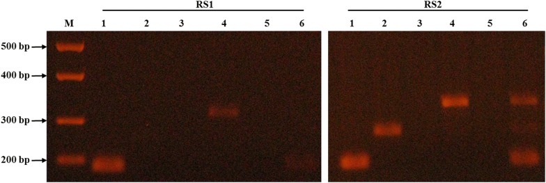
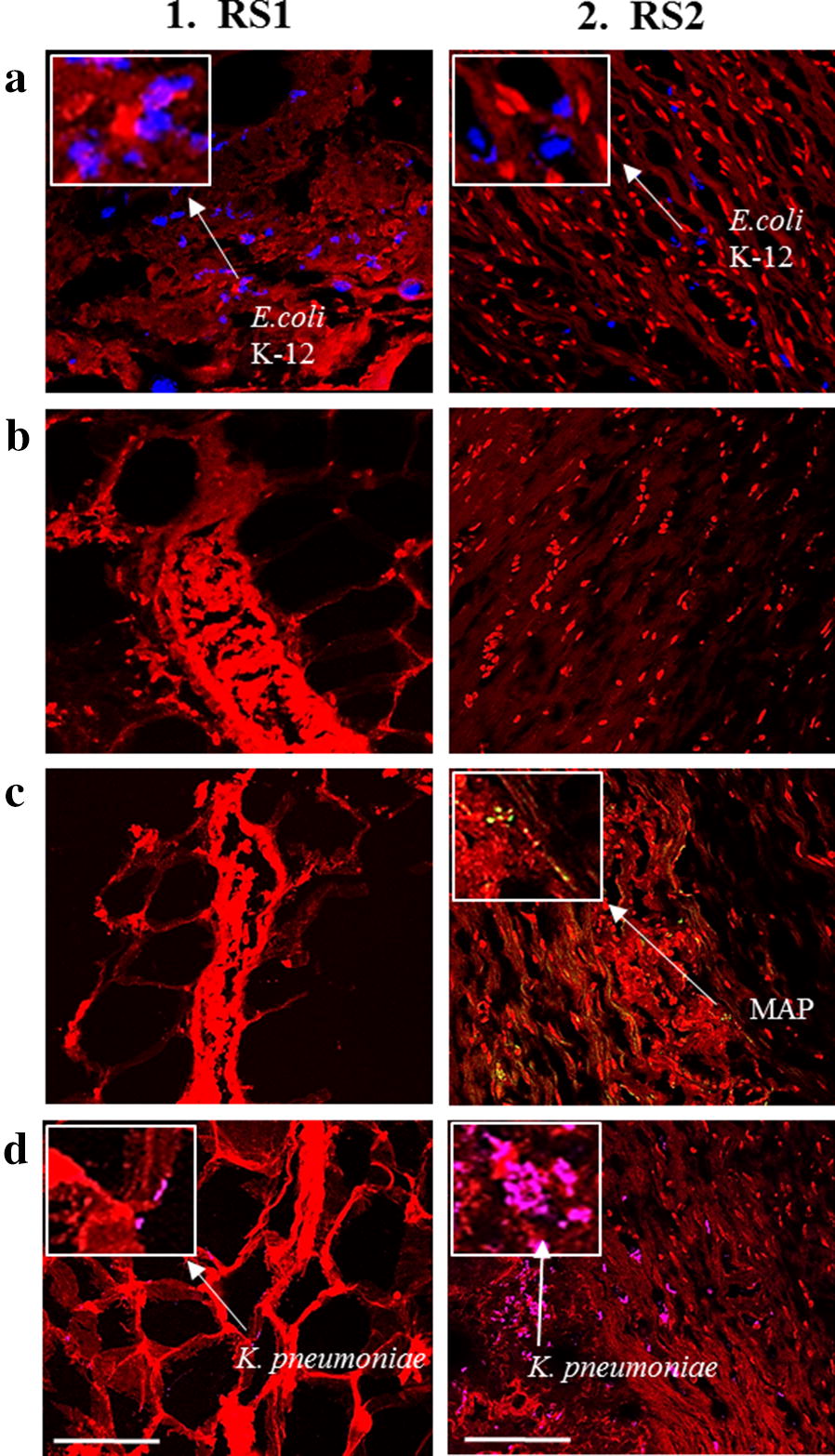
Results: Following several attempts, the concentration of the oligonucleotide primers and DNA templates and the PCR annealing temperatures were optimized. Multiplex PCR analyses revealed excellent amplification signal in trials where a single primer set and combinations of two and three primers sets were tested against a mixture of DNA from three different bacteria or a mixture of three bacterial cultures mixed in one tube before DNA extraction. Slides with individual and mixtures of bacterial cultures and intestinal tissue sections from IBD patients were tested by m-FISH and the CSLM images verified multiplex PCR results detected on 3% agarose gel.

Conclusion: We developed a 4-h multiplex PCR protocol, which was validated by m-FISH images, capable of detecting up to four genes from major pathogens associated with CD. The new protocol should serve as an excellent tool to support efforts to study multi-pathogens involved in CD and other autoimmune disease.

Keywords: Crohn’s disease; MAP; Multiplex PCR; Ulcerative colitis; m-FISH.


Figures

Fig. 1
Fig. 1
Specificity and sensitivity of the IS900 nPCR in DNA extracts from the modified DNAzol® and the standard phenol/chloroform/isoamyl-alcohol DNA extraction protocols. nPCR based on the IS900 specific to MAP was performed on DNA template extracted by the standard phenol/chloroform/isoamyl-alcohol DNA extraction (I) and the modified DNAzol® DNA extraction technique (II). A 298 bp fragment on 2% agarose gel is positive for MAP. a (1) Non-pathogenic E. coli strain K-12; (2) S. aureus; (3) L. monocytogenes; (4) K. pneumoniae; (5) M. smegmatis; (6) M. avium subspecies avium; (7) M. xenopi; (8) M. fortuitum subspecies fortuitum; (9) MAP Clinical Strain JF7. b (1) MAP Strain 1; (2) MAP Strain 3; (3) MAP Strain 8B; (4) MAP Para 18; (5) MAP UCF3; (6) MAP UCF5; (7) MAP UCF7; (8) MAP Linda; (9) MAP MS137. c Serial dilution of MAP UCF4 DNA concentrations were analyzed by nPCR. (1) 31.7 ng/μL; (2) 3.17 ng/μL; (3) 317 pg/μL; (4) 31.7 pg/μL; (5) 3.17 pg/μL; (6) 317 fg/μL; (7) 31.7 fg/μL; (8) 3.17 fg/μL; (9) 317 ag/μL; (10) 31.7 ag/μL. +: MAP UCF4; N: No DNA; M: molecular weight marker
Fig. 2
Fig. 2
Validation of multiplex PCR using individual and combined oligonucleotide primer sets and DNA extracts from mixed bacterial culture. Multiplex PCR was performed on DNA extracts from mixed bacterial cultures. DNA template was extracted using the modified DNAzol®. (1) Non-pathogenic E. coli strain K-12 18s primers were used (171 bp); (2) MAP UCF4 IS900 AV1/AV2 primers were used (298 bp); (3) K. pneumoniae 23s primers were used (493 bp); (4) Mycobacterium avium complex (MAC) IS1311 primers were used (534 bp); (5) a cocktail of the 4 primer sets mentioned above were used. M: DNA molecular weight marker
Fig. 3
Fig. 3
Validation of multiplex PCR using individual and combined oligonucleotide primer sets and DNA extracts from IBD tissue. Multiplex PCR was performed on DNA extracts from intestinal tissue samples. DNA template was extracted using the modified DNAzol®. RS1: ulcerative colitis (UC) patient; RS2: Crohn’s disease (CD) patient; (1) Non-pathogenic E. coli strain K-12 18s primers were used (171 bp); (2) MAP UCF4 IS900 AV1/AV2 primers were used (298 bp); (3) AIEC strain LF82 gipA primers were used (357 bp); (4) K. pneumoniae 23s primers were used (493 bp); (5) Mycobacterium avium complex (MAC) IS1311 primers were used (534 bp); (6) a cocktail of the 5 primer sets mentioned above were used. M: DNA molecular weight marker
Fig. 4
Fig. 4
Gram stain, acid-fast stain and m-FISH detection of bacterial cultures. Gram stain (1), acid-fast stain (2), and oligonucleotide m-FISH (3) images of bacterial cultures: a, b Non-pathogenic E. coli strain K-12; c MAP UCF4; and d K. pneumoniae. For -m-FISH detection: am-FISH using non-pathogenic E. coli K-12 18s probe labeled with AF647 fluorophore; bm-FISH using AIEC strain LF82 gipA probe labeled with AF568 fluorophore; cm-FISH using MAP UCF4 IS900 AV1 probe labeled with AF488 fluorophore; and d3 m-FISH using K. pneumoniae 23s probe labeled with AF546 fluorophore. All microscopic images were obtained at 1000 × magnification. White measurement bar found in D3 represents 100 μm. All m-FISH images were obtained using CSLM
Fig. 5
Fig. 5
Use of m-FISH using individual probes to detect multiple pathogens in intestinal tissue. Oligonucleotide m-FISH analysis in tissue sections from patients with UC (RS1) and CD (RS2) was done targeting for: a Non-pathogenic E. coli strain K-12; b AIEC strain LF82; c MAP; and d K. pneumoniae. Images illustrating DAPI are in red. The individual m-FISH probes that were used were: a non-pathogenic E. coli strain K-12 18s probe labeled with AF647 fluorophore; b AIEC strain LF82 gipA probe labeled with AF568 fluorophore; c MAP UCF4 IS900 AV1 probe labeled with AF488 fluorophore; and d K. pneumoniae 23s probe labeled with AF546 fluorophore. White measurement bars found in D1 and D2 represents 20 μm. All m-FISH images were obtained using CSLM
Fig. 6
Fig. 6
Use of m-FISH using combined probes to detect multiple pathogens in intestinal tissue from CD. Oligonucleotide m-FISH images of intestinal tissue sections from four CD patients (a RS3, b RS4, c RS5, and d RS6). A combined probes mixture including (2) EC647 (non-pathogenic E. coli strain K-12 18s probe labeled with AF647 fluorophore) and (3) MAP488 (MAP UCF4 IS900 AV1 probe labeled with AF488 fluorophore) were used in all tissue sections. (1) DAPI staining and (4) merged images between DAPI and the corresponding probe. White measurement bar located in B3 represents 20 μm. All m-FISH images were obtained using CSLM

References

    1. Khor B, Gardet A, Xavier RJ. Genetics and pathogenesis of inflammatory bowel disease. Nature. 2011;474:307–317. doi: 10.1038/nature10209. - DOI - PMC - PubMed
    1. Jostins L, Ripke S, Weersma RK, Duerr RH, McGovern DP, Hui KY, Lee JC, Schumm LP, Sharma Y, Anderson CA, Essers J, Cho JH. Host–microbe interactions have shaped the genetic architecture of inflammatory bowel disease. Nature. 2012;491:119–124. doi: 10.1038/nature11582. - DOI - PMC - PubMed
    1. Cho JH. The genetics and immunopathogenesis of inflammatory bowel disease. Nat Rev Immunol. 2008;8:458–466. doi: 10.1038/nri2340. - DOI - PubMed
    1. Farh KK, Marson A, Zhu J, Kleinewietfeld M, Housley WJ, Beik S, Shoresh N, Whitton H, Ryan RJ, Shishkin AA, Hatan M, Bernstein BE. Genetic and epigenetic fine mapping of causal autoimmune disease variants. Nature. 2015;518:337–343. doi: 10.1038/nature13835. - DOI - PMC - PubMed
    1. Sharp RC, Beg SA, Naser SA. Role of PTPN2/22 polymorphisms in pathophysiology of Crohn’s disease. World J Gastroenterol. 2018;24:657–670. doi: 10.3748/wjg.v24.i6.657. - DOI - PMC - PubMed