The role of the testa during the establishment of physical dormancy in the pea seed
- PMID: 30534972
- PMCID: PMC6526324
- DOI: 10.1093/aob/mcy213
The role of the testa during the establishment of physical dormancy in the pea seed
Abstract
Background: A water-impermeable testa acts as a barrier to a seed's imbibition, thereby imposing dormancy. The physical and functional properties of the macrosclereids are thought to be critical determinants of dormancy; however, the mechanisms underlying the maintenance of and release from dormancy in pea are not well understood.
Methods: Seeds of six pea accessions of contrasting dormancy type were tested for their ability to imbibe and the permeability of their testa was evaluated. Release from dormancy was monitored following temperature oscillation, lipid removal and drying. Histochemical and microscopic approaches were used to characterize the structure of the testa.
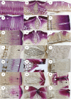
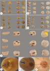
Key results: The strophiole was identified as representing the major site for the entry of water into non-dormant seeds, while water entry into dormant seeds was distributed rather than localized. The major barrier for water uptake in dormant seeds was the upper section of the macrosclereids, referred to as the 'light line'. Dormancy could be released by thermocycling, dehydration or chloroform treatment. Assays based on either periodic acid or ruthenium red were used to visualize penetration through the testa. Lipids were detected within a subcuticular waxy layer in both dormant and non-dormant seeds. The waxy layer and the light line both formed at the same time as the establishment of secondary cell walls at the tip of the macrosclereids.
Conclusions: The light line was identified as the major barrier to water penetration in dormant seeds. Its outer border abuts a waxy subcuticular layer, which is consistent with the suggestion that the light line represents the interface between two distinct environments - the waxy subcuticular layer and the cellulose-rich secondary cell wall. The mechanistic basis of dormancy break includes changes in the testa's lipid layer, along with the mechanical disruption induced by oscillation in temperature and by a decreased moisture content of the embryo.
Keywords: Pisum sativum seed; Hardseedness; light line; macrosclereid; physical dormancy; seed coat; subcuticular lipids; testa; water permeability.
© The Author(s) 2018. Published by Oxford University Press on behalf of the Annals of Botany Company. All rights reserved. For permissions, please e-mail: journals.permissions@oup.com.
Figures


Similar articles
-
Variation in wild pea (Pisum sativum subsp. elatius) seed dormancy and its relationship to the environment and seed coat traits.PeerJ. 2019 Jan 14;7:e6263. doi: 10.7717/peerj.6263. eCollection 2019. PeerJ. 2019. PMID: 30656074 Free PMC article.
-
Anatomy and Histochemistry of Seed Coat Development of Wild (Pisum sativum subsp. elatius (M. Bieb.) Asch. et Graebn. and Domesticated Pea (Pisum sativum subsp. sativum L.).Int J Mol Sci. 2021 Apr 27;22(9):4602. doi: 10.3390/ijms22094602. Int J Mol Sci. 2021. PMID: 33925728 Free PMC article.
-
A Combined Comparative Transcriptomic, Metabolomic, and Anatomical Analyses of Two Key Domestication Traits: Pod Dehiscence and Seed Dormancy in Pea (Pisum sp.).Front Plant Sci. 2017 Apr 25;8:542. doi: 10.3389/fpls.2017.00542. eCollection 2017. Front Plant Sci. 2017. PMID: 28487704 Free PMC article.
-
The role of the testa during development and in establishment of dormancy of the legume seed.Front Plant Sci. 2014 Jul 17;5:351. doi: 10.3389/fpls.2014.00351. eCollection 2014. Front Plant Sci. 2014. PMID: 25101104 Free PMC article. Review.
-
Underlying Biochemical and Molecular Mechanisms for Seed Germination.Int J Mol Sci. 2022 Jul 31;23(15):8502. doi: 10.3390/ijms23158502. Int J Mol Sci. 2022. PMID: 35955637 Free PMC article. Review.
Cited by
-
Variation in wild pea (Pisum sativum subsp. elatius) seed dormancy and its relationship to the environment and seed coat traits.PeerJ. 2019 Jan 14;7:e6263. doi: 10.7717/peerj.6263. eCollection 2019. PeerJ. 2019. PMID: 30656074 Free PMC article.
-
Domestication has altered the ABA and gibberellin profiles in developing pea seeds.Planta. 2023 Jun 23;258(2):25. doi: 10.1007/s00425-023-04184-2. Planta. 2023. PMID: 37351659 Free PMC article.
-
Genetic and transcriptomic analysis of lentil seed imbibition and dormancy in relation to its domestication.Plant Genome. 2025 Jun;18(2):e70021. doi: 10.1002/tpg2.70021. Plant Genome. 2025. PMID: 40164967 Free PMC article.
-
Anatomy and Histochemistry of Seed Coat Development of Wild (Pisum sativum subsp. elatius (M. Bieb.) Asch. et Graebn. and Domesticated Pea (Pisum sativum subsp. sativum L.).Int J Mol Sci. 2021 Apr 27;22(9):4602. doi: 10.3390/ijms22094602. Int J Mol Sci. 2021. PMID: 33925728 Free PMC article.
-
Analysis of Elymus nutans seed coat development elucidates the genetic basis of metabolome and transcriptome underlying seed coat permeability characteristics.Front Plant Sci. 2022 Aug 18;13:970957. doi: 10.3389/fpls.2022.970957. eCollection 2022. Front Plant Sci. 2022. PMID: 36061807 Free PMC article.
References
-
- Agbo GN, Hosfield MA, Uebersax MA, Klomparens K. 1987. Seed microstructure and its relationship to water uptake in isogenic lines and a cultivar of dry beans (Phaseolus vulgaris L.). Food Microstructure 6: 91–102.
-
- Baskin CC. 2003. Breaking physical dormancy in seeds: focussing on the lens. New Phytologist 158: 229–232.
-
- Baskin JM, Baskin CC. 2000. Evolutionary considerations of claims for physical dormancy-break by microbial action and abrasion by soil particles. Seed Science Research 10: 409–413.
-
- Baskin CC, Baskin JM. 2014. Seeds: ecology, biogeography and evolution of dormancy and germination, 2nd edn. New York: Academic Press.
-
- Baskin JM, Baskin CC, Li X. 2000. Taxonomy, anatomy and evolution of physical dormancy in seeds. Plant Species Biology 15: 139–152.
Publication types
MeSH terms
Substances
LinkOut - more resources
Full Text Sources

