. 2019 Mar 1;22(3):232-246.
doi: 10.1093/ijnp/pyy098.

Small Extracellular Vesicles in Rat Serum Contain Astrocyte-Derived Protein Biomarkers of Repetitive Stress

Affiliations

Small Extracellular Vesicles in Rat Serum Contain Astrocyte-Derived Protein Biomarkers of Repetitive Stress

Cristóbal Gómez-Molina et al. Int J Neuropsychopharmacol. .

Abstract

Background: Stress precipitates mood disorders, characterized by a range of symptoms present in different combinations, suggesting the existence of disease subtypes. Using an animal model, we previously described that repetitive stress via restraint or immobilization induced depressive-like behaviors in rats that were differentially reverted by a serotonin- or noradrenaline-based antidepressant drug, indicating that different neurobiological mechanisms may be involved. The forebrain astrocyte protein aldolase C, contained in small extracellular vesicles, was identified as a potential biomarker in the cerebrospinal fluid; however, its specific origin remains unknown. Here, we propose to investigate whether serum small extracellular vesicles contain a stress-specific protein cargo and whether serum aldolase C has a brain origin.

Methods: We isolated and characterized serum small extracellular vesicles from rats exposed to restraint, immobilization, or no stress, and their proteomes were identified by mass spectrometry. Data available via ProteomeXchange with identifier PXD009085 were validated, in part, by western blot. In utero electroporation was performed to study the direct transfer of recombinant aldolase C-GFP from brain cells to blood small extracellular vesicles.

Results: A differential proteome was identified among the experimental groups, including aldolase C, astrocytic glial fibrillary acidic protein, synaptophysin, and reelin. Additionally, we observed that, when expressed in the brain, aldolase C tagged with green fluorescent protein could be recovered in serum small extracellular vesicles.

Conclusion: The protein cargo of serum small extracellular vesicles constitutes a valuable source of biomarkers of stress-induced diseases, including those characterized by depressive-like behaviors. Brain-to-periphery signaling mediated by a differential molecular cargo of small extracellular vesicles is a novel and challenging mechanism by which the brain might communicate health and disease states to the rest of the body.

Keywords: biomarkers; exosomes; stress subtypes.


Figures

Figure 1.
Figure 1.
Characterization of small extracellular vesicles (sEVs). (a) Experimental design: rats were habituated in their home cages for 7 days. Then, stress by restraint or immobilization or no stress was applied for 10 days. (b) Images of sEVs obtained by electron microscopy. Bar scale = 100 nm. (c) sEV size and concentration detected by the nanoparticle tracking analysis in 4 to 6 independent samples per animal group. (d) Western blots and (e) the corresponding densitometric quantification of changes in the content of the indicated proteins in the experimental conditions: no stress (NS), restraint (R), or immobilization (I). Equal amounts of proteins were loaded per lane (including astrocyte homogenates, AH). The data represent mean ± SEM. n = 10 for CD-63, n = 8 and 5 for flotillin after restraint and immobilization, respectively; n = 7 for TSG-101. #P < .05 in the Mann-Whitney test (used to compare pairs of data, i.e., restraint vs immobilization). *P < .05, **P < .01 in a Wilcoxon-signed rank test (to compare with a hypothetical value of 0 (no change).
Figure 2.
Figure 2.
Proteomic analysis of extracellular vesicles (EVs) show that after restraint, proteins that are expressed in the brain are increased. (a) Venn analysis of the identified proteins in the experimental conditions. (b) Venn analysis of the identified proteins including the proteome of astrocyte small extracellular vesicles (sEVs). (c) The percentage of expression of the identified proteins in different body organs is shown (obtained with DAVID Bioinformatics Resources 6.8 [NIAID, NIH]).
Figure 3.
Figure 3.
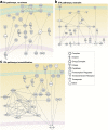
Network analysis of proteins identified exclusively in one of the experimental conditions (no stress, restraint, or immobilization). Dotted lines indicate indirect interactions; solid lines represent direct interactions between proteins. (a) Network analysis of the proteins identified uniquely in the small extracellular vesicles (sEVs) of the no stress group indicated the convergence of survival pathways/cell protection and intracellular trafficking. ABCB8, ATP-binding cassette, subfamily B (MDR/TAP), member 8; aconitase, aconitate hydratase; actin, G-actin; Akt, AKT1/2/3; AP1B1, adaptor protein complex AP-1, β 1 subunit; ARF1, ADP-ribosylation factor 1; CLTA, clathrin, light chain A; DBNL, drebrin-like protein; DNM3, dynamin 3; dynamin, dynamin GTPase; FYB, FYN binding protein; NUCB2, nucleobindin 2; PARK7, parkinson protein 7; PBXIP1, pre-B-cell leukemia transcription factor interacting protein 1; RPS20, ribosomal protein S20; SCAMP1, secretory carrier membrane protein 1; SKAP2, src kinase associated phosphoprotein 2; SLC2A4, solute carrier family 2 (facilitated glucose transporter), member 4; SMPD3, sphingomyelin phosphodiesterase 3; SNAP23, synaptosomal-associated protein 23; STX4, syntaxin 4; STXBP2, syntaxin binding protein 2; Tpm1, tropomyosin 1α; Tpm2, tropomyosin 2β; Tpm3, tropomyosin 3; VAMP7, vesicle-associated membrane protein 7; ZYX, zyxin. (b) Network analysis of proteins identified uniquely in the sEVs after restraint indicated proteins related to cellular stress responses. 26 s Proteasome, Proteasome; BAHCC1, BAH domain and coiled-coil containing 1; C1QTNF5, C1q and tumor necrosis factor related protein 5; CEP250, centrosomal protein 2; CHD8, chromodomain helicase DNA binding protein 8; CLIC1, chloride intracellular channel 1; ERK, p42/44 MAPK; HECTD1, HECT domain-containing E3 ubiquitin protein ligase 1; Histone h3, Histone H3B; HK1, hexokinase 1; Hsp90, Heat shock protein 90 kDa; HSPA2, heat shock 70 kDa protein 2; HSPA8, Heat Shock 70kD Protein 8; IDH1, isocitrate dehydrogenase 1; KHSRP, KH-type splicing regulatory protein; MAP1B, microtubule-associated protein 1B; MAPT, microtubule-associated protein tau; MTMR1, Myotubularin Related Protein 1; NCL, nucleolin; NOA1, nitric oxide associated 1; POLR1A, polymerase (RNA) I polypeptide A; Rnr, 47S Pre-rRNA, Ribosomal; RPS23, ribosomal protein S23; SPAG9, sperm associated antigen 9; SYPL2, synaptophysin-like 2; TNRC6A, trinucleotide repeat containing 6A; TUFM, Tu translation elongation factor. (c) Network analysis of the proteins identified uniquely in the sEVs after immobilization paradigm indicated protein components related to cancer and hematological and immunological diseases. 20 S proteasome, 20 S Core Complex; ABCC1, ATP-binding cassette, subfamily C (CFTR/MRP); CCT3:, chaperonin-containing TCP1, subunit 3 (gamma); CLIP2, CAP-GLY domain-containing linker protein 2; elastase, serine elastase; OC100360846/Psmb6, proteasome subunit β 6; MYH14, myosin, heavy chain 14; MYH7, beta cardiac myosin heavy chain; MYH8, myosin, heavy chain 8; NFkB (complex), transcription factor nuclear factor κ b; PGRMC1, progesterone receptor membrane component 1; PRKCB, protein kinase C β II; PSMA3, proteasome subunit α 3; PSMA5, proteasome subunit α 5; PSMA6, proteasome subunit α 6; PSMB1, proteasome subunit β 1; PSMB3, proteasome subunit β 3; PSMB4, proteasome subunit β 3; PSMB5, poteasome subunit β 5; PSMB7, proteasome subunit β 7; PSME1, Proteasome activator pa28 α subunit; SERPINA6, Corticosteroid-binding globulin, serine (or cysteine) peptidase inhibitor; SERPINB10, serine (or cysteine) peptidase inhibitor, clade B; SERPINB6, serine (or cysteine) peptidase inhibitor, clade B, member 6; TUBB2A, tubulin, β 2A; TUBB4B, tubulin, β 4B; TUBB6, tubulin, β 6.
Figure 4.
Figure 4.
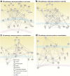
Network analysis of proteins in serum small extracellular vesicles (sEVs) and in astrocyte sEVs. Dotted lines indicate indirect interactions; solid lines represent direct interaction between proteins. (a) Network analysis of proteins obtained from serum sEVs of no stress group also present in astrocytes sEVs indicated protein components related to metabolic disease, endocrine system disorders, and gastrointestinal disease. A2 M, Alpha-2-microglobulin; AHSG, alpha-2-HS-GLYCOPROTEIN; ALB, Albumin 1; Alpha 1 antitrypsin, Alpha 1 antitrypsin; ANXA6, Annexin VI; APOA1, apolipoprotein A-I; ATP1A3, Na+/K+ ATPase alpha3; B2 M, beta-2-MICROGLOBULIN; CLU, clusterin; ERK1/2, p42/44 MAPK; HABP2, hyaluronic acid binding protein 2; Hba1/Hba2, hemoglobin, α 1, hemoglobin, α 2; HBB, Beta-globin; HDL, high-density lipoprotein; HP, haptoglobin; HPX, hemopexin; ITIH1, inter-alpha trypsin inhibitor, heavy chain 1; ITIH3, inter-alpha trypsin inhibitor, heavy chain 3; ITIH4, inter-alpha trypsin inhibitor, heavy chain 4; LDL, low-density lipoprotein; MVP, major vault protein; RAB7A, member RAS oncogene family, RAB7A; SERPINA1, serine (or cysteine) peptidase inhibitor, clade A; SERPINA3, serine (or cysteine) peptidase inhibitor, clade A, member 3 M; SERPINC1, serine (or cysteine) peptidase inhibitor, clade C, member 1; TTR, Transthyretin isomer 1. (b) Network analysis of the proteins obtained from serum sEVs after restraint also present in astrocytic sEVs indicating the protein components related to metabolic disease, neurological disease, and psychological disorders. A2 M, alpha-2-macroglobulin; AHSG, alpha-2-HS-GLYCOPROTEIN; Akt, AKT1/2/3; ALB, Albumin 1; APOA1, apolipoprotein A-I; APOE, Apolipoprotein E; CLU, clusterin; CP, ceruloplasmin; CPN1, carboxypeptidase N; FGA, Fibrinogen A α; Hba1/Hba2, hemoglobin, α 1, hemoglobin, α 2; HBB, Beta-globin; HDL, high-density lipoprotein; HPX, hemopexin; ITIH1, inter-alpha trypsin inhibitor, heavy chain 1; ITIH3, inter-alpha trypsin inhibitor, heavy chain 3; ITIH4, inter-alpha trypsin inhibitor, heavy chain 4; LDL, low-density lipoprotein; MFGE8, milk fat globule-EGF factor 8 protein; PLG, plasminogen; RAB7A, member RAS oncogene family, RAB7A; SERPINA1, serine (or cysteine) peptidase inhibitor, clade A; SERPINC1, serine (or cysteine) peptidase inhibitor, clade C, member 1; TTR, Transthyretin isomer 1. (c) Network analysis of the proteins obtained from the serum sEVs after immobilization paradigm also present in astrocytic sEVs. Network related to neurological and metabolic diseases. A2 M, alpha-2-macroglobulin; AFM, serum albumin, α-Alb; Akt, AKT1/2/3; ALB, albumin 1; APOA1, apolipoprotein A-I; CLU, clusterin; CP, ceruloplasmin; FGA, alpha-fibrinogen; Hba1/Hba2, hemoglobin, α 1, hemoglobin, α 2; HBB, Beta-globin; HDL, high-density lipoprotein; hemoglobin, hemoglobin; HP, haptoglobin; HPX, hemopexin; ITIH1, inter-alpha trypsin inhibitor, heavy chain 1; ITIH3, inter-alpha trypsin inhibitor, heavy chain 3; ITIH4, inter-alpha trypsin inhibitor, heavy chain 4; SERPINA1, serine (or cysteine) peptidase inhibitor, clade A, member 1; SERPINC1, serine (or cysteine) peptidase inhibitor, clade C; SERPINF1, serine (or cysteine) peptidase inhibitor, clade F, member 1; TTR, transthyretin isomer 1. (d) Network related to inflammatory response. ACTB, actin β; Actin, G-actin; ANXA1, annexin A1; ANXA4, annexin A4; ANXA5, annexin A5; B2 M, beta-2-MICROGLOBULIN; CFL1, cofilin 1; CKB, Creatine kinase b chain; Cofilin; EMILIN1, elastin microfibril interfacer 1; ERK1/2, p42/44 MAPK; Erm; F Actin, Filamentous actin; GSN, Gelsolin; MSN, moesin; MYADM, myeloid-associated differentiation marker; PFN1, profilin 1; PPIA, peptidylprolyl isomerase A; PPIB, peptidylprolyl isomerase B; PRDX5, Peroxiredoxin 5; RAB7A, member RAS oncogene family, RAB7A; WDR1, WD repeat domain 1.
Figure 5.
Figure 5.
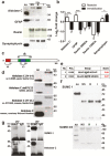
Proteins expressed in astrocytes (aldolase C and glial fibrillary acid protein [GFAP]) are differentially present in small extracellular vesicles (sEVs) obtained after restraint or immobilization. (a) Representative western blots of the indicated proteins and (b) corresponding densitometric quantification. (c) Representation of the antigenic peptides that were used for generation of the Santa Cruz antibodies (A and C), while B indicates the antigenic peptide that was used for the generation of the Abcam antibody. Finally, a monoclonal antibody generated against a crude homogenate of cerebellum and electrosensory lateral line lobe from a weakly electric fish was used (gift from Dr. Richard Hawkes, University of Calgary). (d) Western blots of aldolase C using antibodies of the different sources and directed against different antigenic peptides in the protein. (e) Possible small ubiquitin-like modifier (SUMO)ylating residues of aldolase C are indicated. (f) Western blots of sEVs using anti-SUMO antibodies. (g) Western blots of immunoprecipitated aldolase C (left) and SUMO (right) using SUMO antibody (upper) or aldolase C antibody (lower). The IgG lane indicates the respective control conditions. AH, astrocyte homogenates; I, stress by immobilization; NS, no stress; R, stress by restraint. N = 5–7; #P < .05; ##P < .0 in Mann-Whitney tests (to compare pairs of data, i.e., restraint vs immobilization); *P < .05 in a Wilcoxon-signed rank test (to compare fold changes with a hypothetical value of 1 (no change).
Figure 6.
Figure 6.
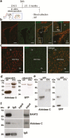
Aldolase C tagged with green fluorescent protein (C-GFP) expressed in forebrain astrocytes is detected in extracellular vesicles (EVs) isolated from the blood. (a) Scheme of the in utero electroporation. Forebrain astrocytes were transduced by in utero electroporation with aldolase C-GFP or GFP. Plasmids were injected into the left lateral ventricle on embryonic day 18.5. The orientation of the electrodes used to apply the voltage pulse is shown. (b) Immunohistofluorescent detection of glial fibrillary acid protein (GFAP) and GFP in coronal brain slices indicated that cells positive for both proteins were detected in the borders of the lateral ventricles (LV, indicated by arrows), and (c) in the hilus of the dentate gyrus (DG). Gr, granule cells. (d) Aldolase C (left) or GFP (right) was detected in astrocyte homogenates (AH) electroporated with aldolase C-GFP or in sEVs isolated from the serum of these animals. Note that in the sEVs, the modified form of aldolase C was detected (~ 55 kDa), as well as the recombinant protein (~ 70 kDa), which was also visible with the GFP antibody. (e) Aldolase C (left) or GFP (right) was detected in the astrocyte homogenates (AH) that were electroporated with GFP or in extracellular vesicles (sEVs) isolated from the serum of these animals. Note that in sEVs, the modified form of aldolase C was detected (~ 55 kDa), while in these animals, no GFP could be detected in the EVs. Observations were conducted in n = 5 independent animal groups (n = 4 rats per group for blood collection). (f) EVs bearing the glial glutamate transporter EAAT2 in their membrane are enriched in aldolase C. EVs were immunoisolated in nondenaturating conditions with an antibody directed against an extracellular epitope of EAAT2. The input lane was loaded with 8% of the EVs used for the precipitation procedure (i.e., 20 µg). Note that EAAT2 can only be detected in the input after overexposure of the same blot (right lanes), revealing a huge enrichment of EAAT2-bearing vesicles in the isolated material that in turn also contains higher aldolase C levels compared with the input EVs.

References

    1. Akil H, Gordon J, Hen R, Javitch J, Mayberg H, McEwen B, Meaney MJ, Nestler EJ (2018) Treatment resistant depression: a multi-scale, systems biology approach. Neurosci Biobehav Rev 84:272–288. - PMC - PubMed
    1. Ampuero E, Luarte A, Santibañez M, Varas-Godoy M, Toledo J, Diaz-Veliz G, Cavada G, Rubio FJ, Wyneken U (2015) Two chronic stress models based on movement restriction in rats respond selectively to antidepressant drugs: aldolase C as a potential biomarker. Int J Neuropsychopharmacol 18:pyv038. - PMC - PubMed
    1. Aydın EP, Genç A, Dalkıran M, Uyar ET, Deniz İ, Özer ÖA, Karamustafalıoğlu KO (2018) Thioredoxin is not a marker for treatment-resistance depression but associated with cognitive function: an rTMS study. Prog Neuropsychopharmacol Biol Psychiatry 80:322–328. - PubMed
    1. Bruni JE. (1998) Ependymal development, proliferation, and functions: a review. Microsc Res Tech 41:2–13. - PubMed
    1. Cai L, Tu J, Song L, Gao Z, Li K, Wang Y, Liu Y, Zhong F, Ge R, Qin J, Ding C, He F (2017a) Proteome-wide mapping of endogenous SUMOylation sites in mouse testis. Mol Cell Proteomics 16:717–727. - PMC - PubMed

Publication types

MeSH terms