Comparative Study
. 2019 Jan 1;11(1):295-318.
doi: 10.1093/gbe/evy265.

Delegating Sex: Differential Gene Expression in Stolonizing Syllids Uncovers the Hormonal Control of Reproduction

Affiliations
Comparative Study

Delegating Sex: Differential Gene Expression in Stolonizing Syllids Uncovers the Hormonal Control of Reproduction

Patricia Álvarez-Campos et al. Genome Biol Evol. .

Abstract

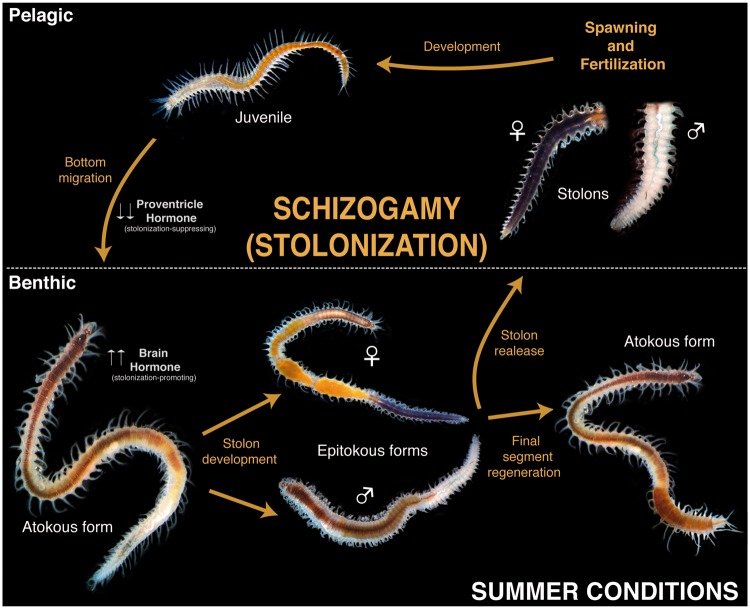
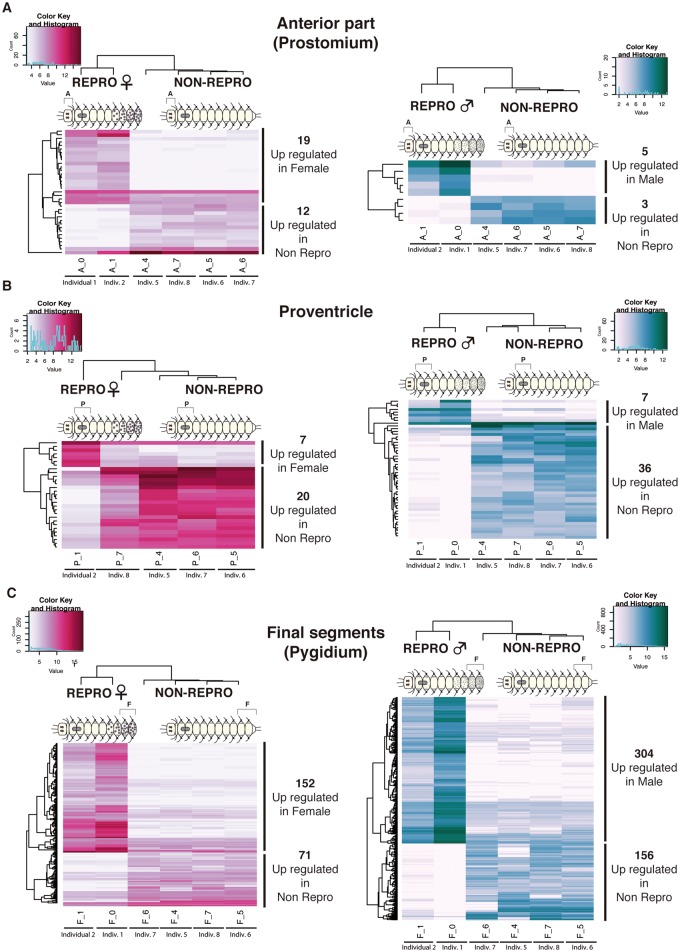
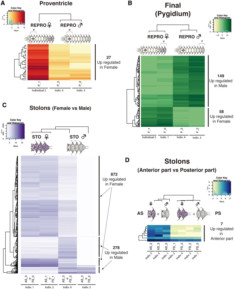
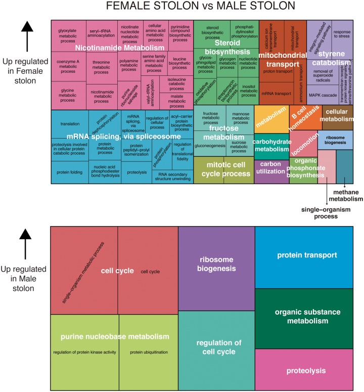
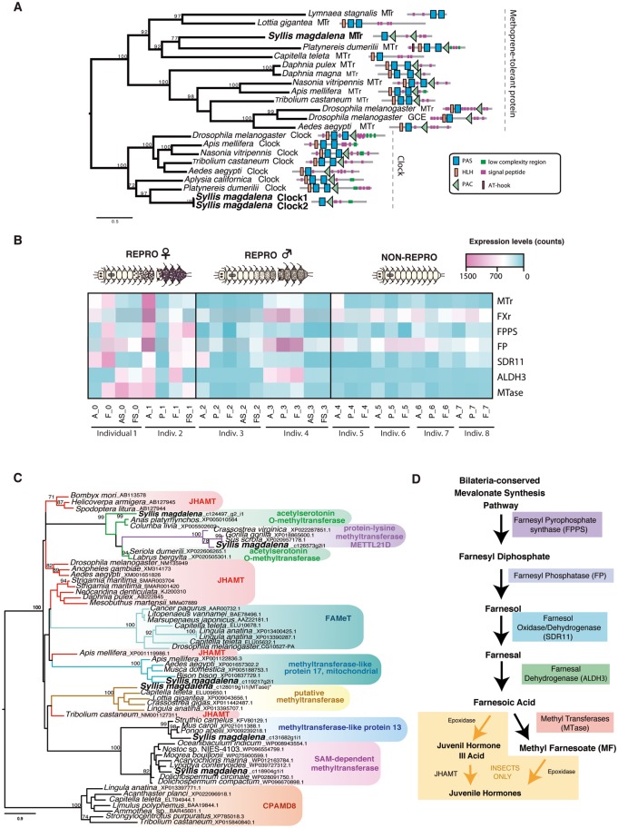
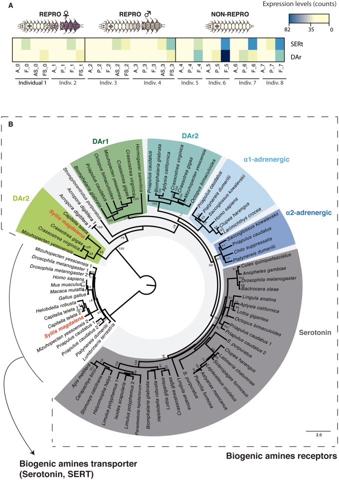
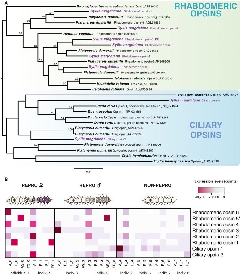
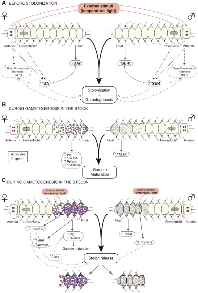
Stolonization in syllid annelids is a unique mode of reproduction among animals. During the breeding season, a structure resembling the adult but containing only gametes, called stolon, is formed generally at the posterior end of the animal. When stolons mature, they detach from the adult and gametes are released into the water column. The process is synchronized within each species, and it has been reported to be under environmental and endogenous control, probably via endocrine regulation. To further understand reproduction in syllids and to elucidate the molecular toolkit underlying stolonization, we generated Illumina RNA-seq data from different tissues of reproductive and nonreproductive individuals of Syllis magdalena and characterized gene expression during the stolonization process. Several genes involved in gametogenesis (ovochymase, vitellogenin, testis-specific serine/threonine-kinase), immune response (complement receptor 2), neuronal development (tyrosine-protein kinase Src42A), cell proliferation (alpha-1D adrenergic receptor), and steroid metabolism (hydroxysteroid dehydrogenase 2) were found differentially expressed in the different tissues and conditions analyzed. In addition, our findings suggest that several neurohormones, such as methyl farnesoate, dopamine, and serotonin, might trigger stolon formation, the correct maturation of gametes and the detachment of stolons when gametogenesis ends. The process seems to be under circadian control, as indicated by the expression patterns of r-opsins. Overall, our results shed light into the genes that orchestrate the onset of gamete formation and improve our understanding of how some hormones, previously reported to be involved in reproduction and metamorphosis processes in other invertebrates, seem to also regulate reproduction via stolonization.


Figures

<sc>Fig</sc>. 1.
Fig. 1.
—Syllinae schizogamous reproductive cycle (stolonization) using light microscope pictures of Syllis magdalena.
<sc>Fig</sc>. 2.
Fig. 2.
—Light microscopy pictures of Syllis magdalena stolonizing female (A) and male (B). Confocal micrographs of S. magdalena stolonizing female (C), male (D), female stolon (E), and male stolons (F). Arrows in (A)–(D) pointing to the eyes of stolons (e). Arrows in (E) pointing to antennae (a). Arrow in (F) pointing to the regeneration of the final segments in the stock (rfs).
<sc>Fig</sc>. 3.
Fig. 3.
—Light and electron microscopy pictures of the anterior part of the female and male stolons of Syllis magdalena. (A, B) Location of antennae (a) and the two pairs of eyes (e) in the female stolon. (C) Transmission electron micrographs of the epithelium of the female stolon showing the muscle fibers (mf), granular cells (gc), and epithelial cells (ec). (D) Developing oocytes showing nucleolate (nu) nucleus (n), ooplasm filled with yolk platelets, and microvilli (m) contacting close oocytes. Note the muscle fibers (mf), nurse cells (nc), and the digestive epithelium (dc) surrounding the germinal epithelium. (EF) Germinal epithelium (ge) in the male stolon. The stolonal epithelium is comprised by a layer of epithelial cells (ec) with interspersed granular cells (gc), and a layer of muscle fibers (mf); spermatogonia develop in the germinal epithelium (gc) below. The digestive cells (dc) lay below the germinal epithelium.
<sc>Fig</sc>. 4.
Fig. 4.
—Heatmaps of differentially expressed genes (annotated and not annotated genes) from pairwise comparisons of somatic tissues between reproductive (both female and male) and nonreproductive individuals. Anterior part tissue comparisons (A), proventricle comparisons (B), and final segments comparisons (C). Different colors indicate relative expression levels based on raw read counts (see color key and histogram on each). Similarity in expression patterns between genes and individuals is represented by clustering. A, anterior part; P, proventricle; F, final segments.
<sc>Fig</sc>. 5.
Fig. 5.
—Heatmaps based on differentially expressed genes (annotated and not annotated genes) from pairwise comparisons of somatic tissues between females and males (A, B) and reproductive tissues (stolons) (C, D). Proventricle comparisons (A), final segments comparisons (B), female and male stolons comparisons (C), and anterior and posterior parts of stolons (female and male together) (D). Different colors indicate relative expression levels based on raw read counts (see color key and histogram on each). Similarity in expression patterns between genes and individuals is represented by clustering. A, anterior part; P, proventricle; F, final segments; AS, anterior half of stolon; FS, posterior part of stolon.
<sc>Fig</sc>. 6.
Fig. 6.
—Gene ontology treemaps for annotated differentially expressed genes in female stolons versus male stolons. The GO terms downregulated in female stolons are upregulated accordingly in male stolons.
<sc>Fig</sc>. 7.
Fig. 7.
—(A) Phylogenetic reconstruction of the protein alignment for methoprene-tolerant receptor (MTr) found in our samples. (B) Heatmap showing the relative levels of expression in the different tissues and conditions analyzed of the transcripts that putatively may be involved in the synthesis of the neurohormone methyl farnesoate (MF): MTr, Farnesol oxidase/dehydrogenase (SDR11), Farnesal dehydrogenases (ALDHE3), the differentially expressed transcript Farnesyl pyrophosphate synthase (FPPS) and putative methyl transferase (Mtase). Different colors indicate relative expression levels based on raw read counts (see color key and histogram on each). (C) Phylogenetic reconstruction of the differentially expressed MTases in the female stolon. (D) Synthesis pathway of MF and JH in arthropods. A, anterior part; P, proventricle; F, final segments; AS, anterior half of stolon; FS, posterior part of stolon.
<sc>Fig</sc>. 8.
Fig. 8.
—Phylogenetic reconstruction (A) and heatmap of relative levels of expression in all the tissues and conditions (B) of the genes dopamine receptor (DAr) and serotonin transporter (SERT). Different colors indicate relative expression levels based on raw read counts (see color key and histogram on each). A, anterior part; P, proventricle; F, final segments; AS, anterior half of stolon; FS, posterior part of stolon.
<sc>Fig</sc>. 9.
Fig. 9.
—Phylogenetic reconstruction of the protein alignment for the different opsin genes (rhabdomeric and ciliary) found in our samples (A) and levels of expression of all of them in the different tissues and conditions analyzed (B). Rhabdomeric opsin 5 appeared differentially expressed in the anterior part of stolons. A, anterior part; P, proventricle; F, final segments; AS, anterior half of stolon; FS, posterior part of stolon. Different colors indicate relative expression levels based on raw read counts (see color key and histogram on each).
<sc>Fig</sc>. 10.
Fig. 10.
—Proposed multihormonal model for stolonization control. During the breeding season, DA and SER levels increase in response to external stimuli triggering gamete production in the final segments (up-regulation of DAr and SERt) (A). Once stolonization has begun, a variety of other hormones and proteins are produced for the correct development and maturation of gametes (up-regulation of Vtg, OVOCH Relaxin, Follistatin, and TSSK) (B). Finally, when gametes are completely mature and also as a response to external stimuli (up regulation of r-opsins), MF or a similar hormone (up-regulation of FPP and Mtransf) is produced to allow stolon release (C). Dashed lines represent hypothesized involvement of molecules, whereas solid lines represent molecule expression results observed in our study.

References

    1. Abascal F, Zardoya R, Posada D.. 2005. ProtTest: selection of best-fit models of protein evolution. Bioinformatics 21(9):2104–2105. - PubMed
    1. Abeloos M. 1950. Régénération et stolonisation épigame chez l’Annélide Syllis prolifera Krohn. C R Acad Sci (Comptes rendus de l'Academie des Sciences). 230:1899–1900. - PubMed
    1. Agassiz A. 1863. On alternate generation in annelids, and the embryology of Autolytus cornutus. Boston J Nat Hist. 7:384–409.
    1. Altschul SF, et al. 1997. Gapped Blast and PSIBlast: a new generation of protein database search programs. Nucleic Acids Res. 25(17):3389–3402. - PMC - PubMed
    1. Anders S, Huber W.. 2010. Differential expression analysis for sequence count data. Genome Biol. 11(10):R106.. - PMC - PubMed

Publication types