

Expression levels and co‑targets of miRNA‑126‑3p and miRNA‑126‑5p in lung adenocarcinoma tissues: Αn exploration with RT‑qPCR, microarray and bioinformatic analyses
- PMID: 30535503
- PMCID: PMC6313014
- DOI: 10.3892/or.2018.6901
Expression levels and co‑targets of miRNA‑126‑3p and miRNA‑126‑5p in lung adenocarcinoma tissues: Αn exploration with RT‑qPCR, microarray and bioinformatic analyses
Abstract
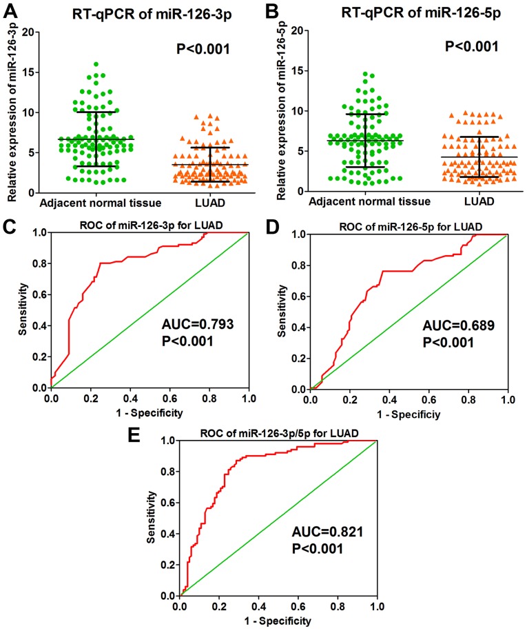
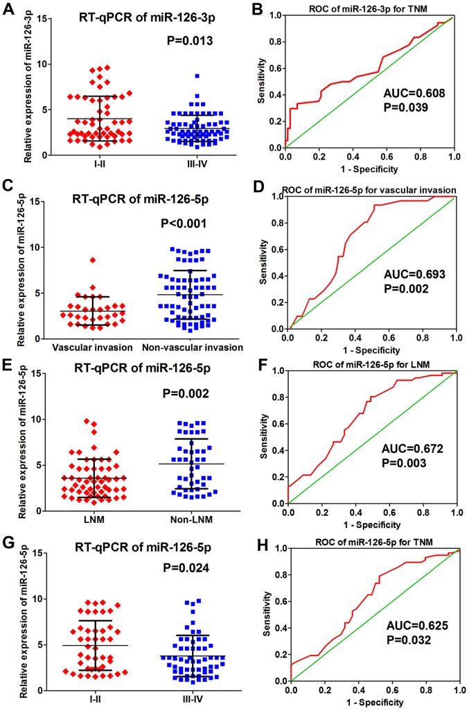
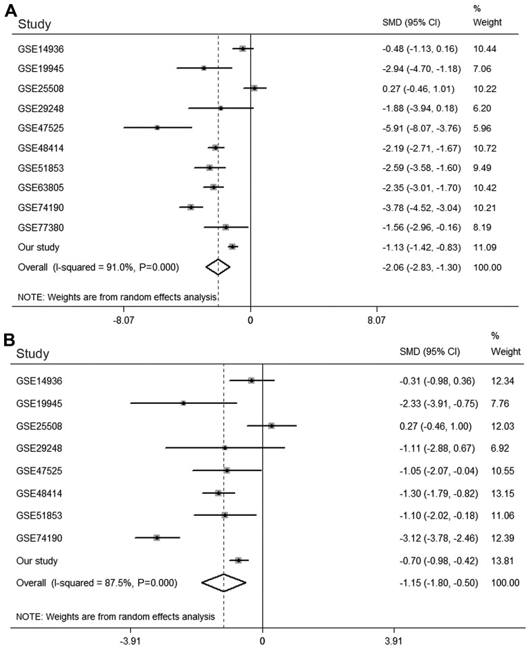
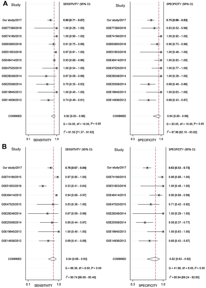
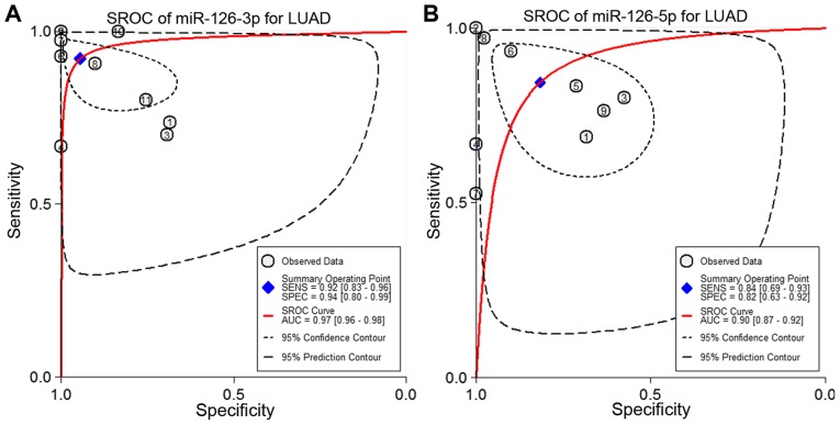
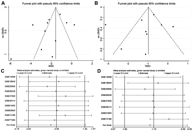
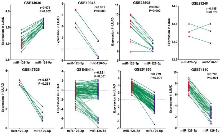
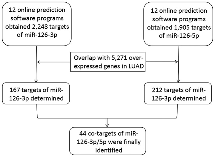
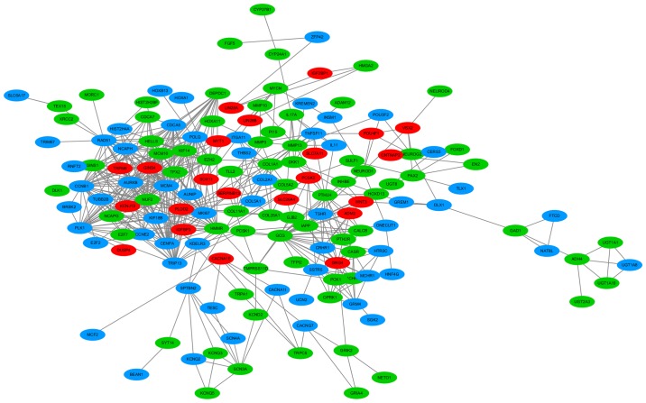
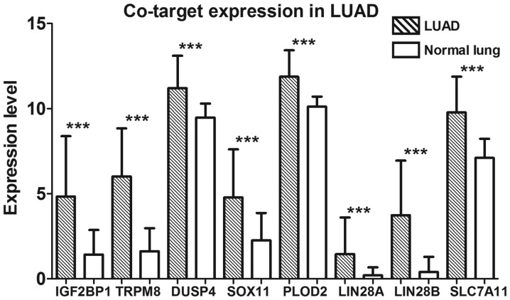
Lung adenocarcinoma (LUAD) is the most common histological subtype of lung cancer. Previous studies have found that many microRNAs (miRNAs), including miRNA‑126‑3p, may play a critical role in the development of LUAD. However, no study of LUAD has researched the synergistic effects and co‑targets of both miRNA‑126‑3p and miRNA‑126‑5p. The present study used real‑time quantitative polymerase chain reaction (RT‑qPCR) to explore the expression values of miRNA‑126‑3p and miRNA‑126‑5p in 101 LUAD and 101 normal lung tissues. Ten relevant microarray datasets were screened to further validate the expression levels of miRNA‑126‑3p and ‑5p in LUAD. Twelve prediction tools were employed to obtain potential targets of miRNA‑126‑3p and miRNA‑126‑5p. The results showed that both miRNA‑126‑3p and ‑5p were expressed significantly lower in LUAD. A significant positive correlation was also present between miRNA‑126‑3p and ‑5p expression in LUAD. In addition, lower expression of miRNA‑126‑3p and ‑5p was indicative of vascular invasion, lymph node metastasis (LNM), and a later tumor/node/metastasis (TNM) stage of LUAD. The authors obtained 167 targets of miRNA‑126‑3p and 212 targets of miRNA‑126‑5p; 44 targets were co‑targets of both. Eight co‑target genes (IGF2BP1, TRPM8, DUSP4, SOX11, PLOD2, LIN28A, LIN28B and SLC7A11) were initially identified as key genes in LUAD. The results of the present study indicated that the co‑regulation of miRNA‑126‑3p and miRNA‑126‑5p plays a key role in the development of LUAD, which also suggests a fail‑proof mode between miRNA‑3p and miRNA‑126‑5p.
Figures
Similar articles
-
Downregulation of HOXA3 in lung adenocarcinoma and its relevant molecular mechanism analysed by RT-qPCR, TCGA and in silico analysis.Int J Oncol. 2018 Oct;53(4):1557-1579. doi: 10.3892/ijo.2018.4508. Epub 2018 Jul 30. Int J Oncol. 2018. PMID: 30066858 Free PMC article.
-
Identifying the miRNA signature associated with survival time in patients with lung adenocarcinoma using miRNA expression profiles.Sci Rep. 2017 Aug 8;7(1):7507. doi: 10.1038/s41598-017-07739-y. Sci Rep. 2017. PMID: 28790336 Free PMC article.
-
miR-9-5p Promotes Lung Adenocarcinoma Cell Proliferation, Migration and Invasion by Targeting ID4.Technol Cancer Res Treat. 2021 Jan-Dec;20:15330338211048592. doi: 10.1177/15330338211048592. Technol Cancer Res Treat. 2021. PMID: 34723712 Free PMC article.
-
MiRNAs in Lung Adenocarcinoma: Role, Diagnosis, Prognosis, and Therapy.Int J Mol Sci. 2023 Aug 27;24(17):13302. doi: 10.3390/ijms241713302. Int J Mol Sci. 2023. PMID: 37686110 Free PMC article. Review.
-
[Progress in Research on the Cribriform Component in Lung Adenocarcinoma].Zhongguo Fei Ai Za Zhi. 2020 Jul 20;23(7):621-625. doi: 10.3779/j.issn.1009-3419.2020.101.19. Epub 2020 May 26. Zhongguo Fei Ai Za Zhi. 2020. PMID: 32450628 Free PMC article. Review. Chinese.
Cited by
-
Potential upstream lncRNA-miRNA-mRNA regulatory network of the ferroptosis-related gene SLC7A11 in renal cell carcinoma.Transl Androl Urol. 2023 Jan 30;12(1):33-57. doi: 10.21037/tau-22-663. Epub 2023 Jan 11. Transl Androl Urol. 2023. PMID: 36760866 Free PMC article.
-
The Role of Thyroid Hormone Synthesis Gene-Related miRNAs Profiling in Structural and Functional Changes of The Thyroid Gland Induced by Excess Iodine.Biol Trace Elem Res. 2024 Feb;202(2):580-596. doi: 10.1007/s12011-023-03691-3. Epub 2023 May 27. Biol Trace Elem Res. 2024. PMID: 37243879
-
miR-126-5p enhances radiosensitivity of lung adenocarcinoma cells by inhibiting EZH2 via the KLF2/BIRC axis.J Cell Mol Med. 2022 May;26(9):2529-2542. doi: 10.1111/jcmm.17135. Epub 2022 Mar 24. J Cell Mol Med. 2022. PMID: 35322532 Free PMC article.
-
Linc00426 accelerates lung adenocarcinoma progression by regulating miR-455-5p as a molecular sponge.Cell Death Dis. 2020 Dec 11;11(12):1051. doi: 10.1038/s41419-020-03259-2. Cell Death Dis. 2020. PMID: 33311443 Free PMC article.
-
Tumor-suppressive action of miR-30a-5p in lung adenocarcinoma correlates with ABL2 inhibition and PI3K/AKT pathway inactivation.Clin Transl Oncol. 2024 Feb;26(2):398-413. doi: 10.1007/s12094-023-03255-w. Epub 2023 Jul 21. Clin Transl Oncol. 2024. PMID: 37479901
References
-
- Blanco-Prieto S, Barcia-Castro L, Páez de la Cadena M, Rodríguez-Berrocal FJ, Vázquez-Iglesias L, Botana-Rial MI, Fernández-Villar A, De Chiara L. Relevance of matrix metalloproteases in non-small cell lung cancer diagnosis. BMC Cancer. 2017;17:823. doi: 10.1186/s12885-017-3842-z. - DOI - PMC - PubMed
Publication types
MeSH terms
Substances
LinkOut - more resources
Full Text Sources
Medical
Research Materials
Miscellaneous
