

Periodontal Ehlers-Danlos syndrome is associated with leukoencephalopathy
- PMID: 30535813
- PMCID: PMC6411670
- DOI: 10.1007/s10048-018-0560-x
Periodontal Ehlers-Danlos syndrome is associated with leukoencephalopathy
Abstract
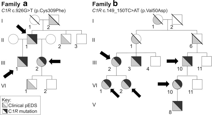
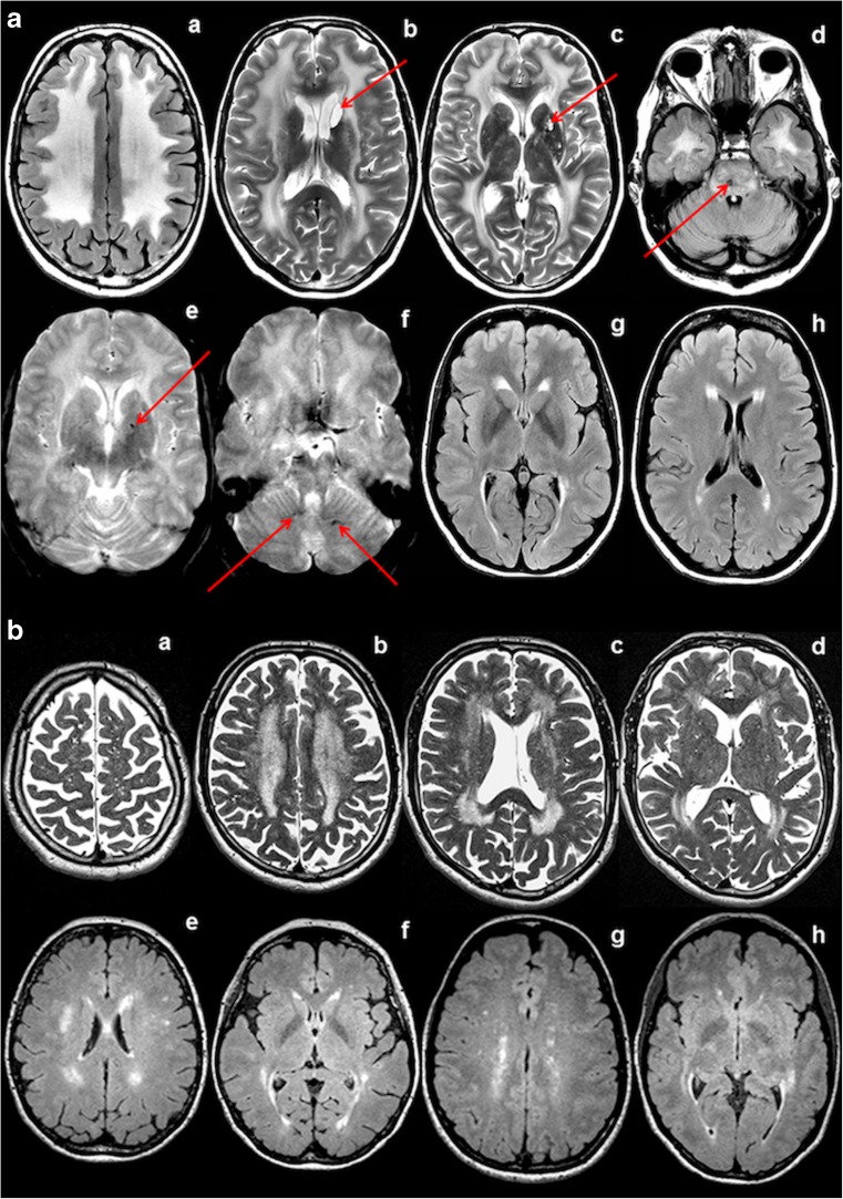
Here, we report brain white matter alterations in individuals clinically and genetically diagnosed with periodontal Ehlers-Danlos syndrome, a rare disease characterized by premature loss of teeth and connective tissue abnormalities. Eight individuals of two families clinically diagnosed with periodontal Ehlers-Danlos syndrome were included in the present study and underwent general physical, dental, and neurological examination. Whole exome sequencing was performed, and all patients included in the study underwent MRI of the brain. Whole exome sequencing revealed heterozygous C1R mutations c.926G>T (p.Cys309Phe, Family A) and c.149_150TC>AT (p.Val50Asp, Family B). All adult individuals (n = 7; age range 31 to 68 years) investigated by MRI had brain white matter abnormalities. The MRI of one investigated child aged 8 years was normal. The MRI pattern was suggestive of an underlying small vessel disease that is progressive with age. As observed in other leukoencephalopathies related to microangiopathies, the extent of the white matter changes was disproportionate to the neurologic features. Medical history revealed recurrent headaches or depression in some cases. Neurological examination was unremarkable in all individuals but one had mild cognitive decline and ataxia and experienced a seizure. The observation that periodontal Ehlers-Danlos syndrome caused by missense mutations in C1R is consistently associated with a leukoencephalopathy opens a new pathogenic link between the classical complement pathway, connective tissue, brain small vessels, and brain white matter abnormalities.
Keywords: Complement 1; Ehlers–Danlos; Leukoencephalopathy; Periodontitis; Small vessel disease.
Conflict of interest statement
Disclosures
The authors report no disclosures relevant to the manuscript.
Figures
References
-
- Kapferer-Seebacher I, Pepin M, Werner R, Aitman TJ, Nordgren A, Stoiber H, Thielens N, Gaboriaud C, Amberger A, Schossig A, Gruber R, Giunta C, Bamshad M, Bjorck E, Chen C, Chitayat D, Dorschner M, Schmitt-Egenolf M, Hale CJ, Hanna D, Hennies HC, Heiss-Kisielewsky I, Lindstrand A, Lundberg P, Mitchell AL, Nickerson DA, Reinstein E, Rohrbach M, Romani N, Schmuth M, Silver R, Taylan F, Vandersteen A, Vandrovcova J, Weerakkody R, Yang M, Pope FM, Molecular Basis of Periodontal EDSC. Byers PH, Zschocke J. Periodontal Ehlers-Danlos syndrome is caused by mutations in C1R and C1S, which encode subcomponents C1r and C1s of complement. Am J Hum Genet. 2016;99(5):1005–1014. - PMC - PubMed
-
- Lacroix M, Tessier A, Dumestre-Perard C, Vadon-Le Goff S, Gout E, Bruckner-Tuderman L, Kiritsi D, Nystrom A, Ricard-Blum S, Moali C, Hulmes DJS, Thielens NM. Interaction of complement defence collagens C1q and mannose-binding lectin with BMP-1/tolloid-like proteinases. Sci Rep. 2017;7(1):16958. - PMC - PubMed
-
- Echaniz-Laguna A, de Saint-Martin A, Lafontaine AL, Tasch E, Thomas P, Hirsh E, Marescaux C, Andermann F. Bilateral focal polymicrogyria in Ehlers-Danlos syndrome. Arch Neurol. 2000;57(1):123–127. - PubMed
-
- Ezzeddine H, Sabouraud P, Eschard C, El Tourjuman O, Bednarek N, Motte J. Bilateral frontal polymicrogyria and Ehlers-Danlos syndrome. Arch Pediatr. 2005;12(2):173–175. - PubMed
Publication types
MeSH terms
Substances
Grants and funding
LinkOut - more resources
Full Text Sources
Medical
Research Materials
Miscellaneous
