. 2019:1877:61-76.
doi: 10.1007/978-1-4939-8861-7_4.

BH3 Profiling: A Functional Assay to Measure Apoptotic Priming and Dependencies

Affiliations

BH3 Profiling: A Functional Assay to Measure Apoptotic Priming and Dependencies

Cameron Fraser et al. Methods Mol Biol. 2019.

Abstract

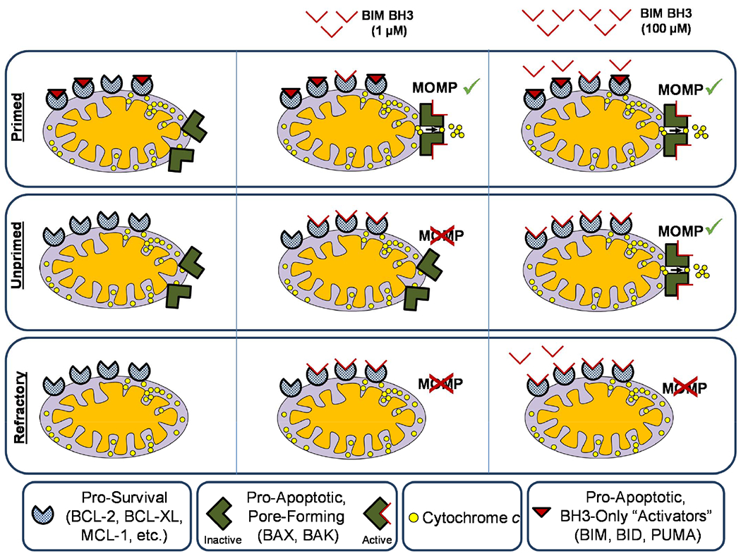
Apoptosis (programmed cell death) is activated by a wide variety of cellular stresses or insults and is vital for proper mammalian development as well as the maintenance of organismal homeostasis. The apoptosis pathway is also engaged by many common types of anticancer therapies and ionizing radiation, which contributes to the regressions of tumors or the toxic side effects of treatment. Due to the importance of maintaining healthy cell survival or the efficient clearance of cancer cells, the BH3 profiling assay was developed to functionally measure the state of the apoptosis pathway in any given cells. This assay involves the exposure of cellular mitochondria, where the BCL-2 family of proteins resides and controls the commitment to apoptosis, to proapoptotic BH3 peptides that mimic the activity of endogenous proapoptotic proteins. By using either activator or sensitizer peptides, the level of mitochondrial apoptotic priming (proximity to the threshold at which a cell commits to cell death) or dependence on prosurvival BCL-2 family proteins can be determined. Described here are two methods of BH3 profiling that can enable a user to make these functional measurements, which can be useful for predicting cellular responses to proapoptotic stressors or therapeutics (BH3 mimetics) that inhibit the activity of prosurvival proteins.

Keywords: BCL-2 family; Chemotherapeutics; MOMP; Mitochondrial apoptotic priming.


Figures

Figure 1:
Figure 1:. The mitochondrial apoptosis pathway.
In this simplified schematic, cellular stress or damage signals [1] unleash pro-apoptotic proteins (BH3-only “activators” of apoptosis) [2], which can either be bound and sequestered by pro-survival proteins such as BCL-2, BCL-XL or MCL-1 [3] or activate BAX and/or BAK [4]. Activation of BAX or BAK causes mitochondrial outer membrane permeabilization (MOMP), resulting in the release of cytochrome c from mitochondria and consequent activation of caspases [5] for dismantling of the cell.
Figure 2:
Figure 2:. Potential states of apoptotic priming and competence as determined by BH3 Profiling.
In the BH3 Profiling assay, cells are permeabilized and treated with titrated doses of pro-apoptotic peptides derived from the BH3 domains of BH3-only proteins. Cytochrome c staining is used to monitor mitochondrial permeabilization, which occurs upon activation of BAX or BAK and consequent release of cytochrome c to trigger apoptosis. Mitochondria that have a small reserve of unbound pro-survival proteins are quickly and fully depolarized (loss of cytochrome c) by even low doses of peptides (1-3μM) during the 1 hour assay and are thus considered primed for apoptosis. Mitochondria that have a large reserve are unprimed and only respond to large doses of peptides (100μM). Mitochondria that lack BAX or BAK are apoptosis incompetent. The pretreatment level of apoptotic priming can determine cell fate in response to pro-apoptotic signaling.
Figure 3:
Figure 3:. Binding pattern for BCL-2 family interactions.
Binding affinities for interactions between BH3 peptides derived from activator or sensitizer BH3-only proteins and their pro-survival and pro-apoptotic partners. (syn) designates a synthetic peptide.
Figure 4:
Figure 4:. Diagnosing dependencies on pro-survival BCL-2 family proteins.
Using the BH3-only sensitizer peptides, the BH3 profiling assay can detect if a cell has a particular dependence on one or multiple anti-apoptotic BCL-2 family proteins for survival. The sensitizer peptides can selectively inhibit particular anti-apoptotic proteins. If a cell is primed, but does not express a particular anti-apoptotic protein at high levels, the sensitizer peptides will have little effect. If a cell is primed and expresses BCL-2 or MCL-1 at a high level, treatment with the BAD or NOXA peptides respectively will inhibit the anti-apoptotic and allow for the activation of Bax/Bak. Treatment with BAD on an MCL-1 dependent cell will have no effect, as will treatment with NOXA on a BCL-2 dependent cell.
Figure 5:
Figure 5:. JC1 BH3 profiling workflow.
Cells are collected and prepared into a single-cell suspension. Cells are then permeabilized with digitonin and stained with JC1. Cells are loaded into a 384 well plate that contains BH3 peptides and fluorescence is monitored in a plate reader over 180 minutes.
Figure 6:
Figure 6:. JC1 BH3 profiles of parental HeLa and BAX/BAK knockout HeLa cells.
Each line on the three-hour time course (left) reflects the fluorescence for a single treatment. The corresponding bar in the bar graph (right) is the difference between the AUC of the negative control and the AUC of the treatment from the three-hour time course. The HeLa parental cell lines are an example of a moderately primed cell line. When BAX and BAK are knocked out from that cell line, the cells become apoptosis refractory, no longer responding to BH3-only peptides BIM and BID.
Figure 7:
Figure 7:. Flow cytometry BH3 profiling (iBH3) workflow.
Cells are collected and prepared into a single-cell suspension. Cells of interest are then labeled with fluorescent antibodies and washed. Cells are then permeabilized with digitonin and loaded into a 384 well plate that contains BH3 peptides and incubated for 60 minutes. Cells are then fixed and stained for cytochrome c. Cells are analyzed by flow cytometry.
Figure 8:
Figure 8:. Flow cytometry-based iBH3 profiling analysis.
Single cell analysis enables the user to gate on cellular content, specifically singlets, using forward and side-scatter variables. Specific subtypes of cells can then be identified and gated on for further analysis. After selecting the population of interest, the differences in cytochrome c negative cells can be examined between different treatments and populations. In this example, only DAPI positive cells (permeabilized cells with full DNA content) are included in the cytochrome c panel. The negative control treatment (DMSO) population is mostly cytochrome c positive. Increasing dosage of the BIM-BH3 peptide results in increased cytochrome c negativity. Alamethicin causes total cytochrome c loss.
Figure 9:
Figure 9:
Effective peptide concentration decreases as cell concentration increases.
Figure 10:
Figure 10:
Increased incubation temperature, time increases cytochrome c release.

References

    1. Moldoveanu T, Follis AV, Kriwacki RW, Green DR (2014) Many players in BCL-2 family affairs. Trends Biochem Sci 1–11 . doi: 10.1016/j.tibs.2013.12.006 - DOI - PMC - PubMed
    1. Tait SWG, Green DR (2013) Mitochondrial regulation of cell death. Cold Spring Harb Perspect Biol 5: . doi: 10.1101/cshperspect.a008706 - DOI - PMC - PubMed
    1. Czabotar PE, Westphal D, Dewson G, et al. (2013) Bax Crystal Structures Reveal How BH3 Domains Activate Bax and Nucleate Its Oligomerization to Induce Apoptosis. Cell 152:519–531 . doi: 10.1016/j.cell.2012.12.031 - DOI - PubMed
    1. Sarosiek K, Chi X, Bachman J, et al. (2013) BID Preferentially Activates BAK while BIM Preferentially Activates BAX, Affecting Chemotherapy Response. Mol Cell 51: . doi: 10.1016/j.molcel.2013.08.048 - DOI - PMC - PubMed
    1. Antignani A, Youle RJ (2006) How do Bax and Bak lead to permeabilization of the outer mitochondrial membrane? Curr Opin Cell Biol 18:685–9 . doi: 10.1016/j.ceb.2006.10.004 - DOI - PubMed

Substances

LinkOut - more resources