Bilateral projections to the thalamus from individual neurons in the inferior colliculus
- PMID: 30536721
- PMCID: PMC6368862
- DOI: 10.1002/cne.24600
Bilateral projections to the thalamus from individual neurons in the inferior colliculus
Abstract
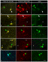
The medial geniculate body (MG) receives a large input from the ipsilateral inferior colliculus (IC) and a smaller but substantial input from the contralateral IC. Both crossed and uncrossed inputs comprise a large percentage of glutamatergic cells and a smaller percentage of GABAergic cells. We used double labeling with fluorescent retrograde tracers to identify individual IC cells that project bilaterally to the MGs in adult guinea pigs. We also used immunohistochemistry for glutamic acid decarboxylase to distinguish GABAergic from glutamatergic cells that project bilaterally to the MG. We found cells in the IC that contained both retrograde tracers, indicating that they project bilaterally. Across cases, the bilaterally projecting cells constituted up to 37% of the cells that project to the ipsilateral MG and up to 73% of the cells that project to the contralateral MG. GABAergic cells averaged 20% of the bilaterally-projecting population. We conclude that a population of IC cells sends branching axonal projections to innervate the MG bilaterally. Most of the neurons in this population are glutamatergic, with a minority that are GABAergic. A mixed projection, with glutamatergic cells outnumbering GABAergic cells, originates from each of the major IC subdivisions (central nucleus, dorsal cortex, and lateral cortex). The bilaterally projecting cells are likely to serve functions different from the larger unilateral projections, perhaps synchronizing activity on the two sides of the auditory brain.
Keywords: GABA; collateral; inferior colliculus; medial geniculate body; synchronization, RRID:AB_2278725, RRID:AB_260754.
© 2018 Wiley Periodicals, Inc.
Conflict of interest statement
Figures


Similar articles
-
Cholinergic cells in the tegmentum send branching projections to the inferior colliculus and the medial geniculate body.Neuroscience. 2011 Apr 14;179:120-30. doi: 10.1016/j.neuroscience.2011.01.044. Epub 2011 Jan 26. Neuroscience. 2011. PMID: 21277952 Free PMC article.
-
Separate projections from the inferior colliculus to the cochlear nucleus and thalamus in guinea pigs.Hear Res. 2004 May;191(1-2):67-78. doi: 10.1016/j.heares.2004.01.009. Hear Res. 2004. PMID: 15109706
-
Unilateral and bilateral projections from cortical cells to the inferior colliculus in guinea pigs.Brain Res. 2005 Apr 25;1042(1):62-72. doi: 10.1016/j.brainres.2005.02.015. Brain Res. 2005. PMID: 15823254
-
Subtypes of GABAergic cells in the inferior colliculus.Hear Res. 2019 May;376:1-10. doi: 10.1016/j.heares.2018.10.001. Epub 2018 Oct 4. Hear Res. 2019. PMID: 30314930 Free PMC article. Review.
-
Anatomy and physiology of binaural hearing.Audiology. 1991;30(3):125-34. doi: 10.3109/00206099109072878. Audiology. 1991. PMID: 1953442 Review.
Cited by
-
Neuropeptide Y mRNA expression in the aging inferior colliculus of fischer brown norway rats.Front Aging Neurosci. 2025 Jul 23;17:1626021. doi: 10.3389/fnagi.2025.1626021. eCollection 2025. Front Aging Neurosci. 2025. PMID: 40771198 Free PMC article.
-
A novel class of inferior colliculus principal neurons labeled in vasoactive intestinal peptide-Cre mice.Elife. 2019 Apr 18;8:e43770. doi: 10.7554/eLife.43770. Elife. 2019. PMID: 30998185 Free PMC article.
-
Hierarchical differences in the encoding of sound and choice in the subcortical auditory system.J Neurophysiol. 2023 Mar 1;129(3):591-608. doi: 10.1152/jn.00439.2022. Epub 2023 Jan 18. J Neurophysiol. 2023. PMID: 36651913 Free PMC article.
-
Ototoxicity-related changes in GABA immunolabeling within the rat inferior colliculus.Hear Res. 2024 Oct;452:109106. doi: 10.1016/j.heares.2024.109106. Epub 2024 Aug 21. Hear Res. 2024. PMID: 39181061
-
Cholinergic modulation in the vertebrate auditory pathway.Front Cell Neurosci. 2024 Jun 19;18:1414484. doi: 10.3389/fncel.2024.1414484. eCollection 2024. Front Cell Neurosci. 2024. PMID: 38962512 Free PMC article. Review.
References
-
- Aitkin LM, Dickhaus H, Schult W, & Zimmermann M (1978). External nucleus of inferior colliculus: auditory and spinal somatosensory afferents and their interactions. Journal of Neurophysiology, 41(4), 837–847. - PubMed
-
- Aitkin LM, Gates GR, & Phillips SC (1984). Responses of neurons in inferior colliculus to variations in sound-source azimuth. Journal of Neurophysiology, 52(1), 1–17. - PubMed
Publication types
MeSH terms
Grants and funding
LinkOut - more resources
Full Text Sources

