. 2018 Dec 11;19(1):218.
doi: 10.1186/s13059-018-1594-y.

A novel FLI1 exonic circular RNA promotes metastasis in breast cancer by coordinately regulating TET1 and DNMT1

Affiliations

A novel FLI1 exonic circular RNA promotes metastasis in breast cancer by coordinately regulating TET1 and DNMT1

Naifei Chen et al. Genome Biol. .

Abstract

Background: Friend leukemia virus integration 1 (FLI1), an ETS transcription factor family member, acts as an oncogenic driver in hematological malignancies and promotes tumor growth in solid tumors. However, little is known about the mechanisms underlying the activation of this proto-oncogene in tumors.

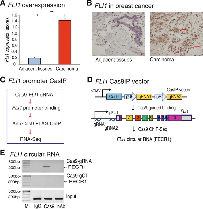
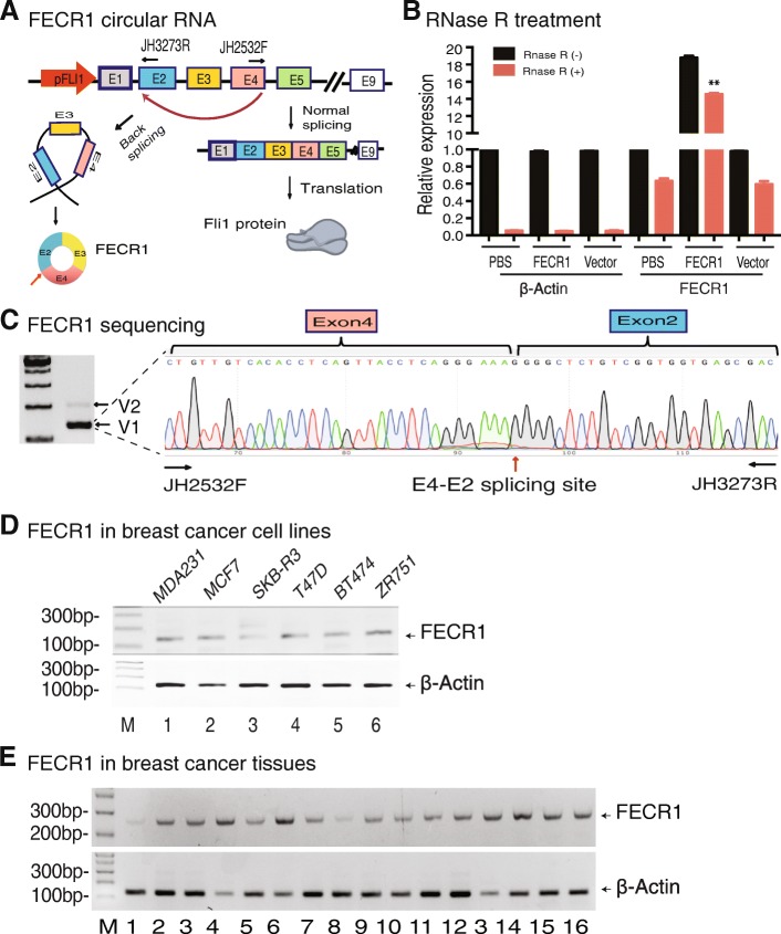
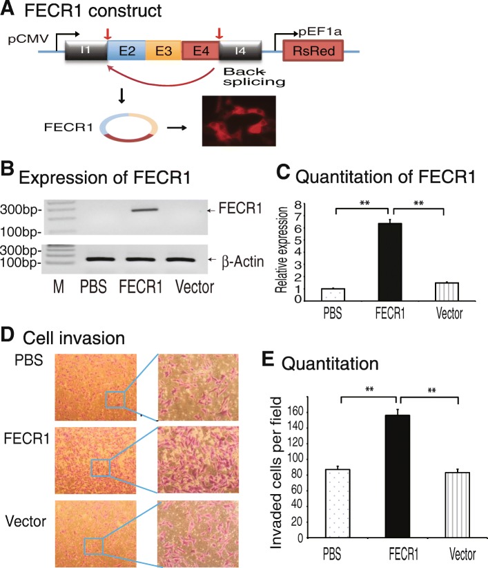
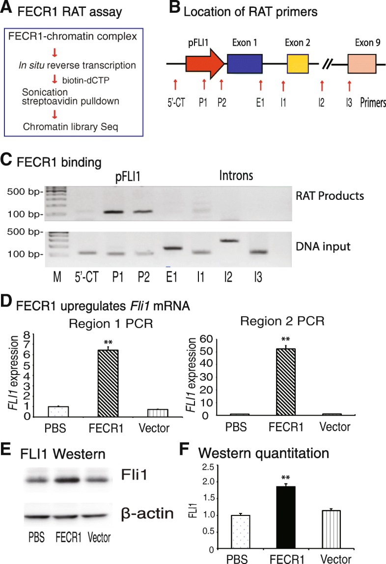
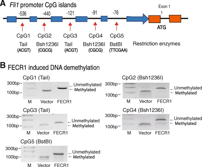
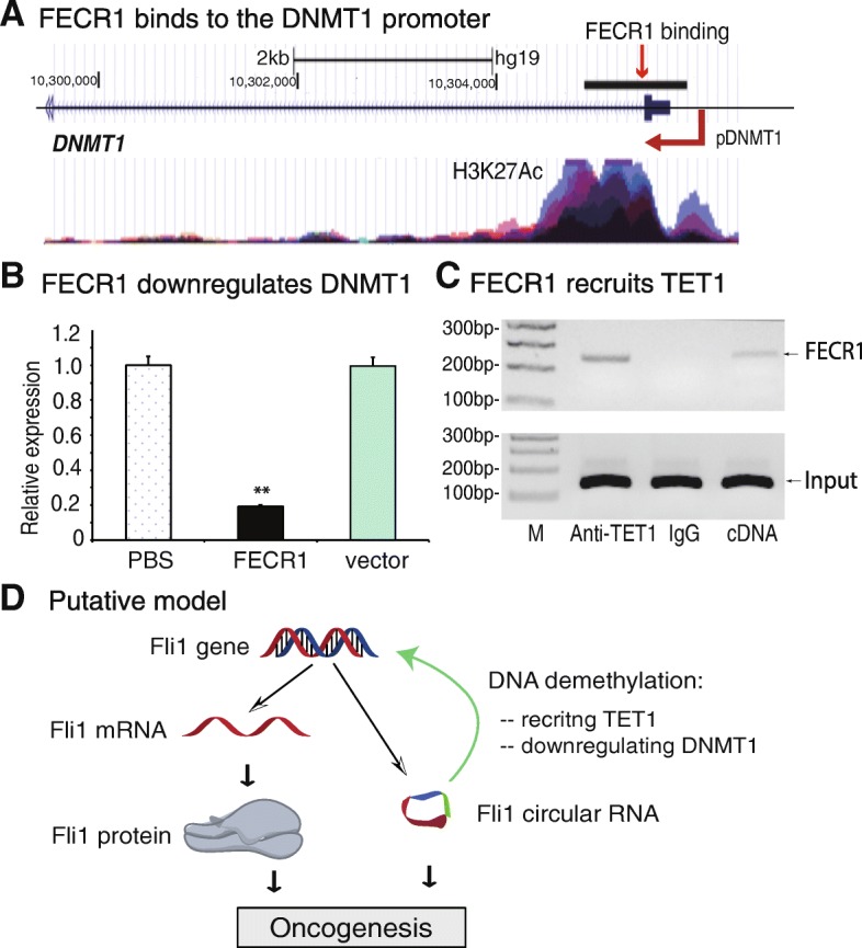
Results: Immunohistochemical staining showed that FLI1 is aberrantly overexpressed in advanced stage and metastatic breast cancers. Using a CRISPR Cas9-guided immunoprecipitation assay, we identify a circular RNA in the FLI1 promoter chromatin complex, consisting of FLI1 exons 4-2-3, referred to as FECR1.Overexpression of FECR1 enhances invasiveness of MDA-MB231 breast cancer cells. Notably, FECR1 utilizes a positive feedback mechanism to activate FLI1 by inducing DNA hypomethylation in CpG islands of the promoter. FECR1 binds to the FLI1 promoter in cis and recruits TET1, a demethylase that is actively involved in DNA demethylation. FECR1 also binds to and downregulates in trans DNMT1, a methyltransferase that is essential for the maintenance of DNA methylation.

Conclusions: These data suggest that FECR1 circular RNA acts as an upstream regulator to control breast cancer tumor growth by coordinating the regulation of DNA methylating and demethylating enzymes. Thus, FLI1 drives tumor metastasis not only through the canonical oncoprotein pathway, but also by using epigenetic mechanisms mediated by its exonic circular RNA.

Keywords: Breast cancer; Circular RNA; DNA methylation; DNMT1; FLI1; TET1; Tumor.


Conflict of interest statement

Ethics approval and consent to participate

The study was conducted in compliance with the Helsinki Declaration. The study protocol was approved by the Research Ethics Board of the First Hospital of Jilin University (IRB 2015-260). Informed consent was obtained from each participant in the study.

Consent for publication

Not applicable.

Competing interests

The authors declare that they have no competing interests.

Publisher’s Note

Springer Nature remains neutral with regard to jurisdictional claims in published maps and institutional affiliations.

Figures

Fig. 1
Fig. 1
Identification of FLI1 circular RNA by Cas9IP. a Overexpression of FLI1 in breast cancer tissues. FLI1 expression was quantitated by immunohistochemical staining and was evaluated as the expression score. **p < 0.01 in breast carcinoma tissues as compared with their adjacent tissues. b High expression of FLI1 in carcinoma as compared with adjacent tissues. Red arrow: dark brown immunohistochemical staining of FLI1 oncoprotein. c CRISPR Cas9-guided chromatin immunoprecipitation (CasIP). Cas9, CRISPR Cas9; FLI1 gRNA, Cas9 guiding RNAs that target the FLI1 promoter. Cas9 binds to the FLI1 promoter through a mechanism of base pairing between the gRNA and target DNA. After fixation, the Cas9-FLI1 promoter chromatin complex was immunoprecipitated by an anti-Cas9 antibody. The CasIP-captured RNAs were sequenced to identify the RNA components that regulate the gene activity in breast cancers. d Identification of FLI1 circular RNA by CasIP. In the FLI1 Cas9-gRNA cassette vector, two Cas9 gRNAs are transcribed by U6 and H1 promoters, respectively, and guide Cas9 to the FLI1 promoter. The CasIP sequencing identifies a novel FLI1 exonic circular RNA that interacts with the gene promoter. e Enrichment of FECR1 in the FLI1 promoter. After CasIP, the captured RNAs were reverse transcribed to quantitate the abundance of FECR1 in the Cas9-captured promoter complex. M, 100 bp marker; IgG, ChIP with antibody control; Cas9, ChIP with anti-Cas9 antibody; nAb, the ChIP negative control, in which the anti dCas9-FLAG antibody was replaced by the equal amount of albumin protein
Fig. 2
Fig. 2
FECR1 is associated with the development of breast cancer. a Formation of FLI1 exonic circular RNA. FECR1 is formed by back splicing of FLI1 exon 4 to exon 2 and is composed of exons 4-2-3. b Detection of FECR1 circular RNA after RNase R digestion. RNA samples were treated with RNase R to remove linear RNAs. For qPCR normalization, the abundance of FECR1 was calculated by standardizing over the spike DNA control and setting the PBS control as 1. **p < 0.01 as compared with PBS and vector controls. c Sequencing of FECR1. Two FECR1 variants were detected by RT-PCR (left panel). V2 was expressed at very low level. We thus focused on the major form V1. The circular RNA (V1) was amplified with a forward primer (JH2532F) that is located at the end of exon 4 and a reverse primer (JH3273R) at the 5′-region of exon 2. Red arrow: back-splicing site. d Expression of FECR1 in breast cancer cell lines. e Expression of FECR1 in breast cancer tissues. Beta-actin was used as the PCR control
Fig. 3
Fig. 3
FLI1 circular RNA promotes invasion of breast cancer cells. a Ectopic expression of FECR1 in MDA-MB231 breast cancer cells. The FECR1 expression cassette is composed of FLI1 exons 4-2-3 and the intron fragments containing the back-splicing elements. pCMV, CMV promoter; pEF1a, EF1a promoter. DsRed fluorescent marker was used to track the transfection. b RT-PCR of FECR1. After stable transfection, cells were collected and FECR1 was amplified by PCR. PBS and vector, control groups. c Quantitation of FECR1 by qPCR. **p < 0.01 as compared with PBS and vector controls. d FECR1 promotes cell invasion. Cells that crossed through the collagen-coated membrane of the transwell were stained and photographed. e Quantitation of invading cells. All data shown are mean ± SEM from three independent experiments. **p < 0.01 as compared with PBS and vector control groups
Fig. 4
Fig. 4
FECR1 upregulates FLI1. a Diagram of the RNA reverse transcription-associated trap (RAT) assay. FECR1 was in situ reverse transcribed using circular RNA-specific primers in the presence of biotin-dCTP. The FECR1-interacting chromatin DNAs were isolated for library sequencing. b Location of PCR primers to detect the interaction of FECR1 at the FLI1 locus. 5′-CT, 5′-upstream control site. c Binding of FECR1 in the FLI1 locus. The FECR1 RAT-captured chromatin DNAs were amplified by PCR using primers covering the FLI1 locus. Note the binding of FECR1 in the FLI1 promoter (P1, P2). d Activation of FLI1 by FECR1. Expression of FLI1 was quantitated by qPCR using two pairs of primers that cover different regions of FLI1. Region 1, the PCR product covers exon 4 to exon 6; region 2, the PCR product covers exon 3 to exon 4. **p < 0.01 as compared with PBS and vector control groups. Both qPCR data show that the overexpressed FECR1 upregulates the linear FLI1 mRNA. e Western blot of FLI1 protein. Cells that were stably transfected with FECR1-overexpression vector, vector control, and PBS were collected for Western blotting. f Quantitation of FLI1 oncoprotein Western blot. **p < 0.01 as compared with PBS and vector control groups
Fig. 5
Fig. 5
FECR1 induces DNA demethylation in the FLI1 promote. a CpG islands in the FLI1 promoter. In order to detect the status of DNA methylation in FECR1-expressing cells, we designed five pairs of methylation-specific primers located at each CpG island. Three restriction enzymes were used to separate methylated and unmethylated DNAs. b FECR1 induces DNA demethylation. DNA methylation was measured by combined bisulfite restriction analysis (COBRA). PCR products from FECR1-expressing cells and vector control cells were digested by TaiI, Bsh1236I, and BstB1 to separate the unmethylated and methylated DNAs. They recognize and digest the methylated ACGT, CGCG, and TTCGAA sites, respectively. After treatment with sodium bisulfate, unmethylated cytosines were converted to uracils, and the ATGT, TGTG, and TTTGAA sites are not digested by these enzymes. After digestion, unmethylated and methylated DNAs were separated on 3% agarose gels. Note the uncut demethylated bands in FECR1-expressing cells
Fig. 6
Fig. 6
FECR1 coordinately regulates DNMT1 and TET1. a Binding of FECR1 to the DNMT1 promoter. After RAT sequencing, the FECR1 binding sequences were blasted to the human genome at the UCSC website. Histone 3 lysine 27 (H3K27) acetylation signal was also shown correspondingly. b FECR1 downregulates DNMT1. Expression of DNMT1 was measured by qPCR. **p < 0.01 as compared with PBS and vector control groups. c FECR1 recruits TET1 enzyme. RNA-chromatin immunoprecipitation (RIP) was performed to identify FECR1-TET1 binding. The TET-FECR1 chromatin complex was immunoprecipitated with an antibody against TET1. After removal of crosslinking, the immunoprecipitated RNAs were reverse transcribed, and the TET-interacting FECR1 was measured by PCR. IgG was use as the antibody control, and cDNA was used as the positive control. d Putative model of FECR1 in breast cancer. In addition to the conventional FLI1 mRNA-oncoprotein model, FLI1 also produces circular RNA FECR1. Through the interaction with FLI1 promoter, FECR1 recruits TET1 demethylase and induces extensive DNA demethylation in the CpG islands. In addition, FECR1 also inhibits DNMT1, the critical enzyme that maintains DNA demethylation during DNA replication. Working together, FECR1 activates FLI1, which in turn promotes tumor cell invasion in breast cancers

References

    1. Li Y, Luo H, Liu T, Zacksenhaus E, Ben-David Y. The ets transcription factor Fli-1 in development, cancer and disease. Oncogene. 2015;34:2022–2031. doi: 10.1038/onc.2014.162. - DOI - PMC - PubMed
    1. Maroulakou IG, Bowe DB. Expression and function of Ets transcription factors in mammalian development: a regulatory network. Oncogene. 2000;19:6432–6442. doi: 10.1038/sj.onc.1204039. - DOI - PubMed
    1. Oikawa T. ETS transcription factors: possible targets for cancer therapy. Cancer Sci. 2004;95:626–633. doi: 10.1111/j.1349-7006.2004.tb03320.x. - DOI - PMC - PubMed
    1. Ben-David Y, Giddens EB, Bernstein A. Identification and mapping of a common proviral integration site Fli-1 in erythroleukemia cells induced by Friend murine leukemia virus. Proc Natl Acad Sci U S A. 1990;87:1332–1336. doi: 10.1073/pnas.87.4.1332. - DOI - PMC - PubMed
    1. Hart A, Melet F, Grossfeld P, Chien K, Jones C, Tunnacliffe A, Favier R, Bernstein A. Fli-1 is required for murine vascular and megakaryocytic development and is hemizygously deleted in patients with thrombocytopenia. Immunity. 2000;13:167–177. doi: 10.1016/S1074-7613(00)00017-0. - DOI - PubMed

Publication types

MeSH terms