. 2018 Dec 11;51(1):56.
doi: 10.1186/s40659-018-0203-6.

MicroRNA-153-3p enhances cell radiosensitivity by targeting BCL2 in human glioma

Affiliations

MicroRNA-153-3p enhances cell radiosensitivity by targeting BCL2 in human glioma

Deyu Sun et al. Biol Res. .

Abstract

Background: Glioma is the most prevalent malignant tumor in human central nervous systems. Recently, the development of resistance to radiotherapy in glioma patients markedly vitiates the therapy outcome. MiR-153-3p has been reported to be closely correlated with tumor progression, but its effect and molecular mechanism underlying radioresistance remains unclear in glioma.

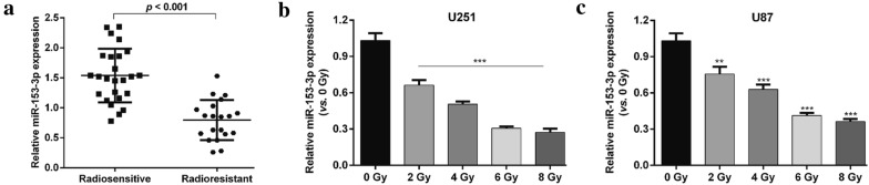
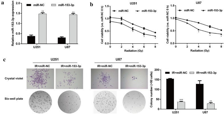
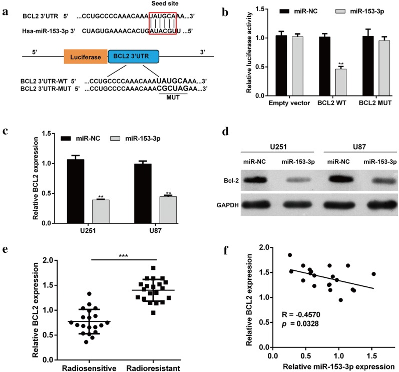
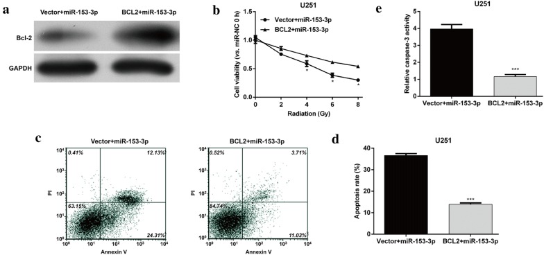
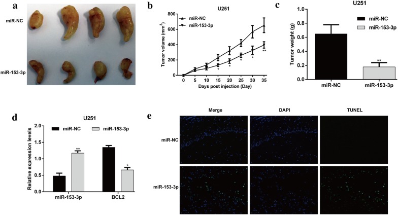
Methods: The expression of miR-153-3p was determined in radioresistant glioma clinical specimens as well as glioma cell lines exposed to irradiation (IR) using quantitative real-time PCR. Cell viability, proliferation and apoptosis were then evaluated by MTT assay, colony formation assay, Flow cytometry analysis and caspase-3 activity assay in glioma cells (U87 and U251). Tumor forming was evaluated by nude mice model in vivo. TUNEL staining was used to detect cell apoptosis in nude mice model. The target genes of miR-153-3p were predicted and validated using integrated bioinformatics analysis and a luciferase reporter assay.

Results: Here, we found that miR-153-3p was down-regulated in radioresistant glioma clinical specimens as well as glioma cell lines (U87 and U251) exposed to IR. Enhanced expression of miR-153-3p promoted the radiosensitivity, promoted apoptosis and elevated caspase-3 activity in glioma cells in vitro, as well as the radiosensitivity in U251 cell mouse xenografs in vivo. Mechanically, B cell lymphoma-2 gene (BCL2) was identified as the direct and functional target of miR-153-3p. Moreover, restoration of BCL2 expression reversed miR-153-3p-induced increase of radiosensitivity, apoptosis and caspase-3 activity in U251 cells in vitro. In addition, clinical data indicated that the expression of miR-153-3p was significantly negatively associated with BCL2 in radioresistance of glioma samples.

Conclusions: Our findings suggest that miR-153-3p is a potential target to enhance the effect of radiosensitivity on glioma cells, thus representing a new potential therapeutic target for glioma.

Keywords: BCL2; Glioma; Radiosensitivity; miR-153-3p.


Figures

Fig. 1
Fig. 1
MiR-153-3p expression in radioresistant glioma tissues and cell lines treated with irradiation. a The expression profiles of miR-153-3p in radiosensitive (n = 25) and radioresistant (n = 20) glioma patient tumors; MiR-153-3p expression in b U251 cells and c U87 cells after irradiation treatment at different doses (0 Gy, 2 Gy, 4 Gy, 6 Gy and 8 Gy) by quantitative real-time PCR analysis. Data represent mean ± SD. **p < 0.01, ***p < 0.001 versus 0 Gy
Fig. 2
Fig. 2
MiR-153-3p regulated glioma cell viability and proliferation after IR exposure. a The expression of miR-153-3p in U251 and U87 cells transfected with miR-153-3p or miR-NC was determined using quantitative real-time PCR. b Transfected U251 and U87 cells were exposed to different doses of IR, followed by cell viability evaluation by MTT assay. c The transfected U251 and U87 cells were treated with IR at dose of 4 Gy, followed by the measurement of colony formation. Data represent mean ± SD. *p < 0.05, **p < 0.01, ***p < 0.001 versus miR-NC or IR + miR-NC; IR: irradiation
Fig. 3
Fig. 3
MiR-153-3p enhanced IR-mediated apoptosis in glioma cells. a, b U251 and U87 cells with miR-153-3p or miR-NC transfection were subjected to 4 Gy IR, followed by the measurement of cells apoptosis rate by flow cytometry analysis. c Caspase-3 activity was measured in transfected U251 and U87 cells after 4 Gy IR treatment. Data represent mean ± SD. *p < 0.05, **p < 0.01, ***p < 0.001
Fig. 4
Fig. 4
BCL2 was a downstream target of miR-153-3p in glioma cells. a The sequence alignment of human miR-153-3p with the 3′UTR of BCL2 is shown. The luciferase reporter constructs, BCL2 3′UTR-WT and BCL2 3′UTR-MUT were made using the seed sequence of miR-153-3p matching the 3′UTR of BCL2 mRNA. b HEK293 cells were co-transfected with the indicated plasmids and miR-153-3p or miR-NC for 48 h, and then they were subjected to the luciferase assay. U251 and U87 cells were transfected with miR-153-3p or miR-NC. The BCL2 mRNA (c) and protein (d) levels were determined by quantitative real-time PCR and Western blotting, respectively. Data represent mean ± SD. **p < 0.01 versus miR-NC; e the relative BCL2 mRNA levels in radioresistant glioma tumors (n = 20) and radiosensitive tumors (n = 25) were assessed using quantitative real-time PCR. ***p < 0.001 versus radiosensitive tumors; f correlation between the expression levels of BCL2 and miR-153-3p in radioresistant glioma tumors
Fig. 5
Fig. 5
Restoration of BCL2 expression inhibited miR-153-3p-mediated radiosensitivity of glioma cells. U251 cells with miR-153-3p overexpression were transfected with empty vector or BCL2 vector. a The expression of BCL2 protein was determined using western blot analysis. b After exposure to different doses of IR, MTT assay was used to determine the cell viability. After exposure to 4 Gy IR, cell apoptosis and caspase-3 activity were detected using c, d flow cytometry analysis and e caspase-3 activity assay, respectively. Data represent mean ± SD. *p < 0.05, ***p < 0.001 versus Vector + miR-153-3p
Fig. 6
Fig. 6
MiR-153-3p enhanced the tumor suppression capability of IR in vivo. U251/miR-NC or U251/miR-153-3p cells were subcutaneously injected into nude mice. The mice were exposed to IR at a dose of 8 Gy after 10 days injection. a Representative images of tumors formed in the mice in which miR-153-3p or miR-NC-infected U251 cells were implanted. b Tumor volumes were measured every 5 days after injection. c Tumors were dissected, and the tumor weights were measured on day 35 after inoculation. d Expression of miR-153-3p and BCL2 in miR-153-3p or miR-NC-expressing U251 cells-derived tumors by quantitative real-time PCR analysis. Data represent mean ± SD. e The TUNEL staining detected cell apoptosis in xenograft tumors. Blue staining represents the nucleus, and green staining indicates TUNEL-positive cells. *p < 0.05, **p < 0.01 versus miR-NC

References

    1. DeWitt JC, Jordan JT, Frosch MP, Samore WR, Iafrate AJ, Louis DN, Lennerz JK. Cost-effectiveness of IDH testing in diffuse gliomas according to the 2016 WHO classification of tumors of the central nervous system recommendations. Neuro Oncol. 2017;19:1640–1650. doi: 10.1093/neuonc/nox120. - DOI - PMC - PubMed
    1. Ostrom QT, Gittleman H, Fulop J, Liu M, Blanda R, Kromer C, Kruchko C, Barnholtz-Sloan JS, Kromer C. CBTRUS statistical report: primary brain and central nervous system tumors diagnosed in the United States in 2008–2012. Neuro Oncol. 2015;17(Suppl 4):iv1–iv62. doi: 10.1093/neuonc/nov189. - DOI - PMC - PubMed
    1. Naidu MD, Mason JM, Pica RV, Fung H, Pena LA. Radiation resistance in glioma cells determined by DNA damage repair activity of Ape1/Ref-1. J Radiat Res. 2010;51:393–404. doi: 10.1269/jrr.09077. - DOI - PubMed
    1. Wiencke JK, Koestler DC, Salas LA, Wiemels JL, Roy RP, Hansen HM, Rice T, McCoy LS, Bracci PM, Molinaro AM, et al. Immunomethylomic approach to explore the blood neutrophil lymphocyte ratio (NLR) in glioma survival. Clin Epigenet. 2017;9:10. doi: 10.1186/s13148-017-0316-8. - DOI - PMC - PubMed
    1. Besse A, Sana J, Fadrus P, Slaby O. MicroRNAs involved in chemo- and radioresistance of high-grade gliomas. Tumour Biol. 2013;34:1969–1978. doi: 10.1007/s13277-013-0772-5. - DOI - PubMed

MeSH terms