. 2018 Dec 26;115(52):13234-13239.
doi: 10.1073/pnas.1810058115. Epub 2018 Dec 11.

Cofactors are essential constituents of stable and seeding-active tau fibrils

Affiliations

Cofactors are essential constituents of stable and seeding-active tau fibrils

Yann Fichou et al. Proc Natl Acad Sci U S A. .

Abstract

Amyloid fibrils are cross-β-rich aggregates that are exceptionally stable forms of protein assembly. Accumulation of tau amyloid fibrils is involved in many neurodegenerative diseases, including Alzheimer's disease (AD). Heparin-induced aggregates have been widely used and assumed to be a good tau amyloid fibril model for most biophysical studies. Here we show that mature fibrils made of 4R tau variants, prepared with heparin or RNA, spontaneously depolymerize and release monomers when their cofactors are removed. We demonstrate that the cross-β-sheet assembly formed in vitro with polyanion addition is unstable at room temperature. We furthermore demonstrate high seeding capacity with transgenic AD mouse brain-extracted tau fibrils in vitro that, however, is exhausted after one generation, while supplementation with RNA cofactors resulted in sustained seeding over multiple generations. We suggest that tau fibrils formed in brains are supported by unknown cofactors and inhere higher-quality packing, as reflected in a more distinct conformational arrangement in the mouse fibril-seeded, compared with heparin-induced, tau fibrils. Our study suggests that the role of cofactors in tauopathies is a worthy focus of future studies, as they may be viable targets for diagnosis and therapeutics.

Keywords: DEER; aggregate cofactors; amyloid aggregates; seeding; tau.


Conflict of interest statement

The authors declare no conflict of interest.

Figures
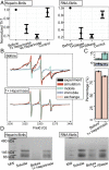
Fig. 1.
Fig. 1.
Fibrils depolymerize upon cofactor digestion. (A) ThT fluorescence of heparin-induced tau fibrils and RNA-induced tau fibrils before and after incubation with different concentrations of heparinase/RNase. ThT fluorescence value measured before enzyme addition was used as the normalization value. (B) cw-EPR spectra (black) of tau heparin fibrils spin-labeled at site 322, before and after incubation with heparinase. The simulation spectra (red) are composed of mobile (green), immobile (purple), and spin-exchange components (orange). (C) Population of each component extracted from cw-EPR spectrum analysis. (D) BNPAGE of heparin and RNA fibrils before and after incubation. Freshly prepared tau monomers were loaded as reference (soluble). In all panels, 1× heparinase: 1 U enzyme per 1 μg heparin; 1× RNase: 2.5 μg/mL. Error bars show SD (n ≥ 3).
Fig. 2.
Fig. 2.
Cofactors are bound to fibrils and partially digested. (A) Schematic diagram of experimental procedures. Digested soluble cofactors were separated from tau fibrils via dialysis (RNA fibrils) or filtration (heparin fibrils) before measuring their concentration in the flow-through by cw-EPR (heparin) or UV absorbance (RNA). (B, Left) The concentration of spin-labeled heparin in the flow-through of a 0.2-μm filter for heparin and heparin fibrils before and after digestion. The concentration is given as the percentage of the concentration before filtration. (B, Right) RNA concentration outside a dialysis bag (12 kDa molecular mass cutoff) at equilibrium was measured and the percentage of RNA that flowed through is given on the y axis. Error bars show SD of three independent repeats.
Fig. 3.
Fig. 3.
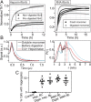
Refibrillization and conformation recovery of depolymerized tau monomers. (A, Left) ThT fluorescence of non/predigested heparin fibrils after heparin addition at time t = 0 h. (A, Right) Aggregation of monomers purified from digested RNA fibrils compared with fresh monomers. Curves of the same color show independent repeats. (B) DEER time-domain signal (Left) and corresponding distance distributions (Right), extracted with the SVD method, of tau labeled at residues 272 and 285 before and after digestion, compared with soluble monomer. (C) Quantification of in vivo seeding experiments. The ratios of cells that contain fluorescent puncta are reported. Digst, digested; fib, fibrils; Hep, heparin.
Fig. 4.
Fig. 4.
In vitro seeding of tau fibrils. (A) ThT fluorescence of recombinant tau187C291S seeded with heparin fibrils or mouse-derived fibrils (mouse-derived) in the presence or absence of cofactor (polyU RNA). In first-generation seeding (Left), 5% of preaggregated fibrils were added to fresh recombinant tau and incubated at 37 °C while shaking. In the second generation (Right), 10% of the end products of first-generation samples were added to fresh monomers. Control refers to the seeds with cofactor incubated without fresh tau. Error bars shows SD (n = 3). (B) DEER distance distribution between residues 272 and 285 of heparin fibrils and mouse-derived seeded fibrils (seeded fibrils) obtained from the SVD method. (C) ThT fluorescence before and after RNase addition to the first and second generations of mouse-derived seeded fibrils. Error bars shows SD (n ≥ 3). gen, generation.

References

    1. Greenwald J, Riek R. Biology of amyloid: Structure, function, and regulation. Structure. 2010;18:1244–1260. - PubMed
    1. Jahn TR, Radford SE. The yin and yang of protein folding. FEBS J. 2005;272:5962–5970. - PubMed
    1. Knowles TP, et al. Role of intermolecular forces in defining material properties of protein nanofibrils. Science. 2007;318:1900–1903. - PubMed
    1. Mandelkow EM, Mandelkow E. Biochemistry and cell biology of tau protein in neurofibrillary degeneration. Cold Spring Harb Perspect Med. 2012;2:a006247. - PMC - PubMed
    1. Fitzpatrick AWP, et al. Cryo-EM structures of tau filaments from Alzheimer’s disease. Nature. 2017;547:185–190. - PMC - PubMed

Publication types