. 2018 Dec 26;115(52):13264-13269.
doi: 10.1073/pnas.1812717115. Epub 2018 Dec 11.

Crystal structure of the human NK1 tachykinin receptor

Affiliations

Crystal structure of the human NK1 tachykinin receptor

Jie Yin et al. Proc Natl Acad Sci U S A. .

Abstract

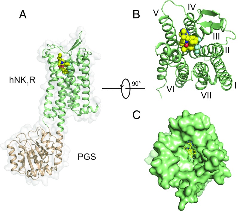
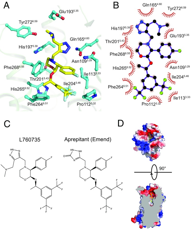
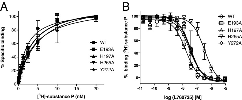
The NK1 tachykinin G-protein-coupled receptor (GPCR) binds substance P, the first neuropeptide to be discovered in mammals. Through activation of NK1R, substance P modulates a wide variety of physiological and disease processes including nociception, inflammation, and depression. Human NK1R (hNK1R) modulators have shown promise in clinical trials for migraine, depression, and emesis. However, the only currently approved drugs targeting hNK1R are inhibitors for chemotherapy-induced nausea and vomiting (CINV). To better understand the molecular basis of ligand recognition and selectivity, we solved the crystal structure of hNK1R bound to the inhibitor L760735, a close analog of the drug aprepitant. Our crystal structure reveals the basis for antagonist interaction in the deep and narrow orthosteric pocket of the receptor. We used our structure as a template for computational docking and molecular-dynamics simulations to dissect the energetic importance of binding pocket interactions and model the binding of aprepitant. The structure of hNK1R is a valuable tool in the further development of tachykinin receptor modulators for multiple clinical applications.

Keywords: GPCR; drug design; ligand recognition; substance P; tachykinin receptor.


Conflict of interest statement

The authors declare no conflict of interest.

Figures

Fig. 1.
Fig. 1.
Structural features of hNK1R; (A) Global structure of the hNK1R-PGS fusion protein from side view. The hNK1R is represented as a green cartoon, with the PGS domain (wheat) fused between TMs 5 and 6. L760735 is shown as spheres with yellow carbons. (B) Extracellular view of hNK1R, with PGS domain removed. (C) Surface representation as in B.
Fig. 2.
Fig. 2.
Stereoview of polder OMIT map (46) for the ligand L760735 (yellow sticks). The contour level is 3.1σ.
Fig. 3.
Fig. 3.
L760735 interaction with hNK1R. (A) All residues within 4 Å of L760735 are depicted with cyan carbons. (B) Two-dimensional schematic of contacts between L760735 and hNK1R, produced in Ligplot (65); (C) Chemical structures of aprepitant and L760735. (D, Top) Extracellular view, receptor surface is colored according to electrostatic potential. (D, Bottom) Side view, cut through the receptor.
Fig. 4.
Fig. 4.
Binding of substance P and L760735 to wild-type hNK1R and point mutants. (A) Saturation binding isotherms for [3H]-substance P. (B) Competition binding assay for L760735, using [3H]-substance P as a probe. Error bars denote SE (n = 3 separate assays, each in duplicate).
Fig. 5.
Fig. 5.
Top flexible docking poses for L760735 and aprepitant in both neutral and protonated states. The L760735 ligand in the crystal structure is shown as orange sticks, while the docked ligands are shown as yellow sticks. Residues that form hydrogen bonds (dashed lines) with L760735 and aprepitant are shown as cyan sticks. (A) L760735 (neutral, ΔGglide = −9.50, rmsd = 1.83 Å, best pose); (B) L760735 (charged, ΔGglide = −9.37, rmsd = 1.73 Å, best pose); (C) aprepitant (neutral, ΔGglide = −9.12, rmsd = 1.27 Å, best pose); (D) aprepitant (charged, ΔGglide = −8.91, rmsd = 1.97 Å, second best pose). Glide docking scores are in kcal/mol.

References

    1. Steinhoff MS, von Mentzer B, Geppetti P, Pothoulakis C, Bunnett NW. Tachykinins and their receptors: Contributions to physiological control and the mechanisms of disease. Physiol Rev. 2014;94:265–301. - PMC - PubMed
    1. V Euler US, Gaddum JH. An unidentified depressor substance in certain tissue extracts. J Physiol. 1931;72:74–87. - PMC - PubMed
    1. Michelot R, Mayer M, Magneney S, Thierry J, Potier P. Substance P and Neurokinins. Springer; New York: 1987. Activity of the C-terminal part in tachykinins on guinea pig ileum and trachea preparations; pp. 158–160.
    1. Almeida TA, et al. Tachykinins and tachykinin receptors: Structure and activity relationships. Curr Med Chem. 2004;11:2045–2081. - PubMed
    1. Douglas SD, Leeman SE. Neurokinin-1 receptor: Functional significance in the immune system in reference to selected infections and inflammation. Ann N Y Acad Sci. 2011;1217:83–95. - PMC - PubMed

Publication types

Associated data

LinkOut - more resources