. 2019 Apr;38(14):2658-2674.
doi: 10.1038/s41388-018-0613-5. Epub 2018 Dec 11.

Caspase-11 regulates the tumour suppressor function of STAT1 in a murine model of colitis-associated carcinogenesis

Affiliations

Caspase-11 regulates the tumour suppressor function of STAT1 in a murine model of colitis-associated carcinogenesis

Brian Flood et al. Oncogene. 2019 Apr.

Abstract

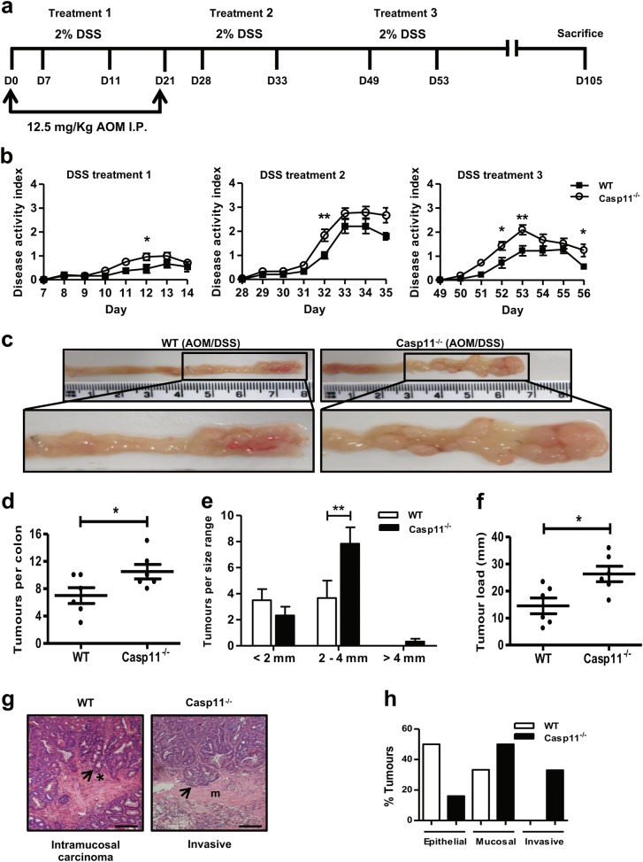
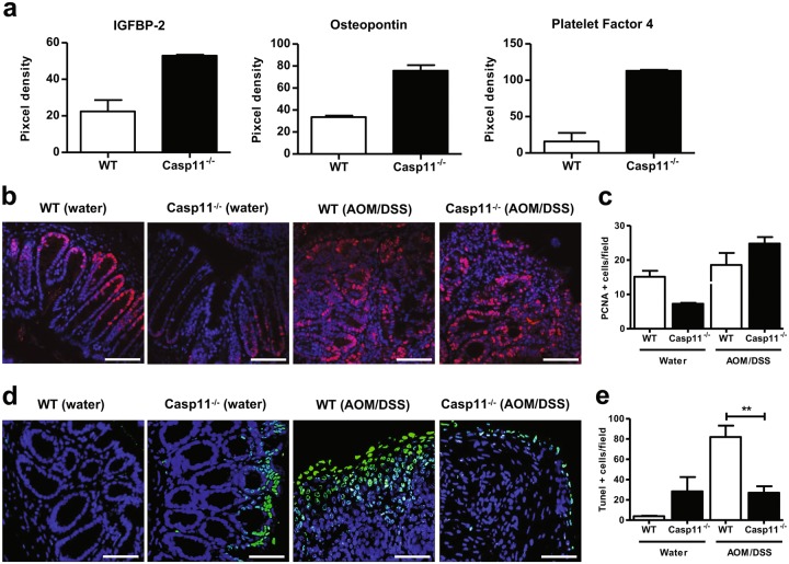
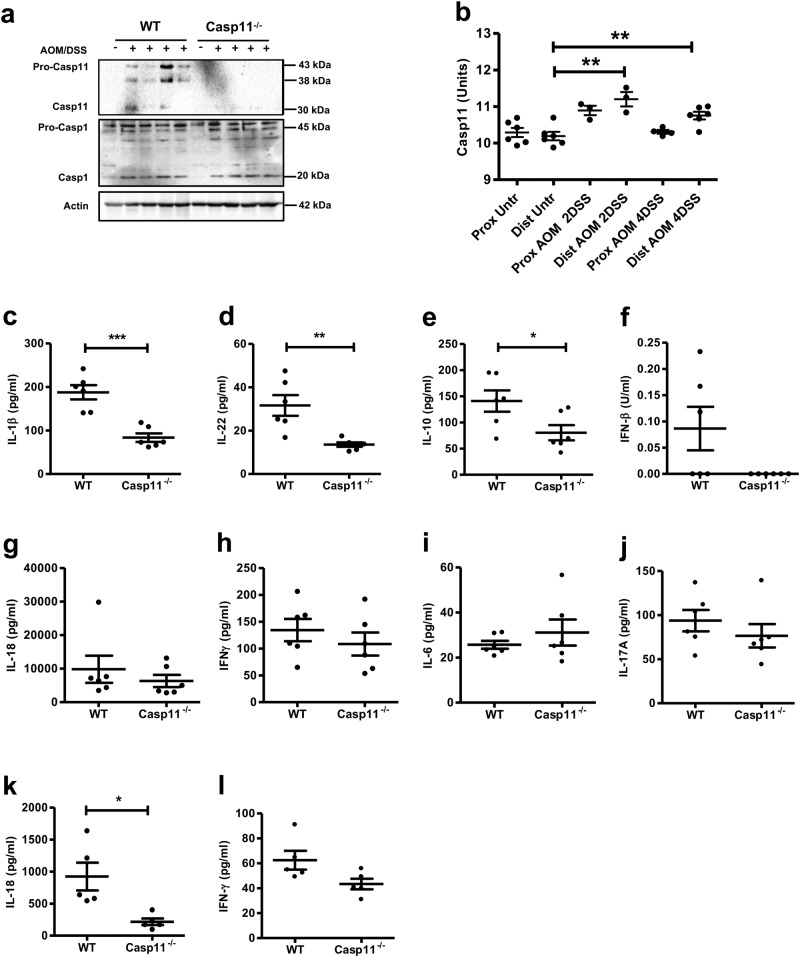
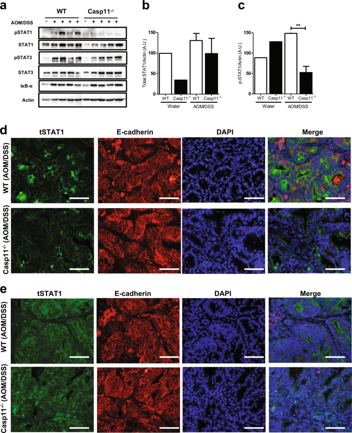
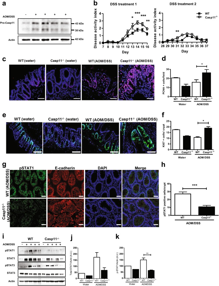
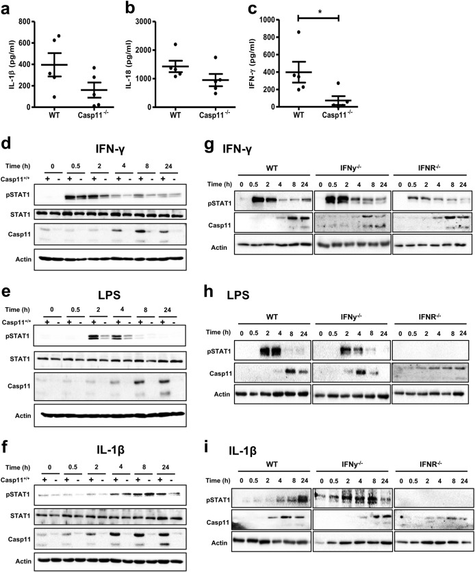
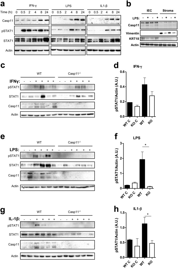
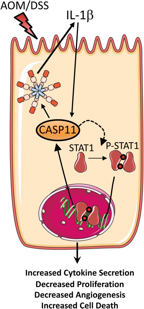
Murine inflammatory caspase-11 has an important role in intestinal epithelial inflammation and barrier function. Activation of the non-canonical inflammasome, mediated by caspase-11, serves as a regulatory pathway for the production of the pro-inflammatory cytokines IL-1β and IL-18, and has a key role in pyroptotic cell death. We have previously demonstrated a protective role for caspase-11 during dextran sulphate sodium (DSS)-induced colitis, however the importance of caspase-11 during colorectal tumour development remains unclear. Here, we show that Casp11-/- mice are highly susceptible to the azoxymethane (AOM)-DSS model of colitis-associated cancer (CAC), compared to their wild type (WT) littermates. We show that deficient IL-18 production occurs at initial inflammation stages of disease, and that IL-1β production is more significantly impaired in Casp11-/- colons during established CAC. We identify defective STAT1 activation in Casp11-/- colons during disease progression, and show that IL-1β signalling induces caspase-11 expression and STAT1 activation in primary murine macrophages and intestinal epithelial cells. These findings uncover an anti-tumour role for the caspase-11 and the non-canonical inflammasome during CAC, and suggest a critical role for caspase-11, linking IL-1β and STAT1 signalling pathways.


Conflict of interest statement

The authors declare that they have no conflict of interest.

Figures

Fig. 1
Fig. 1
Increased susceptibility of Casp11-/- mice to experimental CAC. a Schematic representation of the AOM/DSS administration regime to induce tumour progression. WT and Casp11−/− mice were injected intraperitoneally (IP) with 12.5 mg/kg AOM on days 0 and 21 followed by 3 cycles of 2% DSS (w/v). Mice were culled on day 105. b Disease activity index (combination of weight loss, stool consistency and intestinal bleeding scores) during each DSS administration. Data represent mean ± SEM of n = 6 AOM/DSS-treated mice for both groups; *p< 0.05; **p< 0.01 (two-way ANOVA followed by the Bonferroni post test). c Representative macroscopic view of the AOM/DSS-derived tumours in AOM/DSS-treated WT and Casp11−/− mice, enhanced image magnification facilitates the visualisation of defined adenomas. df Total tumour numbers were macroscopically enumerated in AOM/DSS-treated WT and Casp11−/− whole colons (d); measured and assigned a size range (e); and each mouse was subsequently assigned a tumour load (calculated by the addition of each tumour diameter) (f). Data represent mean ± SEM of n = 6 AOM/DSS-treated mice for both groups; *p< 0.05; **p< 0.01 (two-tailed independent Student t-test). g Representative images of WT and Casp11−/− AOM/DSS-treated H&E stained colon tissue displaying intramucosal carcinoma and invasive carcinoma (scale bar = 200 μm). h Tumours from AOM/DSS-treated mice displaying epithelial, mucosal and invasive carcinomas, as a percentage of total tumours. H&E staining; *muscularis mucosae; m, submucosa
Fig. 2
Fig. 2
Increased angiogenesis and reduced cell death characterise the enhanced CAC phenotype of Casp11-/- mice. a Semi-quantitative IGFBP-2, osteopontin and platelet factor 4 expression in AOM/DSS-treated WT and Casp11−/− colon homogenates 105 days post initial AOM IP injection, as determined using an angiogenesis proteome profiler array and ImageJ densitometry. Data represent mean ± SEM of n = 2 AOM/DSS-treated mice for both groups. be Representative immunofluorescent images of proliferating cell nuclear antigen (PCNA) (b) and TUNEL (d) positive epithelial cells in distal colon tissue sections of AOM/DSS-treated WT and Casp11−/− mice (scale bars = 50 μm (b) and 500 μm (d)). c, e Quantification of PCNA (c) and TUNEL (e) positive epithelial cells shown in b and d
Fig. 3
Fig. 3
Reduced IL-1β production in Casp11-/- mice during advanced stages of CAC. a Western blot analysis of caspase-11 and caspase-1 expression in colon homogenates from AOM/DSS-treated WT and Casp11−/− mice on day 105 post initial AOM IP injection. Each lane represents an individual mouse. b Analysis of relative Casp11 mRNA expression in AOM/DSS-treated C57BL/6 J colon tissue using the published dataset GSE64658. Data represent mean ± SEM; *p< 0.05; **p< 0.01; (two-tailed independent Student t-test). cl Cytokine levels in colon homogenates of AOM/DSS-treated WT and Casp11−/− mice on day 105 (cj) and day 21 (k, l) post initial AOM IP injection, as measured by ELISA. Data represent mean ± SEM of n = 6 (cj) and n = 5 (k, l) AOM/DSS-treated mice for both groups; *p< 0.05; **p< 0.01; ***p< 0.001 (two-tailed independent Student t-test)
Fig. 4
Fig. 4
Caspase-11 mediates STAT1 activation during CAC. a Western blot analysis of phosho-/total STAT1, STAT3, IκBα, and actin (loading control) in colon homogenates from AOM/DSS-treated WT and Casp11−/− mice on day 105 post initial AOM IP injection. Each lane represents an individual mouse. b Total STAT1 and c pSTAT1 expression densitometry (relative to actin) in colon homogenates from AOM/DSS-treated WT and Casp11−/− mice. **p< 0.01 (two-tailed unpaired t-test). Representative immunofluorescent images of d pSTAT1; and e total STAT1; co-stained for the epithelial marker E-cadherin (red) and DAPI (blue) in distal colon sections from AOM/DSS-treated WT and Casp11−/− mice at day 105 (scale bar = 20 μm)
Fig. 5
Fig. 5
Impaired STAT1 activation and increased IEC proliferation are evident in Casp11-/- mice during CAC development. a Western blot revealing caspase-11 expression in colon homogenates of C57BL/6 mice on day 42 post initial AOM IP injection. Each lane represents an individual mouse. b Disease activity index during each DSS administration. Data represent mean ± SEM of n = 6 (WT) and n = 5 (Casp11−/−) AOM/DSS-treated mice; *p< 0.05; **p< 0.01; ***p < 0.001 (two-way ANOVA followed by the Bonferroni post test). cf Representative immunofluorescent images of proliferating cell nuclear antigen (PCNA) (c); and Ki67 (e); positive epithelial cells in distal colon tissue sections of AOM/DSS-treated WT and Casp11−/− mice (scale bars = 50 μm, c and 100 μm e). Quantification of PCNA (d) and Ki67 (f) positive epithelial cells shown in d and f. Data represent mean ± SEM of n = 6 (WT) and n = 5 (Casp11−/−) AOM/DSS-treated mice; *p< 0.05 (two-tailed independent Student t-test). g Representative IF images of distal colon sections from AOM/DSS-treated WT and Casp11−/− mice at day 42, stained for pSTAT1, epithelial marker E-cadherin and DAPI (Scale bar = 20 μm). h Quantification of pSTAT1 positive IECs cells in IF stained colon sections. (i) Western blot analysis of STAT1 and STAT3 activation in colon homogenates from AOM/DSS-treated WT and Casp11−/− mice on day 42 post initial AOM IP injection. Each lane represents an individual mouse. Expression dfensitometry (relative to actin) of j total STAT1 and k pSTAT1; in colon homogenates from AOM/DSS-treated mice on day 42 post initial AOM IP injection. **p< 0.01 (two-tailed unpaired t-test)
Fig. 6
Fig. 6
Caspase-11 regulates STAT1 activation in primary murine macrophages. ac Distal colon explant tissue isolated from AOM/DSS-treated WT and Casp11−/− mice on day 21 post initial AOM IP injection were cultured for 20 h. Supernatants were analysed for the production of IL-1β (a), IL-18 (b), and IFN-γ (c), by ELISA; *p< 0.05 (two-tailed independent Student t-test). df Western blot analysis of pSTAT1, total STAT1 and caspase-11 expression in WT and Casp11−/− BMDMs treated with IFN-γ (d), LPS (e) and IL-1β (f). gi Western blot analysis of STAT1 and caspase-11 expression in WT, Ifnar−/− and Ifn-γ−/− BMDMs treated with IFN-γ (g), LPS (h) and IL-1β (i). Blots are representative of more than three independent experiments
Fig. 7
Fig. 7
Caspase-11 regulates LPS and IL-1β-mediated STAT1 activation in IECs. a Western blot timecourse analysis of caspase-11, pSTAT1 and total STAT1 expression in cell lysates from CT26 colon cancer cells stimulated with IFN-γ, LPS and IL-1β. b Representative western blot showing the relative expression levels of caspase-11, vimentin (mesenchymal cell marker) and keratin 18 (epithelial cell marker) in IECs and mesenchymal cell lysates isolated from WT colon tissue explants, which had been incubated in media or with LPS for 20 h. c, e, g WT and Casp11−/− colon explant tissue were left untreated (n = 2) or treated (n = 4) with IFN-γ (c), LPS (e), and IL-1β (g). Following 20 h incubation, purified IEC lysates were analysed for expression of caspase-11, pSTAT1 and total STAT1 by western blot. d, f, h Densitometric analysis of pSTAT1 (relative to actin) in WT and Casp11−/− IEC lysates following stimulation with IFN-γ (d), LPS (f), and IL-1β (h). Data represent mean ± SEM of n = 4 (untreated) and n = 8 (treated) WT and Casp11−/− mice; *p< 0.05; **p< 0.01 (two-tailed independent Student t-test)
Fig. 8
Fig. 8
Graphical representation—the anti-tumorigenic role of caspase-11 during CAC. Caspase-11 has dual roles during AOM-DSS-induced colitis-associated carcinogenesis: (i) enhancing IL-1β production during tumorigenesis; and (ii) regulating STAT1 activation in IECs in response to IL-1β. The effects of caspase-11 are anti-tumorigenic in this system, causing decreased angiogenesis and increased IEC death

References

    1. Jemal A, Bray F, Center MM, Ferlay J, Ward E, Forman D. Global cancer statistics. CA Cancer J Clin. 2011;61:69–90. doi: 10.3322/caac.20107. - DOI - PubMed
    1. Neurath MF. Cytokines in inflammatory bowel disease. Nat Rev Immunol. 2014;14:329–42. doi: 10.1038/nri3661. - DOI - PubMed
    1. Asquith M, Powrie F. An innately dangerous balancing act: intestinal homeostasis, inflammation, and colitis-associated cancer. J Exp Med. 2010;207:1573–7. doi: 10.1084/jem.20101330. - DOI - PMC - PubMed
    1. Zaki MH, Vogel P, Body-Malapel M, Lamkanfi M, Kanneganti TD. IL-18 production downstream of the Nlrp3 inflammasome confers protection against colorectal tumor formation. J Immunol. 2010;185:4912–20. doi: 10.4049/jimmunol.1002046. - DOI - PMC - PubMed
    1. Mariathasan S, Weiss DS, Newton K, McBride J, O’Rourke K, Roose-Girma M, et al. Cryopyrin activates the inflammasome in response to toxins and ATP. Nature. 2006;440:228–32. doi: 10.1038/nature04515. - DOI - PubMed

Publication types

MeSH terms