Observational Study
. 2018 Nov 21:13:3791-3803.
doi: 10.2147/COPD.S177034. eCollection 2018.

Correlation of serum levels of HIF-1α and IL-19 with the disease progression of COPD: a retrospective study

Affiliations
Observational Study

Correlation of serum levels of HIF-1α and IL-19 with the disease progression of COPD: a retrospective study

Biaoxue Rong et al. Int J Chron Obstruct Pulmon Dis. .

Abstract

Background: The aim of this study was to disclose the correlation between the serum levels of hypoxia-inducible factor 1 alpha (HIF-1α) and IL-19 and stable COPD.

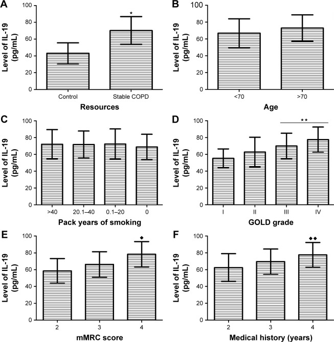
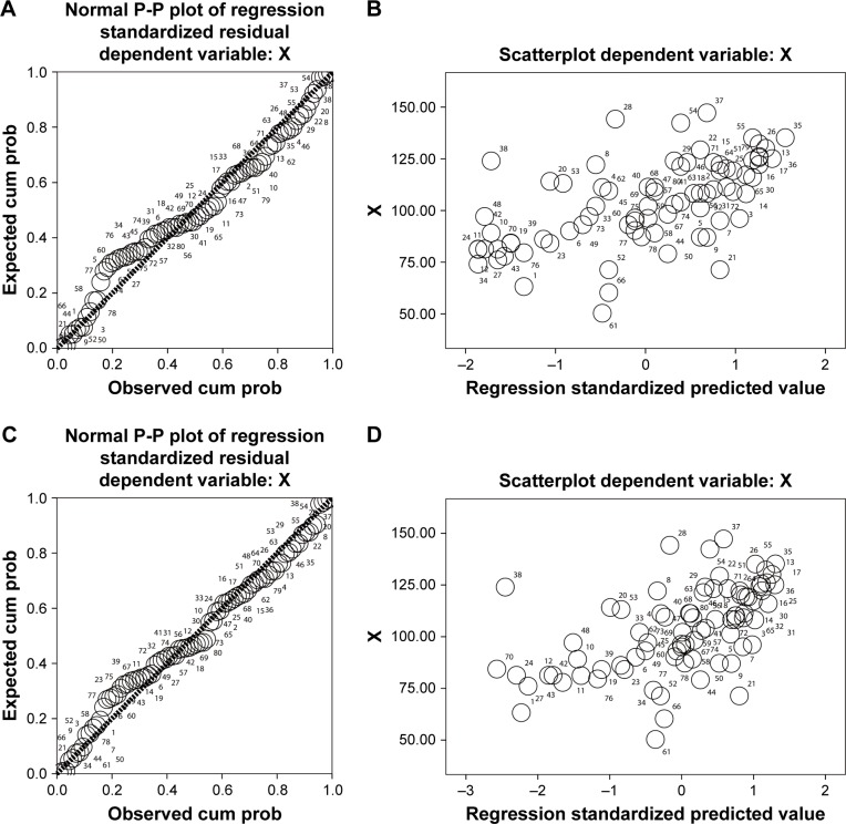
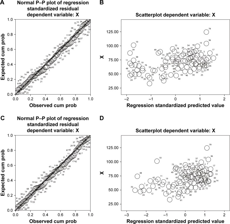
Methods: The serum levels of HIF-1α and IL-19 were tested by ELISA. The relationships between their levels and clinical parameters of stable COPD patients were analyzed by linear regression methods.

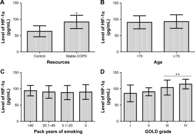
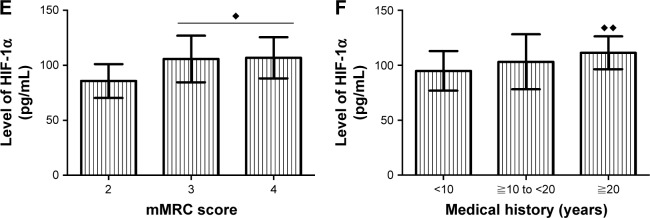
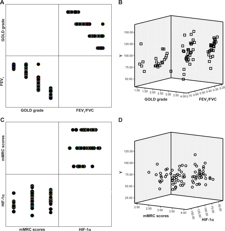
Results: Patients with stable COPD showed higher serum levels of HIF-1α and IL-19 compared with healthy control group (P<0.001), and serum levels of HIF-1α and IL-19 had a positive linear correlation (P<0.05). In stable COPD patients, increased serum levels of HIF-1α and IL-19 were positively correlated with the GOLD grading (P<0.005), modified British Medical Research Council (mMRC) score (P<0.05), and medical history (P<0.05) but negatively related to the pulmonary function (P<0.05). The serum level of HIF-1α (P<0.05) was affected by the patient's FEV1/FVC value and COPD grading, and the serum level of IL-19 was associated with the mMRC scores and the serum level of HIF-1α (P<0.05).

Conclusion: Increased serum levels of HIF-1α and IL-19 correlated with the disease progression of COPD, suggesting that they can be used as indicators to help us understand the COPD.

Keywords: COPD; chronic obstructive pulmonary disease; hypoxia-inducible factor 1 alpha; interleukin-19; pulmonary function; serum.


Conflict of interest statement

Disclosure The authors report no conflicts of interest in this work.

Figures

Figure 1
Figure 1
Relationship between the clinical parameters and serum level of HIF-1α in stable COPD patients. Notes: (A) Patients with stable COPD showed a higher expression of serum HIF-1α compared with control group (P<0.001). (B) Serum HIF-1α expression did correlate with the patient’s age (P=0.612). (C) Serum HIF-1α expression did correlate with the patient’s smoking status (P=0.392). (D) ⋆⋆Serum HIF-1α level of patients with GOLD 3 and 4 was increased compared with that of GOLD 1 and 2 (P<0.001). (E) The patients with mMRC scores of 3 and 4 showed a higher serum level of HIF-1α compared with those with mMRC score of 2 (P=0.007). (F) ♦♦Serum HIF-1α in the patients with longer clinical history was increased compared to those with short history (P=0.009). Abbreviations: HIF-1α, hypoxia-inducible factor 1 alpha; GOLD, Global Initiative for Chronic Obstructive Lung Disease; mMRC, modified British Medical Research Council.
Figure 1
Figure 1
Relationship between the clinical parameters and serum level of HIF-1α in stable COPD patients. Notes: (A) Patients with stable COPD showed a higher expression of serum HIF-1α compared with control group (P<0.001). (B) Serum HIF-1α expression did correlate with the patient’s age (P=0.612). (C) Serum HIF-1α expression did correlate with the patient’s smoking status (P=0.392). (D) ⋆⋆Serum HIF-1α level of patients with GOLD 3 and 4 was increased compared with that of GOLD 1 and 2 (P<0.001). (E) The patients with mMRC scores of 3 and 4 showed a higher serum level of HIF-1α compared with those with mMRC score of 2 (P=0.007). (F) ♦♦Serum HIF-1α in the patients with longer clinical history was increased compared to those with short history (P=0.009). Abbreviations: HIF-1α, hypoxia-inducible factor 1 alpha; GOLD, Global Initiative for Chronic Obstructive Lung Disease; mMRC, modified British Medical Research Council.
Figure 2
Figure 2
Relationship between the clinical parameters and serum level of IL-19 in serum of stable COPD patients. Notes: (A) Patients with stable COPD showed a higher expression of serum IL-19 compared with the control group (P<0.001). (B) Serum IL-19 expression did correlate with the patient’s age (P=0.841). (C) Serum IL-19 expression did correlate with the patient’s smoking status (P=0.110). (D) ⋆⋆Serum IL-19 level of patients with GOLD 3 and 4 of COPD was increased compared with those of GOLD 1 and 2 (P=0.004). (E) The patients with mMRC scores of 3 and 4 showed a higher serum level of IL-19 compared with those with mMRC score of 2 (P<0.001). (F) ♦♦Serum IL-19 in patients with longer medical history was increased compared to those with short history (P=0.002). Abbreviations: GOLD, Global Initiative for Chronic Obstructive Lung Disease; mMRC, modified British Medical Research Council.
Figure 3
Figure 3
Relationship between serum levels of HIF-1α and IL-19 in stable COPD patients. Notes: (A) The histogram of normal curve showed that the normalized residuals are normal distributions (P>0.05). (B) The cumulative probability plot of the observations suggested that the two variables have a feature of normal distribution (P<0.05). (C) The dependent variable is approximately linear with the standardized predictive value, indicating that HIF-1α and IL-19 have a linear correlation (P>0.05). (D) The vast majority of normalized residuals did not exceed 3, suggesting no specific value was found. Abbreviations: cum prob, cumulative probability; HIF-1α, hypoxia-inducible factor 1 alpha.
Figure 4
Figure 4
Relationship between serum level of HIF-1α and pulmonary function in patients with stable COPD. Notes: (A) The scatter point plot for HIF-1α and FEV1/FVC indicated that the two variables belonged to normal distribution (P>0.05). (B) The dependent variable is approximately linear with the standardized predictive value, indicating that serum HIF-1α and FEV1/FVC of patients have a linear correlation (P<0.05). (C) The scatter point plot for HIF-1α and FEV1% predicted indicated that the two variables belonged to normal distribution (P>0.05). (D) The dependent variable is approximately linear with the standardized predictive value, indicating that serum HIF-1α and FEV1% predicted of patients have a linear correlation (P<0.05). Abbreviations: cum prob, cumulative probability; HIF-1α, hypoxia-inducible factor 1 alpha.
Figure 5
Figure 5
Relationship between the serum level of IL-19 and pulmonary function in patients with stable COPD. Notes: (A) The scatter point plot for IL-19 and FEV1/FVC indicated that the two variables belonged to normal distribution (P>0.05). (B) The dependent variable is approximately linear with the standardized predictive value, indicating that serum IL-19 and FEV1/FVC of patients had a linear correlation (P<0.05). (C) The scatter point plot for IL-19 and FEV1% predicted indicated that the two variables belonged to normal distribution (P>0.05). (D) The dependent variable is approximately linear with the standardized predictive value, indicating that serum IL-19 and FEV1% predicted that patients had a linear correlation (P<0.05). Abbreviation: cum prob, cumulative probability.
Figure 6
Figure 6
The clinical factors that affect the serum levels of HIF-1α and IL-19 in stable COPD. Notes: (A) The patient’s FEV1/FVC and COPD grading correlated with the expression of HIF-1α (P<0.05). (B) Scatterplot suggested that the influence of FEV1/FVC on the expression of HIF-1α was greater than that of GOLD grading. (C) The patient’s mMRC scores and HIF-1α correlated with the expression of IL-19 (P<0.05). (D) Scatterplot suggested that the influence of HIF-1α on the expression of IL-19 was greater than that of mMRC scores. Abbreviations: GOLD, Global Initiative for Chronic Obstructive Lung Disease; HIF-1α, hypoxia-inducible factor 1 alpha; mMRC, modified British Medical Research Council.

References

    1. Halpin DM, Miravitlles M, Metzdorf N, Celli B. Impact and prevention of severe exacerbations of COPD: a review of the evidence. Int J Chron Obstruct Pulmon Dis. 2017;12:2891–2908. - PMC - PubMed
    1. Cortopassi F, Gurung P, Pinto-Plata V. Chronic obstructive pulmonary disease in elderly patients. Clin Geriatr Med. 2017;33(4):539–552. - PubMed
    1. Zinellu E, Zinellu A, Fois AG, Carru C, Pirina P. Circulating biomarkers of oxidative stress in chronic obstructive pulmonary disease: a systematic review. Respir Res. 2016;17(1):150. - PMC - PubMed
    1. Adcock IM, Marwick J, Casolari P, et al. Mechanisms of corticosteroid resistance in severe asthma and chronic obstructive pulmonary disease (COPD) Curr Pharm Des. 2010;16(32):3554–3573. - PubMed
    1. Barnes PJ. Cellular and molecular mechanisms of asthma and COPD. Clin Sci. 2017;131(13):1541–1558. - PubMed

Publication types