. 2018 Nov 20:13:7771-7787.
doi: 10.2147/IJN.S187089. eCollection 2018.

Nitric oxide-releasing nanoparticles improve doxorubicin anticancer activity

Affiliations

Nitric oxide-releasing nanoparticles improve doxorubicin anticancer activity

Houman Alimoradi et al. Int J Nanomedicine. .

Abstract

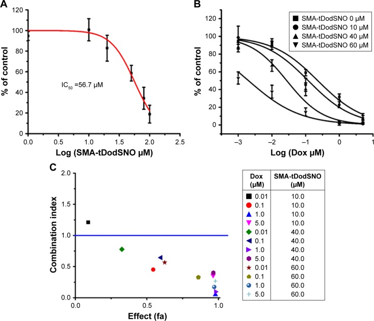
Purpose: Anticancer drug delivery systems are often limited by hurdles, such as off-target distribution, slow cellular internalization, limited lysosomal escape, and drug resistance. To overcome these limitations, we have developed a stable nitric oxide (NO)-releasing nanoparticle (polystyrene-maleic acid [SMA]-tert-dodecane S-nitrosothiol [tDodSNO]) with the aim of enhancing the anticancer properties of doxorubicin (Dox) and a Dox-loaded nanoparticle (SMA-Dox) carrier.

Materials and methods: Effects of SMA-tDodSNO and/or in combination with Dox or SMA-Dox on cell viability, apoptosis, mitochondrial membrane potential, lysosomal membrane permeability, tumor tissue, and tumor growth were studied using in vitro and in vivo model of triple-negative breast cancer (TNBC). In addition, the concentrations of SMA-Dox and Dox in combination with SMA-tDodSNO were measured in cells and tumor tissues.

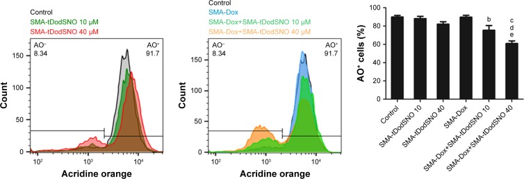
Results: Combination of SMA-tDodSNO and Dox synergistically decreased cell viability and induced apoptosis in 4T1 (TNBC cells). Incubation of 4T1 cells with SMA-tDodSNO (40 µM) significantly enhanced the cellular uptake of SMA-Dox and increased Dox concentration in the cells resulting in a twofold increase (P<0.001). Lysosomal membrane integrity, evaluated by acridine orange (AO) staining, was impaired by 40 µM SMA-tDodSNO (P<0.05 vs control) and when combined with SMA-Dox, this effect was significantly potentiated (P<0.001 vs SMA-Dox). Subcutaneous administration of SMA-tDodSNO (1 mg/kg) to xenografted mice bearing 4T1 cells showed that SMA-tDodSNO alone caused a twofold decrease in the tumor size compared to the control group. SMA-tDodSNO in combination with SMA-Dox resulted in a statistically significant 4.7-fold reduction in the tumor volume (P<0.001 vs control), without causing significant toxicity as monitored through body weight loss.

Conclusion: Taken together, these results suggest that SMA-tDodSNO can be used as a successful strategy to increase the efficacy of Dox and SMA-Dox in a model of TNBC.

Keywords: SMA-tDodSNO; biologic barriers; doxorubicin; nanoparticles; nitric oxide; synergistic cytotoxicity.


Conflict of interest statement

Disclosure The authors report no conflicts of interest in this work.

Figures

Figure 1
Figure 1
The effect of SMA-tDodSNO and/or Dox on the 4T1 cell proliferation. Notes: (A) SMA-tDodSNO showed cytotoxicity with an IC50 value of 56 µM. (B) Addition of SMA-tDodSNO to Dox potentiated cell toxicity of Dox and the IC50 shifted from 205±34 to 1.79±0.51 nM (P<0.001). (C) CI vs the cytotoxic effect of the treatments. The cytotoxicity of the cells treated with Dox (0.01, 0.1, 1, and 5 µM) and/or SMA-tDodSNO (10, 40, and 60 µM) was used for analysis of CI effect using Chou-Talay methodology. Apart from combination at the lowest concentrations (Dox 0.01 µM and SMA-tDodSNO 10 µM), all other doses showed a synergistic activity. Abbreviations: CI, combination index; Dox, doxorubicin; SMA, polystyrene-maleic acid; tDodSNO, tert-dodecane S-nitrosothiol.
Figure 2
Figure 2
Flow cytometric analysis of cell cycle parameters. 4T1 cells were incubated with Dox (0.1 µM) and/or SMA-tDodSNO (10 or 40 µM) for 48 h. Notes: The treatments caused significant increase of the cell population in subG1 phase. Data are expressed as mean values ± SD (n=3). aP<0.05, bP<0.05, cP<0.001 vs control. dP<0.05, pP<0.001 vs Dox, and eP<0.05, fP<0.01, gP<0.001 vs respective SMA-tDodSNO group. Abbreviations: Dox, doxorubicin; SMA, polystyrene-maleic acid; tDodSNO, tert-dodecane S-nitrosothiol.
Figure 3
Figure 3
Flow cytometric analysis of apoptosis in 4T1 cells following incubation with Dox (0.1 µM) and/or SMA-tDodSNO (10 or 40 µM) for 48 hours and Annexin V/PI double staining. Notes: The percentage of viable cells significantly decreased (P<0.001) and apoptotic cell population (Annexin V+/PI-) significantly enhanced by Dox-SMA-tDodSNO 40 µM (P<0.05). Data are expressed as mean values ± SD (n=3). aP<0.05, cP<0.001 vs control. dP<0.05 vs Dox and eP<0.05 vs respective SMA-tDodSNO group. Abbreviations: Dox, doxorubicin; PI, propidium iodide; SMA, polystyrene-maleic acid; tDodSNO, tert-dodecane S-nitrosothiol.
Figure 4
Figure 4
Mitochondrial membrane depolarization was measured using TMRE staining of 4T1 cells treated with Dox (0.1 µM) and/or SMA-tDodSNO (10 and 40 µM) for 48 hours. Notes: Data are expressed as mean values ± SD (n=3). bP<0.01, cP<0.001 vs control, dP<0.05 vs Dox. Abbreviations: Dox, doxorubicin; SMA, polystyrene-maleic acid; tDoDSNO, tert-dodecane S-nitrosothiol; TMRE, tetramethyl-rhodamine ethyl ester.
Figure 5
Figure 5
Effect of SMA-tDodSNO on SMA-Dox endocytosis. Notes: Cells were treated with SMA-Dox (1 µM) and SMA-tDodSNO (10 or 40 µM) for 4 hours. The combination resulted in a significant increase in the SMA-Dox uptake in the cells. Data are expressed as mean values ± SD (n=3). dP<0.01 and pP <0.001 vs SMA-Dox group. Abbreviations: Dox, doxorubicin; SMA, polystyrene-maleic acid; tDoDSNO, tert-dodecane S-nitrosothiol.
Figure 6
Figure 6
SMA-tDodSNO enhances Dox concentration in 4T1 cells. Cells were treated with Dox (0.1 μM) with or without SMA-tDodSNO (10 or 40 μM) for 48 hours. Notes: Data are expressed as mean values ± SD (n=3). pP,0.001 vs Dox group. Abbreviations: Dox, doxorubicin; SMA, polystyrene-maleic acid; tDoDSNO, tert-dodecane S-nitrosothiol.
Figure 7
Figure 7
SMA-tDodSNO treatment impaired lysosomal membrane permeability. Notes: The cells were treated with SMA-Dox (1 µM) and/or SMA-tDodSNO (10 and 40 µM) for 4 hours, then stained by AO. The cells with high fluorescent intensity were named as AO+. Treatment of the cells with SMA-tDodSNO (40 µM) decreased significantly the percentage of AO+ cells. In addition, the combination of SMA-tDodSNO and SMA-Dox resulted in a significant decrease in the AO+ cells compared to either treatment alone. Data are expressed as mean values ± SD (N=3). bP<0.01 and cP<0.001 vs control, dP<0.05 and eP<0.05 vs SMA-Dox and SMA-tDodSNO (40 µM), respectively. Abbreviations: AO, acridine orange; Dox, doxorubicin; SMA, polystyrene-maleic acid; tDodSNO, tert-dodecane S-nitrosothiol.
Figure 8
Figure 8
Effect of SMA-tDodSNO on lysosomal membrane permeability. Notes: The cells were treated with Dox (0.1 µM) and/or SMA-tDodSNO (10 and 40 µM) for 48 hours, then stained by AO. In normal cells, the lysosomal compartments (red dots) have a small size and are evenly disbursed throughout the cell. In Dox- and SMA-tDodSNO-treated cells, the total red fluorescence of the cells decreased, and some of the fluorescence areas were larger in size and localized to few parts of the cells. Abbreviations: AO, acridine orange; Dox, doxorubicin; SMA, polystyrene-maleic acid; tDodSNO, tert-dodecane S-nitrosothiol.
Figure 9
Figure 9
Effect of subcutaneous administration of SMA-tDodSNO on the concentration of SMA-Dox in tumor tissue. Notes: To mice, intravenous SMA-Dox (5 mg/kg) or intravenous SMA-Dox (5 mg/kg)+ subcutaneous SMA-tDodSNO (1 mg/kg) were injected, and 24 hours later, the concentration of SMA-Dox in tumor tissue was evaluated. SMA-tDodSNO increased the tumor concentration of Dox; however, it was not statistically significant. Results are expressed as the mean ± SD (n=4). Abbreviations: Dox, doxorubicin; SMA, polystyrene-maleic acid; tDodSNO, tert-dodecane S-nitrosothiol.
Figure 10
Figure 10
Effect of SMA-tDodSNO and SMA-Dox on tumor growth. Notes: SMA-tDodSNO (1 mg/kg; subcutaneous) and SMA-Dox (5 mg/kg; intravenous) were administered alone and in combination to mice bearing mammary 4T1 tumors at days 0 and 8 of the study. The normalized tumor volume was evaluated as a function of time. The difference between the combination and either treatment alone (A) at day 6 was statistically significant (P=0.0406 for SMA-tDodSNO, and vs SMA-Dox P=0.023). (B) Body weight was evaluated as an indicator of general acute toxicity normalized. Data are expressed as mean ± SD (n=5). aP<0.001 vs control. Abbreviations: Dox, doxorubicin; SMA, polystyrene-maleic acid; tDodSNO, tert-dodecane S-nitrosothiol.
Figure 11
Figure 11
Possible mechanisms by which SMA-tDodSNO potentiates the anticancer efficacy of SMA-Dox and Dox. Notes: Due to sustained NO release, the vessels can be dilated and blood supply to the tumor can be enhanced, rendering vessels permeable to SMA-Dox. Endocytosis of SMA-Dox could also be enhanced when combined with SMA-tDodSNO. Endosomal escape of SMA-Dox could be facilitated, given the higher endosomal membrane permeability caused by SMA-tDodSNO treatment. In addition, SMA-tDodSNO as an NO-releasing agent can sensitize cancer cells to chemotherapeutic drugs such as Dox. Abbreviations: Dox, doxorubicin; EPR, enhanced permeability and retention; NO, nitric oxide; RNS, reactive nitrogen species; ROS, reactive nitrogen species; SMA, polystyrene-maleic acid; tDodSNO, tert-dodecane S-nitrosothiol.

References

    1. Greish K. Enhanced permeability and retention of macromolecular drugs in solid tumors: a royal gate for targeted anticancer nanomedicines. J Drug Target. 2007;15(7–8):457–464. - PubMed
    1. Maeda H, Nakamura H, Fang J. The EPR effect for macromolecular drug delivery to solid tumors: improvement of tumor uptake, lowering of systemic toxicity, and distinct tumor imaging in vivo. Adv Drug Deliv Rev. 2013;65(1):71–79. - PubMed
    1. Maeda H, Wu J, Sawa T, Matsumura Y, Hori K. Tumor vascular permeability and the EPR effect in macromolecular therapeutics: a review. J Control Release. 2000;65(1–2):271–284. - PubMed
    1. Alimoradi H, Matikonda SS, Gamble AB, Giles GI, Greish K. Hypoxia responsive drug delivery systems in tumor therapy. Curr Pharm Des. 2016;22(19):2808–2820. - PubMed
    1. Barua S, Mitragotri S. Challenges associated with penetration of nanoparticles across cell and tissue barriers: a review of current status and future prospects. Nano Today. 2014;9(2):223–243. - PMC - PubMed

MeSH terms