

Spatial shifts in frame-based Gamma Knife radiosurgery: A case for cone beam CT imaging as quality assurance using the Gamma Knife® Icon™
- PMID: 30538892
- PMCID: PMC6255723
Spatial shifts in frame-based Gamma Knife radiosurgery: A case for cone beam CT imaging as quality assurance using the Gamma Knife® Icon™
Abstract
Background: Cone beam CT (CBCT) imaging has been integrated into the most recent version of the Leksell Gamma Knife for the primary purpose to facilitate fractionated therapy.
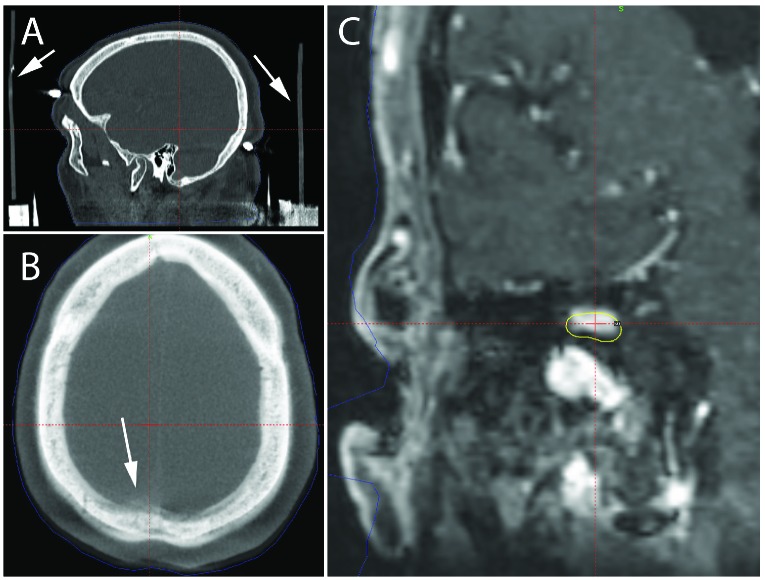
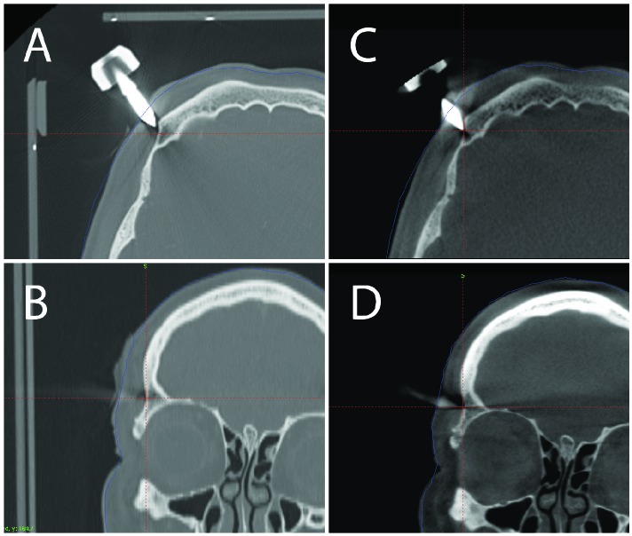
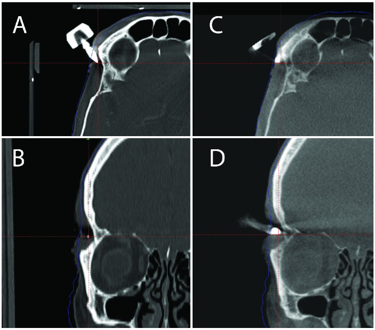
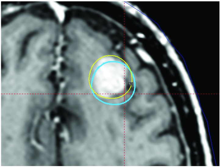
Case description: This case study presents three patients where the CBCT system of the Gamma Knife Icon discovered potentially clinically significant frame shifts. In each case, patients were imaged with volumetric MR prior to stereotactic frame placement. Immediately following frame placement, diagnostic stereotactic CT imaging was acquired with a stereotactic indicator box attached to the frame. Following treatment planning and immediately before radiosurgery, a CBCT was acquired using the on-board imaging functionality of the Gamma Knife Icon, which provides a registration of the patient's anatomy to stereotactic space independent of that provided by the stereotactic frame/fiducials. Co-registration of the CT and CBCT provides an estimate of the difference between these two estimates of stereotactic coordinates. The vector magnitudes of the differences measured at the center of stereotactic space were 0.93mm, 2.64mm and 2.18 mm for Case 1, Case 2 and Case 3 respectively.
Conclusions: Use of the CBCT functionality of the Gamma Knife Icon to verify the consistency of frame placement can prevent clinically significant targeting errors due to frame slippage or frame adapter mounting errors, and allows any required adjustments to be made without interrupting the overall treatment workflow.
Keywords: Gamma Knife; cone beam CT; frame shift; quality assurance; radiosurgery; stereotactic frame.
Conflict of interest statement
Authors’ disclosure of potential conflicts of interest Dr. Schlesinger reports grant from Elekta Instrument, AB, outside the submitted work. Dr. Trifiletti reports other support from Novocure, outside the submitted work. Drs. Dutta, Larner, Peach and Sheehan have nothing to disclose.
Figures


Similar articles
-
Gamma Knife® icon CBCT offers improved localization workflow for frame-based treatment.J Appl Clin Med Phys. 2019 Nov;20(11):95-103. doi: 10.1002/acm2.12745. Epub 2019 Oct 6. J Appl Clin Med Phys. 2019. PMID: 31587520 Free PMC article.
-
Evaluation of stability of stereotactic space defined by cone-beam CT for the Leksell Gamma Knife Icon.J Appl Clin Med Phys. 2017 May;18(3):67-72. doi: 10.1002/acm2.12073. Epub 2017 Apr 17. J Appl Clin Med Phys. 2017. PMID: 28419781 Free PMC article.
-
Quality assurance tests for the Gamma Knife® Icon™ image guidance system.J Appl Clin Med Phys. 2018 Sep;19(5):573-579. doi: 10.1002/acm2.12417. Epub 2018 Aug 3. J Appl Clin Med Phys. 2018. PMID: 30076672 Free PMC article.
-
Frame versus Frameless Leksell Stereotactic Radiosurgery.Prog Neurol Surg. 2019;34:19-27. doi: 10.1159/000493046. Epub 2019 May 16. Prog Neurol Surg. 2019. PMID: 31096212 Review.
-
[Gamma knife versus stereotactic linear accelerator. Utilization, clinical results and cost-benefit relations].Radiologe. 1996 Apr;36(4):345-53. doi: 10.1007/s001170050081. Radiologe. 1996. PMID: 8677327 Review. German.
Cited by
-
Single Fraction Frameless Stereotactic Radiosurgery on the Gamma Knife Icon for Patients With Brain Metastases: Time to Abandon the Frame?Adv Radiat Oncol. 2021 Jun 6;6(5):100736. doi: 10.1016/j.adro.2021.100736. eCollection 2021 Sep-Oct. Adv Radiat Oncol. 2021. PMID: 34646964 Free PMC article.
-
Gamma Knife® icon CBCT offers improved localization workflow for frame-based treatment.J Appl Clin Med Phys. 2019 Nov;20(11):95-103. doi: 10.1002/acm2.12745. Epub 2019 Oct 6. J Appl Clin Med Phys. 2019. PMID: 31587520 Free PMC article.
-
Predictors of Treatment Interruption During Frameless Gamma Knife Icon Stereotactic Radiosurgery.Adv Radiat Oncol. 2020 Jul 1;5(6):1152-1157. doi: 10.1016/j.adro.2020.06.025. eCollection 2020 Nov-Dec. Adv Radiat Oncol. 2020. PMID: 33305076 Free PMC article.
-
Stereotactic radiosurgery for multiple small brain metastases using gamma knife versus single-isocenter VMAT: Normal brain dose based on lesion number and size.J Appl Clin Med Phys. 2025 Jun;26(6):e70065. doi: 10.1002/acm2.70065. Epub 2025 Mar 19. J Appl Clin Med Phys. 2025. PMID: 40108742 Free PMC article.
-
Discrepancies between frame- and CBCT-based stereotactic space definition on the Gamma Knife Icon.J Appl Clin Med Phys. 2022 Jul;23(7):e13637. doi: 10.1002/acm2.13637. Epub 2022 May 30. J Appl Clin Med Phys. 2022. PMID: 35635549 Free PMC article.
References
-
- Wu A. Physics and dosimetry of the gamma knife. Neurosurg Clin N Am 1992;3(1):35-50 - PubMed
-
- Leksell L, Jernberg B. Stereotaxis and tomography. A technical note. Acta Neurochir (Wien) 1980;52(1-2):1-7 - PubMed
-
- Lindquist C, Paddick I. The Leksell Gamma Knife Perfexion and comparisons with its predecessors. Neurosurgery 2007;61(3 Suppl):130-140; discussion 140-131 - PubMed
-
- Zeverino M, Jaccard M, Patin D, Ryckx N, Marguet M, Tuleasca C, Schiappacasse L, Bourhis J, Levivier M, Bochud FO, Moeckli R. Commissioning of the Leksell Gamma Knife(R) Icon. Med Phys 2017;44(2):355-363 - PubMed
LinkOut - more resources
Full Text Sources