

An Introduction to the Performance of Immunohistochemistry
- PMID: 30539453
- PMCID: PMC6749998
- DOI: 10.1007/978-1-4939-8935-5_25
An Introduction to the Performance of Immunohistochemistry
Abstract
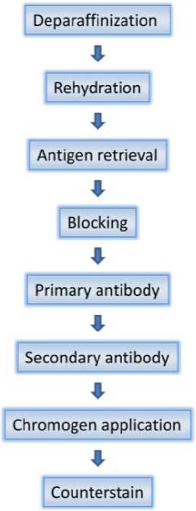
Immunohistochemistry (IHC) is a powerful technique that exploits the specific binding between an antibody and antigen to detect and localize specific antigens in cells and tissue, most commonly detected and examined with the light microscope. A standard tool in many fields in the research setting, IHC has become an essential ancillary technique in clinical diagnostics in anatomic pathology (Lin F, Chen Z. Arch Pathol Lab Med 138:1564-1577, 2014) with the advent of antigen retrieval methods allowing it to be performed conveniently on formalin fixed paraffin embedded (FFPE) tissue (Taylor CR, Shi S-R, Barr NJ. Techniques of immunohistochemistry: principles, pitfalls, and standardization. In: Dabbs DJ (ed) Diagnostic immunohistochemistry: theranostic and genomic applications, 3rd edn. Saunders, Philadelphia, 2010; Shi SR, Key ME, Kalra KL. J Histochem Cytochem 39:741-748, 1991) and automated methods for high volume processing with reproducibility (Prichard J, Hicks D, Hammond E. Automated immunohistochemistry overview. In: Fan L, Jeffrey P (eds) Handbook of practical immunohistochemistry: frequently asked questions, 2nd edn. Springer, New York, 2015). IHC is frequently utilized to assist in the classification of neoplasms, determination of a metastatic tumor's site of origin and detection of tiny foci of tumor cells inconspicuous on routine hematoxylin and eosin (H&E) staining. Furthermore, it is increasingly being used to provide predictive and prognostic information, such as in testing for HER2 amplification in breast cancer (Wolff AC, Hammond MEH, Hicks DG et al. Arch Pathol Lab Med 138:241-256, 2014) in addition to serving as surrogate markers for molecular alterations in neoplasms, including IDH1 and ATRX mutations in brain tumors (Appin CL, Brat DJ. Mol Aspects Med. 45:87-96, 2015). In this chapter we describe the basic methods of immunohistochemical staining which has become an essential tool in the daily practice of anatomic pathology worldwide.
Keywords: Antibodies; Antigen retrieval; Antigens; Immunohistochemistry; Light microscopy.
References
-
- Lin F, Chen Z (2014) Standardization of diagnostic immunohistochemistry: literature review and Geisinger experience. Arch Pathol Lab Med 138:1564–1577 - PubMed
-
- Taylor CR, Shi S-R, Barr NJ (2010) Techniques of immunohistochemistry: principles, pitfalls, and standardization In: Dabbs DJ (ed) Diagnostic immunohistochemistry: theranostic and genomic applications, 3rd edn. Saunders, Philadelphia
-
- Shi SR, Key ME, Kalra KL (1991) Antigen retrieval in formalin-fixed, paraffin-embedded tissues: an enhancement method for immunohistochemical staining based on microwave oven heating of tissue sections. J Histochem Cytochem 39:741–748 - PubMed
-
- Prichard J, Hicks D, Hammond E (2015) Automated immunohistochemistry overview In: Fan L, Jeffrey P (eds) Handbook of practical immunohistochemistry: frequently asked questions, 2nd edn. Springer, New York
Publication types
MeSH terms
Substances
Grants and funding
LinkOut - more resources
Full Text Sources
Other Literature Sources
Research Materials
Miscellaneous
