. 2019 Mar-Apr;41(2):101-111.
doi: 10.1590/1516-4446-2017-0002. Epub 2018 Dec 6.

Topography of 11C-Pittsburgh compound B uptake in Alzheimer's disease: a voxel-based investigation of cortical and white matter regions

Affiliations

Topography of 11C-Pittsburgh compound B uptake in Alzheimer's disease: a voxel-based investigation of cortical and white matter regions

Daniele de P Faria et al. Braz J Psychiatry. 2019 Mar-Apr.

Abstract

Objective: To compare results of positron emission tomography (PET) with carbon-11-labeled Pittsburgh compound B (11C-PIB) obtained with cerebellar or global brain uptake for voxel intensity normalization, describe the cortical sites with highest tracer uptake in subjects with mild Alzheimer's disease (AD), and explore possible group differences in 11C-PIB binding to white matter.

Methods: 11C-PIB PET scans were acquired from subjects with AD (n=17) and healthy elderly controls (n=19). Voxel-based analysis was performed with statistical parametric mapping (SPM).

Results: Cerebellar normalization showed higher 11C-PIB uptake in the AD group relative to controls throughout the cerebral cortex, involving the lateral temporal, orbitofrontal, and superior parietal cortices. With global uptake normalization, greatest cortical binding was detected in the orbitofrontal cortex; decreased 11C-PIB uptake in white matter was found in the posterior hippocampal region, corpus callosum, pons, and internal capsule.

Conclusion: The present case-control voxelwise 11C-PIB PET comparison highlighted the regional distribution of amyloid deposition in the cerebral cortex of mildly demented AD patients. Tracer uptake was highest in the orbitofrontal cortex. Decreased 11C-PIB uptake in white-matter regions in this patient population may be a marker of white-matter damage in AD.


Conflict of interest statement

The authors report no conflicts of interest.

Figures
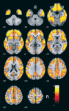
Figure 1
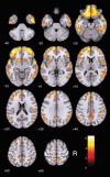
Figure 1. Findings of increased carbon-11 labeled Pittsburgh compound B (11C-PIB) uptake in patients with Alzheimer’s disease (n=17) in comparison with healthy elderly controls (n=19) after cerebellum gray-matter normalization, highlighted in yellow (at the z >3.09 cutoff, corresponding to p < 0.001 uncorrected for multiple comparisons, and using an extent threshold of 10 voxels). Findings are overlaid on axial brain slices spatially normalized into an approximation to the Talairach and Tournoux stereotactic atlas, and the numbers associated with each frame represent standard coordinates in the z -axis. Clusters of increased 11C-PIB uptake are shown throughout the cerebral cortex. R = right side.
Figure 2
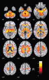
Figure 2. Findings of increased carbon-11 labeled Pittsburgh compound B (11C-PIB) uptake in patients with Alzheimer’s disease (n=17) in comparison with healthy elderly controls (n=19) after global brain normalization, highlighted in yellow (at the z > 3.09 cutoff, corresponding to p < 0.001 uncorrected for multiple comparisons, and using an extent threshold of 10 voxels). Findings are overlaid on axial brain slices spatially normalized into an approximation to the Talairach and Tournoux stereotactic atlas, and the numbers associated with each frame represent standard coordinates in the z-axis. Clusters are shown in several portions of the cerebral cortex, with the greatest size and statistical significance in the orbitofrontal cortex (Table 3). R = right side.
Figure 3
Figure 3. Illustrative positron emission tomography (PET) with carbon-11 labeled Pittsburgh compound B (11C-PIB) images (top) are shown for a healthy control subject (left-side) and an AD patient (right-side) with regional tracer uptake data normalized either to total PIB uptake in the brain (A) or to cerebellar grey-matter uptake (B). The respective time-activity curves are shown at the bottom. AD = Alzheimer’s disease; R = right side.
Figure 4
Figure 4. Findings of decreased carbon-11 labeled Pittsburgh compound B (11C-PIB) uptake in patients with Alzheimer’s disease (n=17) compared with healthy elderly controls (n=19) after global brain uptake normalization, highlighted in yellow (at the z > 3.09 cutoff, uncorrected for multiple comparisons and using an extent threshold of 10 voxels). Findings are overlaid on axial brain slices spatially normalized into an approximation to the Talairach and Tournoux stereotactic atlas, and the numbers associated with each frame represent standard coordinates in the z-axis. Clusters are shown throughout the cerebral white matter. R = right side.

References

    1. Toussaint PJ, Perlbarg V, Bellec P, Desarnaud S, Lacomblez L, Doyon J, et al. Resting state FDG-PET functional connectivity as an early biomarker of Alzheimer's disease using conjoint univariate and independent component analyses. Neuroimage. 2012;63:936–46. - PubMed
    1. Mosconi L, Tsui WH, Herholz K, Puri A, Drzezga A, Lucignani G, et al. Multicenter standardized 18F-FDG PET diagnosis of mild cognitive impairment, Alzheimer’s disease, and other dementias. J Nucl Med. 2008;49:390–8. - PMC - PubMed
    1. Klunk WE, Engler H, Nordberg A, Wang Y, Blomqvist G, Holt DP, et al. Imaging brain amyloid in Alzheimer’s disease with Pittsburgh compound‐B. Ann Neurol. 2004;55:306–19. - PubMed
    1. Price JC, Klunk WE, Lopresti BJ, Lu X, Hoge JA, Ziolko SK, et al. Kinetic modeling of amyloid binding in humans using PET imaging and Pittsburgh compound-B. J Cereb Blood Flow Metab. 2005;25:1528–47. - PubMed
    1. Drzezga A, Grimmer T, Henriksen G, Stangier I, Perneczky R, Diehl-Schmid J, et al. Imaging of amyloid plaques and cerebral glucose metabolism in semantic dementia and Alzheimer’s disease. Neuroimage. 2008;39:619–33. - PubMed