Review
. 2019 Jan 1;99(1):853-892.
doi: 10.1152/physrev.00005.2018.

Evolving and Expanding the Roles of Mitophagy as a Homeostatic and Pathogenic Process

Affiliations
Review

Evolving and Expanding the Roles of Mitophagy as a Homeostatic and Pathogenic Process

Åsa B Gustafsson et al. Physiol Rev. .

Abstract

The central functions fulfilled by mitochondria as both energy generators essential for tissue homeostasis and gateways to programmed apoptotic and necrotic cell death mandate tight control over the quality and quantity of these ubiquitous endosymbiotic organelles. Mitophagy, the targeted engulfment and destruction of mitochondria by the cellular autophagy apparatus, has conventionally been considered as the mechanism primarily responsible for mitochondrial quality control. However, our understanding of how, why, and under what specific conditions mitophagy is activated has grown tremendously over the past decade. Evidence is accumulating that nonmitophagic mitochondrial quality control mechanisms are more important to maintaining normal tissue homeostasis whereas mitophagy is an acute tissue stress response. Moreover, previously unrecognized mitophagic regulation of mitochondrial quantity control, metabolic reprogramming, and cell differentiation suggests that the mechanisms linking genetic or acquired defects in mitophagy to neurodegenerative and cardiovascular diseases or cancer are more complex than simple failure of normal mitochondrial quality control. Here, we provide a comprehensive overview of mitophagy in cellular homeostasis and disease and examine the most revolutionary concepts in these areas. In this context, we discuss evidence that atypical mitophagy and nonmitophagic pathways play central roles in mitochondrial quality control, functioning that was previously considered to be the primary domain of mitophagy.


Conflict of interest statement

No conflicts of interest, financial or otherwise, are declared by the authors.

Figures

FIGURE 1.
FIGURE 1.
Overview of nonselective autophagy and selective autophagy. The phagophore engulfs cargo in the cytosol nonselectively or selectively to form a double membrane autophagosome. The autophagsome fuses with a lysosome leading to the formation of the autolysosome where the cargo is degraded and then recycled.
FIGURE 2.
FIGURE 2.
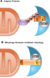
Selective mitophagy. A: an adaptor protein containing a ubiquitin binding domain (UBD) recognizes a polyubiquitinated protein in the outer mitochondrial membrane (OMM). It physically connects the mitochondrion to the autophagosome membrane via its LC3 interacting region (LIR) that binds to lipidated LC3 (GABARAP/Atg8). B: mitophagy receptors, such as Atg32, BNIP3, and Nix, are anchored in the outer mitochondrial membrane via a COOH-terminal transmembrane domain. A LIR motif in the NH2-terminal domain interacts directly with lipidated LC3/GABARAP/Atg8.
FIGURE 3.
FIGURE 3.
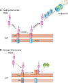
Regulation of PINK1. A: newly synthesized PINK1 is immediately imported into healthy mitochondria with an intact membrane potential (ΔΨ) by the TOM/TIM import machinery. PINK1 undergoes proteolytic cleavage by the mitochondrial intramembrane protease PARL. The cleaved PINK1 retrotranslocates to the cytosol where it is subjected to ubiquitination by E3 ubiquitin ligases UBR1, UBR2, and UBR4 and proteasomal degradation. B: upon mitochondrial damage and loss of ΔΨ, import of PINK1 is abrogated, and it accumulates on the outer mitochondrial membrane which leads to recruitment of Parkin.
FIGURE 4.
FIGURE 4.
Activation of PINK1/Parkin-mediated mitophagy. PINK1 accumulates on the outer mitochondrial membrane (OMM) in the absence of ΔΨ. PINK1 recruits and activates Parkin in a process involving PINK1-mediated phosphorylation of Mfn2, ubiquitin (Ub), and Parkin. Activated Parkin conjugates ubiquitin to various proteins in the OMM. The ubiquitin chains on proteins in the OMM are recognized by autophagy adaptors which in turn tether the ubiquitinated cargo to the autophagosome via binding to lipidated LC3.
FIGURE 5.
FIGURE 5.
Formation of autophagosomes. Activation of the Ulk1/2-Atg13-Fip200 complex by AMP-activated protein kinase (AMPK) initiates the formation of the phagophore through activation of the class III phosphatidylinositol 3-kinase (PI3K) complex composed of Vps34-Vps15-Beclin1-Atg14L. Atg9-positive vesicles contribute membrane to the growing phagophore. The subsequent elongation and closure of the membrane are mediated by two ubiquitin-like conjugation pathways: the Atg5–Atg12 and the microtubule-associated protein 1 light chain 3 (LC3) pathways. 1) The Atg12-Atg5 conjugating system consists of Atg12 (ubiquitin-like module), Atg5 (substrate), Atg7 (E1-like enzyme), and Atg10 (E2-like enzyme). The Atg12–Atg5 conjugate is formed by Atg7 and Atg10. Atg12–Atg5 forms a final complex with Atg16 to form the E3 enzyme for the LC3/Atg8 conjugating system. 2) The LC3 (Atg8) conjugating system consists of LC3 (ubiquitin-like module), Atg4 (a cysteine protease), phosphatidylethanolamine (PE, substrate), Atg7 (E1-like enzyme), Atg3 (E2-like enzyme), and Atg5–Atg12–Atg16 (E3-like enzyme).
FIGURE 6.
FIGURE 6.
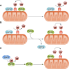
Deubiquitinating enzymes regulate mitophagy. A: USP30 is anchored in the mitochondrial outer membrane and removes the ubiquitin from Parkin substrates to inhibit mitophagy. B: USP35 counteracts Parkin-mediated mitophagy on healthy mitochondria. USP35 dissociates from depolarized mitochondria to allow mitophagy to proceed. C: Parkin activity is inhibited at baseline via auto-ubiquitination. USP8 preferentially removes ubiquitin conjugates from Parkin. This is required for efficient recruitment of Parkin to depolarized mitochondria.
FIGURE 7.
FIGURE 7.
Noncanonical mitophagy pathways. A: alternative Atg5/7-independent autophagy. The autophagosomes are generated from Rab9+ vesicles derived from the trans-Golgi. Double membrane vesicles containing cargo fuse with lysosomes. B: endosomal-mediated elimination of mitochondria. Mitochondria are engulfed into Rab5+ early endosomes that mature into late endosomes before fusing with the lysosomes. C: in microautophagy, mitochondria are directly engulfed by lysosomes.
FIGURE 8.
FIGURE 8.
Generation of mitochondrial-derived vesicles (MDVs) with distinct cargo. Formation of single-membrane vesicles occurs upon mitochondrial exposure to external reactive oxygen species (ROS). In contrast, double-membrane vesicles result from the production of excess ROS inside mitochondria and involve PINK1/Parkin. The MDVs are targeted to the lysosome for degradation of their contents.

References

    1. Abeliovich H, Zarei M, Rigbolt KT, Youle RJ, Dengjel J. Involvement of mitochondrial dynamics in the segregation of mitochondrial matrix proteins during stationary phase mitophagy. Nat Commun 4: 2789, 2013. doi:10.1038/ncomms3789. - DOI - PMC - PubMed
    1. Ahlqvist KJ, Hämäläinen RH, Yatsuga S, Uutela M, Terzioglu M, Götz A, Forsström S, Salven P, Angers-Loustau A, Kopra OH, Tyynismaa H, Larsson NG, Wartiovaara K, Prolla T, Trifunovic A, Suomalainen A. Somatic progenitor cell vulnerability to mitochondrial DNA mutagenesis underlies progeroid phenotypes in Polg mutator mice. Cell Metab 15: 100–109, 2012. doi:10.1016/j.cmet.2011.11.012. - DOI - PubMed
    1. Al Rawi S, Louvet-Vallée S, Djeddi A, Sachse M, Culetto E, Hajjar C, Boyd L, Legouis R, Galy V. Postfertilization autophagy of sperm organelles prevents paternal mitochondrial DNA transmission. Science 334: 1144–1147, 2011. doi:10.1126/science.1211878. - DOI - PubMed
    1. Ali S, Vollaard AM, Widjaja S, Surjadi C, van de Vosse E, van Dissel JT. PARK2/PACRG polymorphisms and susceptibility to typhoid and paratyphoid fever. Clin Exp Immunol 144: 425–431, 2006. doi:10.1111/j.1365-2249.2006.03087.x. - DOI - PMC - PubMed
    1. Aoki Y, Kanki T, Hirota Y, Kurihara Y, Saigusa T, Uchiumi T, Kang D. Phosphorylation of Serine 114 on Atg32 mediates mitophagy. Mol Biol Cell 22: 3206–3217, 2011. doi:10.1091/mbc.e11-02-0145. - DOI - PMC - PubMed

Publication types

LinkOut - more resources