

Cerebrospinal fluid and the early brain development of autism
- PMID: 30541429
- PMCID: PMC6292033
- DOI: 10.1186/s11689-018-9256-7
Cerebrospinal fluid and the early brain development of autism
Abstract
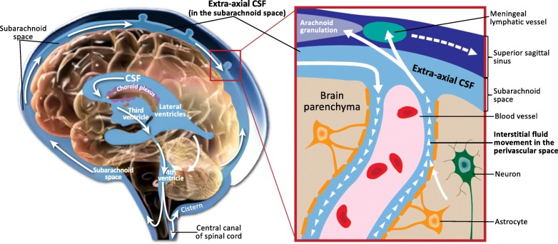
Background: There is currently a renaissance of interest in the many functions of cerebrospinal fluid (CSF). Altered flow of CSF, for example, has been shown to impair the clearance of pathogenic inflammatory proteins involved in neurodegenerative diseases, such as amyloid-β. In addition, the role of CSF in the newly discovered lymphatic system of the brain has become a prominently researched area in clinical neuroscience, as CSF serves as a conduit between the central nervous system and immune system.
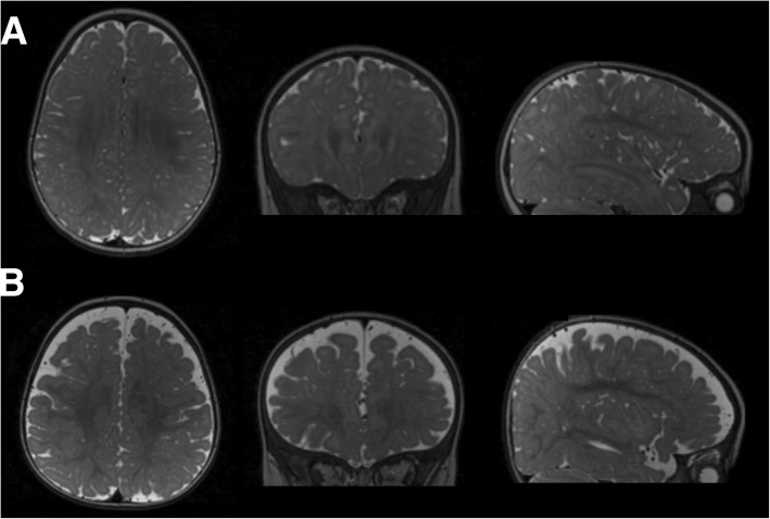
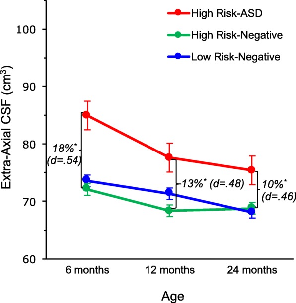
Main body: This article will review the importance of CSF in regulating normal brain development and function, from the prenatal period throughout the lifespan, and highlight recent research that CSF abnormalities in autism spectrum disorder (ASD) are present in infancy, are detectable by conventional structural MRI, and could serve as an early indicator of altered neurodevelopment.
Conclusion: The identification of early CSF abnormalities in children with ASD, along with emerging knowledge of the underlying pathogenic mechanisms, has the potential to serve as early stratification biomarkers that separate children with ASD into biological subtypes that share a common pathophysiology. Such subtypes could help parse the phenotypic heterogeneity of ASD and map on to targeted, biologically based treatments.
Keywords: Autism spectrum disorder; Biomarkers; Brain development; Brain enlargement; Cerebrospinal fluid; Early risk signs; Extra-axial cerebrospinal fluid; Glymphatic system; Heterogeneity; Infancy; Lateral ventricles; Neural meningeal lymphatic system; Neuroinflammation; Stratification biomarker.
Conflict of interest statement
Ethics approval and consent to participate
Not applicable
Consent for publication
Not applicable
Competing interests
The author declares that he has no competing interests.
Publisher’s Note
Springer Nature remains neutral with regard to jurisdictional claims in published maps and institutional affiliations.
Figures
Similar articles
-
Extra-axial cerebrospinal fluid in high-risk and normal-risk children with autism aged 2-4 years: a case-control study.Lancet Psychiatry. 2018 Nov;5(11):895-904. doi: 10.1016/S2215-0366(18)30294-3. Epub 2018 Sep 27. Lancet Psychiatry. 2018. PMID: 30270033 Free PMC article.
-
Increased Extra-axial Cerebrospinal Fluid in High-Risk Infants Who Later Develop Autism.Biol Psychiatry. 2017 Aug 1;82(3):186-193. doi: 10.1016/j.biopsych.2017.02.1095. Epub 2017 Mar 6. Biol Psychiatry. 2017. PMID: 28392081 Free PMC article.
-
The Emerging Clinical Neuroscience of Autism Spectrum Disorder: A Review.JAMA Psychiatry. 2018 May 1;75(5):514-523. doi: 10.1001/jamapsychiatry.2017.4685. JAMA Psychiatry. 2018. PMID: 29590280 Review.
-
Brain and behavior development in autism from birth through infancy.Dialogues Clin Neurosci. 2017 Dec;19(4):325-333. doi: 10.31887/DCNS.2017.19.4/mshen. Dialogues Clin Neurosci. 2017. PMID: 29398928 Free PMC article.
-
Overview of genetic models of autism spectrum disorders.Prog Brain Res. 2018;241:1-36. doi: 10.1016/bs.pbr.2018.10.002. Epub 2018 Nov 7. Prog Brain Res. 2018. PMID: 30447752 Review.
Cited by
-
Looking Back at the Next 40 Years of ASD Neuroscience Research.J Autism Dev Disord. 2021 Dec;51(12):4333-4353. doi: 10.1007/s10803-021-05095-5. Epub 2021 May 27. J Autism Dev Disord. 2021. PMID: 34043128 Free PMC article. Review.
-
Volumetric Analysis of Amygdala and Hippocampal Subfields for Infants with Autism.J Autism Dev Disord. 2023 Jun;53(6):2475-2489. doi: 10.1007/s10803-022-05535-w. Epub 2022 Apr 7. J Autism Dev Disord. 2023. PMID: 35389185 Free PMC article.
-
The development of the social brain in baby siblings of children with autism.Curr Opin Psychiatry. 2020 Mar;33(2):110-116. doi: 10.1097/YCO.0000000000000572. Curr Opin Psychiatry. 2020. PMID: 31815759 Free PMC article. Review.
-
Enlarged Perivascular Spaces in Infancy and Autism Diagnosis, Cerebrospinal Fluid Volume, and Later Sleep Problems.JAMA Netw Open. 2023 Dec 1;6(12):e2348341. doi: 10.1001/jamanetworkopen.2023.48341. JAMA Netw Open. 2023. PMID: 38113043 Free PMC article.
-
Brain perivascular spaces and autism: clinical and pathogenic implications from an innovative volumetric MRI study.Front Neurosci. 2023 Jun 23;17:1205489. doi: 10.3389/fnins.2023.1205489. eCollection 2023. Front Neurosci. 2023. PMID: 37425010 Free PMC article.
