Review
. 2018 Dec 12;11(12):dmm037127.
doi: 10.1242/dmm.037127.

Hypoxia and connectivity in the developing vertebrate nervous system

Affiliations
Review

Hypoxia and connectivity in the developing vertebrate nervous system

Joshua L Bonkowsky et al. Dis Model Mech. .

Abstract

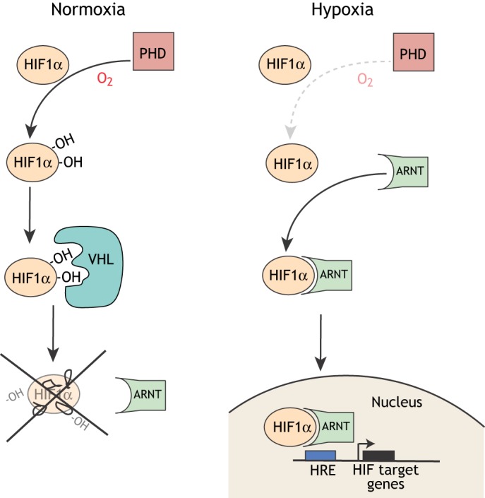
The developing nervous system depends upon precise regulation of oxygen levels. Hypoxia, the condition of low oxygen concentration, can interrupt developmental sequences and cause a range of molecular, cellular and neuronal changes and injuries. The roles and effects of hypoxia on the central nervous system (CNS) are poorly characterized, even though hypoxia is simultaneously a normal component of development, a potentially abnormal environmental stressor in some settings, and a clinically important complication, for example of prematurity. Work over the past decade has revealed that hypoxia causes specific disruptions in the development of CNS connectivity, altering axon pathfinding and synapse development. The goals of this article are to review hypoxia's effects on the development of CNS connectivity, including its genetic and molecular mediators, and the changes it causes in CNS circuitry and function due to regulated as well as unintended mechanisms. The transcription factor HIF1α is the central mediator of the CNS response to hypoxia (as it is elsewhere in the body), but hypoxia also causes a dysregulation of gene expression. Animals appear to have evolved genetic and molecular responses to hypoxia that result in functional behavioral alterations to adapt to the changes in oxygen concentration during CNS development. Understanding the molecular pathways underlying both the normal and abnormal effects of hypoxia on CNS connectivity may reveal novel insights into common neurodevelopmental disorders. In addition, this Review explores the current gaps in knowledge, and suggests important areas for future studies.

Keywords: Connectivity; Hypoxia; Neuroscience; Pathfinding.


Conflict of interest statement

Competing interestsThe authors declare no competing or financial interests.

Figures

Fig. 1.
Fig. 1.
A simplified schematic of the basic cellular response to hypoxia. In normoxia, prolyl hydroxylases (PHDs) hydroxylate HIF1α, which allows the Von Hippel Lindau (VHL) protein to bind HIF1α, leading to degradation of HIF1α. Under hypoxic conditions, HIF1α is not hydroxylated or degraded and dimerizes with ARNT (also known as HIF1β), translocates to the nucleus and, with the transcriptional coactivator CBP/p300, binds genomic DNA at hypoxia response elements (HREs) to activate transcription of target genes.
Fig. 2.
Fig. 2.
The hypoxia experimental system and examples of methods for the analysis of CNS connectivity. (A) Schematic diagram of the hypoxia system for experiments with zebrafish (Stevenson et al., 2012). Embryos are placed in a sealed plexiglass chamber where oxygen levels are manipulated by a controller that monitors oxygen (O2) levels and adjusts nitrogen (N2) flow to displace O2. Altering the timing of hypoxia exposure allows researchers to examine the effects on connectivity at different stages of embryo development, for example, via immunofluorescence axon labeling (experimental procedure shown on the right). (B) Examples of axon pathfinding analysis. The left panel shows the region of the zebrafish CNS imaged in the analysis. The center and right panels show confocal images of the forebrain, both of which are dorsal views with the rostrum at the top. A pan-axonal antibody such as anti-(acetylated) tubulin (central panel, green) labels all of the axon tracts, which permits visualization of significant changes in axon pathfinding upon exposure to hypoxia. An axon reporter expressed in a genetically defined group of axon tracts, for example EGFP-CAAX driven by the foxP2-A.2 enhancer (right panel, red) (Bonkowsky et al., 2008), only labels a subset of axons but allows more precise tracking of axon pathfinding changes. (C) Examples of synapse analysis. Confocal images show lateral views of the zebrafish trunk/spinal cord, dorsal to the top, rostral to the right. A pan-synaptic antibody (anti-PSD95; red signal, white arrowheads) labels all synapses; this makes it difficult to determine what is happening to any specific set of neurons. A genetically targeted synapse label, such as a FingR (Son et al., 2016), shown schematically, can be targeted against PSD95. By expressing the PSD95-FingR (green signal, black arrowheads) under the control of an enhancer or other transgene, researchers can track hypoxia-induced synaptic changes in a genetically defined group of neurons. Scale bars: 50 µm (10 µm in enlarged image).
Fig. 3.
Fig. 3.
Examples of conserved CNS connectivity responses to hypoxia across evolution, including metazoans with bilaterally symmetric nervous systems. There are no HIF homologs in yeast or primitive metazoans. Animals shown are nematodes (C. elegans), fish species including zebrafish, mice and primates/humans.

Similar articles

Cited by

References

    1. Azad P., Stobdan T., Zhou D., Hartley I., Akbari A., Bafna V. and Haddad G. G. (2017). High-altitude adaptation in humans: from genomics to integrative physiology. J. Mol. Med. (Berl.) 95, 1269-1282. 10.1007/s00109-017-1584-7 - DOI - PMC - PubMed
    1. Back S. A. (2014). Cerebral white and gray matter injury in newborns: new insights into pathophysiology and management. Clin. Perinatol. 41, 1-24. 10.1016/j.clp.2013.11.001 - DOI - PMC - PubMed
    1. Baribeau D. A. and Anagnostou E. (2013). A comparison of neuroimaging findings in childhood onset schizophrenia and autism spectrum disorder: a review of the literature. Front. Psychiatry. 4, 175 10.3389/fpsyt.2013.00175 - DOI - PMC - PubMed
    1. Barrett R. D., Bennet L., Davidson J., Dean J. M., George S., Emerald B. S. and Gunn A. J. (2007). Destruction and reconstruction: hypoxia and the developing brain. Birth Defects Res. C Embryo Today 81, 163-176. 10.1002/bdrc.20095 - DOI - PubMed
    1. Bartoszewska S., Kochan K., Piotrowski A., Kamysz W., Ochocka R. J., Collawn J. F. and Bartoszewski R. (2015). The hypoxia-inducible miR-429 regulates hypoxia-inducible factor-1alpha expression in human endothelial cells through a negative feedback loop. FASEB J. 29, 1467-1479. 10.1096/fj.14-267054 - DOI - PMC - PubMed

Publication types

Substances