. 2018 Dec 12;3(6):e00493-18.
doi: 10.1128/mSphere.00493-18.

Contact Challenge of Cattle with Foot-and-Mouth Disease Virus Validates the Role of the Nasopharyngeal Epithelium as the Site of Primary and Persistent Infection

Affiliations

Contact Challenge of Cattle with Foot-and-Mouth Disease Virus Validates the Role of the Nasopharyngeal Epithelium as the Site of Primary and Persistent Infection

Carolina Stenfeldt et al. mSphere. .

Abstract

The pathogenesis of foot-and-mouth disease virus (FMDV) in cattle was investigated through early and late stages of infection by use of an optimized experimental model for controlled contact exposure. Time-limited exposure of cattle to FMDV-infected pigs led to primary FMDV infection of the nasopharyngeal mucosa in both vaccinated and nonvaccinated cattle. In nonvaccinated cattle, the infection generalized rapidly to cause clinical disease, without apparent virus amplification in the lungs prior to establishment of viremia. Vaccinated cattle were protected against clinical disease and viremia; however, all vaccinated cattle were subclinically infected, and persistent infection occurred at similarly high prevalences in both animal cohorts. Infection dynamics in cattle were consistent and synchronous and comparable to those of simulated natural and needle inoculation systems. However, the current experimental model utilizes a natural route of virus exposure and is therefore superior for investigations of disease pathogenesis and host response. Deep sequencing of viruses obtained during early infection of pigs and cattle indicated that virus populations sampled from sites of primary infection were markedly more diverse than viruses from vesicular lesions of cattle, suggesting the occurrence of substantial bottlenecks associated with vesicle formation. These data expand previous knowledge of FMDV pathogenesis in cattle and provide novel insights for validation of inoculation models of bovine FMD studies.IMPORTANCE Foot-and-mouth disease virus (FMDV) is an important livestock pathogen that is often described as the greatest constraint to global trade in animal products. The present study utilized a standardized pig-to-cow contact exposure model to demonstrate that FMDV infection of cattle initiates in the nasopharyngeal mucosa following natural virus exposure. Furthermore, this work confirmed the role of the bovine nasopharyngeal mucosa as the site of persistent FMDV infection in vaccinated and nonvaccinated cattle. The critical output of this study validates previous studies that have used simulated natural inoculation models to characterize FMDV pathogenesis in cattle and emphasizes the importance of continued research of the unique virus-host interactions that occur within the bovine nasopharynx. Specifically, vaccines and biotherapeutic countermeasures designed to prevent nasopharyngeal infection of vaccinated animals could contribute to substantially improved control of FMDV.

Keywords: FMD; FMDV; NGS; cattle; foot-and-mouth disease; foot-and-mouth disease virus; pathogenesis; pigs; transmission; virus.


Figures

FIG 1
FIG 1
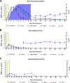
FMDV infection dynamics in pigs and cattle during early infection. (A) Detection of FMDV RNA by qRT-PCR in nasal swabs (green), oropharyngeal fluid (OPF; blue), and serum (red) from pigs (n = 12) from 0 to 72 h post-heel bulb inoculation. The blue-shaded area represents the cumulative lesion score. The FMDV-infected pigs were used as virus donors for cattle during a 24-h exposure period corresponding to 48 to 72 h postinoculation of the pigs (yellow box). (B) Detection of FMDV RNA by qRT-PCR in nasal swabs (green), saliva (blue), and serum (red) from cattle following 24 h of contact exposure to pigs (yellow box). The blue-shaded area represents the cumulative lesion score. There were 12 cattle at the start of the time course study, with 2 animals euthanized at each of 6, 12, 48, and 72 h postexposure (hpe) and 4 cattle euthanized at 24 hpe. The data represent geometric means ± SEM for all animals sampled at each time point.
FIG 2
FIG 2
FMDV infection dynamics in cattle from early to persistent phases of infection. Shown is detection of FMDV RNA by qRT-PCR in nasal swabs, saliva, serum, and OPF from 0 to 35 days postexposure to FMDV-infected pigs (yellow boxes). The blue-shaded area represents the cumulative lesion score, which was recorded up to 10 days postexposure (dpe). There were no FMDV lesions in vaccinated cattle. FMDV carrier status was determined based on sustained detection of FMDV in oropharyngeal fluid (OPF [probang samples]). The graphs represent (A) nonvaccinated carriers (n = 6), (B) vaccinated carriers (n = 5), and (C) a vaccinated noncarrier (n = 1). The data points represent geometric means ± SEM for all animals sampled at each time point.
FIG 3
FIG 3
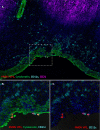
FMDV infection of nasopharyngeal mucosa and palatine tonsils of cattle during early stages of disease. (A) Primary FMDV infection of the dorsal nasopharyngeal mucosa of cattle at 24 h postexposure (hpe [animal ID 16-22]). FMDV VP1 (red) is localized to a focal area of MALT-associated epithelium of the dorsal nasopharynx, overlying a lymphoid follicle (purple). There is a partial-thickness erosion of the surface epithelium in association with the FMDV-infected area and a marked presence of CD11c+, presumed dendritic cells (turquoise). However, FMDV-infected cells are exclusively cytokeratin-positive epithelial cells (green). Magnification, ×20. (B) FMDV replication in an epithelial crypt of the bovine palatine tonsil at 48 hpe (animal ID 16-23). During this clinical stage of infection, large quantities of FMDV VP1 (red) and 3D (turquoise) protein are present within cytokeratin-positive (green) epithelial cells within the tonsil crypt. Magnification, ×10. (C) Higher-magnification and individual channel views of the area of interest boxed in panel B. Magnification, ×40.
FIG 4
FIG 4
Persistent FMDV infection in the bovine nasopharyngeal mucosa. (A) FMDV infection in the dorsal nasopharyngeal mucosa at 35 days postexposure (animal ID 16-04). FMDV VP1 (red) is localized to cytokeratin-positive epithelial cells (green) within a segment of MALT-associated epithelium overlying a subepithelial lymphoid follicle of CD21+ B cells (purple). CD11c+ presumptive dendritic cells are abundant throughout the submucosa and interspersed within the epithelium, rarely colocalizing with virus-infected cells (C). However, there is no structural disruption of the tissue, and no evidence of a marked inflammatory reaction. Magnification, ×10. (B and C) Higher-magnification and individual channel views of the area of interest boxed in panel A. Magnification, ×40.
FIG 5
FIG 5
Detection of FMDV in air filter samples after exposure of cattle to infected pigs. Shown are the FMDV RNA quantities and isolation of infectious FMDV from dry air filters collected from isolation rooms housing vaccinated or nonvaccinated cattle that were exposed to FMDV-infected pigs for 24 h. The y axis represents FMDV RNA quantities (CT values) determined by qRT-PCR. Hexagon symbols indicate samples from which infectious virus was isolated, while X indicates virus isolation-negative samples. Rooms A and B housed nonvaccinated cattle (6 per room) that were euthanized at predetermined time points from 6 to 72 h postexposure. Rooms C and D housed nonvaccinated and vaccinated cattle, respectively (6 cattle per room), which were monitored through 35 days. Each room housed 3 FMDV-infected pigs from 0 to 1 day postexposure (yellow box). All 6 nonvaccinated cattle in room C developed clinical FMD, while none of the 6 vaccinated cattle in room D developed any signs of FMD.
FIG 6
FIG 6
Subconsensus single nucleotide polymorphism (SNP) frequencies in porcine and bovine FMDV samples during early infection. Shown are frequencies (percentages) of SNPs, compared to the inoculum consensus sequence, present at ≥5% in samples obtained from 3 pigs and 4 cattle that were housed in room A. All animals were housed together for 24 h, corresponding to 48 to 72 h postinoculation (hpi) of the pigs and 0 to 24 h postexposure (hpe) of the cattle. Cattle 16-27 and 16-28 were euthanized for tissue harvest at 24 hpe, and cattle 16-29 and 16-30 were euthanized at 72 hpe. Additionally, FMDV sequence was obtained from air samples in the room at 24 and 48 hpe. The numbers in the figure represent the percentages of distinct SNPs in each sample. Color gradients indicate increasing frequency of SNP detection with greater color intensity. Blue, porcine samples; yellow, air samples; red, bovine samples. Syn, synonymous nucleotide change.

Similar articles

Cited by

References

    1. Grubman MJ, Baxt B. 2004. Foot-and-mouth disease. Clin Microbiol Rev 17:465–493. doi:10.1128/CMR.17.2.465-493.2004. - DOI - PMC - PubMed
    1. Knight-Jones TJ, Rushton J. 2013. The economic impacts of foot and mouth disease—what are they, how big are they and where do they occur? Prev Vet Med 112:161–173. doi:10.1016/j.prevetmed.2013.07.013. - DOI - PMC - PubMed
    1. Alexandersen S, Mowat N. 2005. Foot-and-mouth disease: host range and pathogenesis. Curr Top Microbiol Immunol 288:9–42. - PubMed
    1. Arzt J, Juleff N, Zhang Z, Rodriguez LL. 2011. The pathogenesis of foot-and-mouth disease. I. Viral pathways in cattle. Transbound Emerg Dis 58:291–304. doi:10.1111/j.1865-1682.2011.01204.x. - DOI - PubMed
    1. Alexandersen S, Zhang Z, Donaldson AI, Garland AJM. 2003. The pathogenesis and diagnosis of foot-and-mouth disease. J Comp Pathol 129:1–36. doi:10.1016/S0021-9975(03)00041-0. - DOI - PubMed

Publication types

MeSH terms

LinkOut - more resources