. 2019 Feb 5;93(4):e01774-18.
doi: 10.1128/JVI.01774-18. Print 2019 Feb 15.

Development of Stable Rotavirus Reporter Expression Systems

Affiliations

Development of Stable Rotavirus Reporter Expression Systems

Yuta Kanai et al. J Virol. .

Abstract

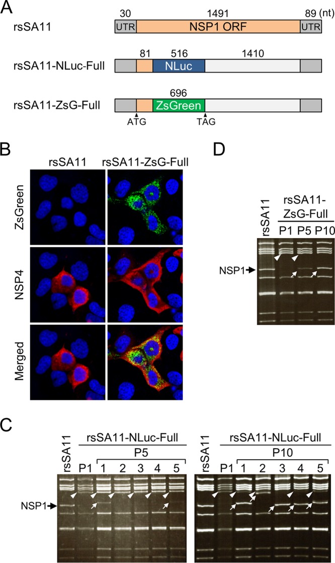
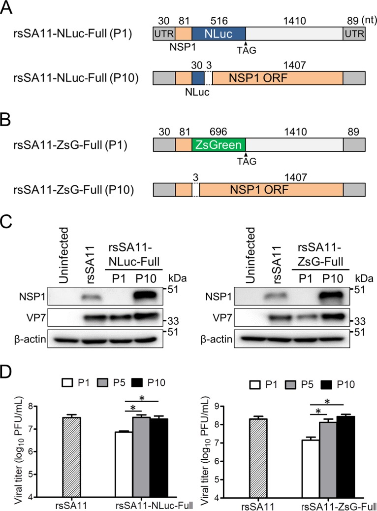
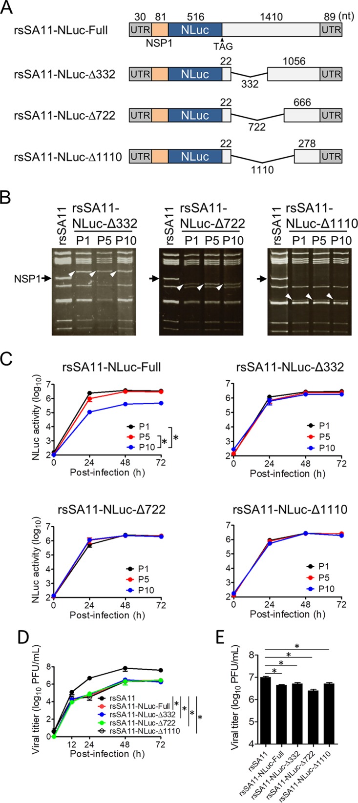
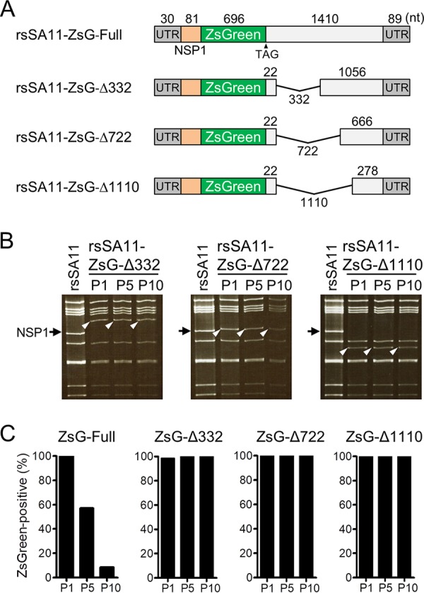
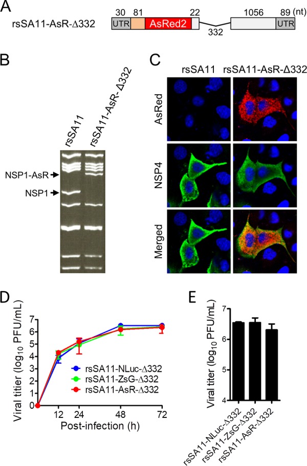
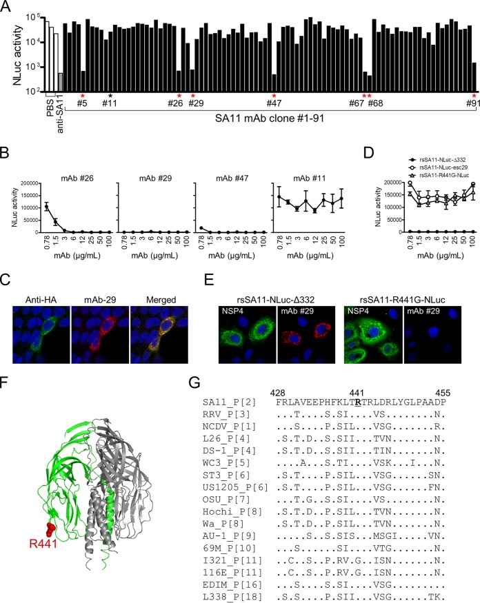
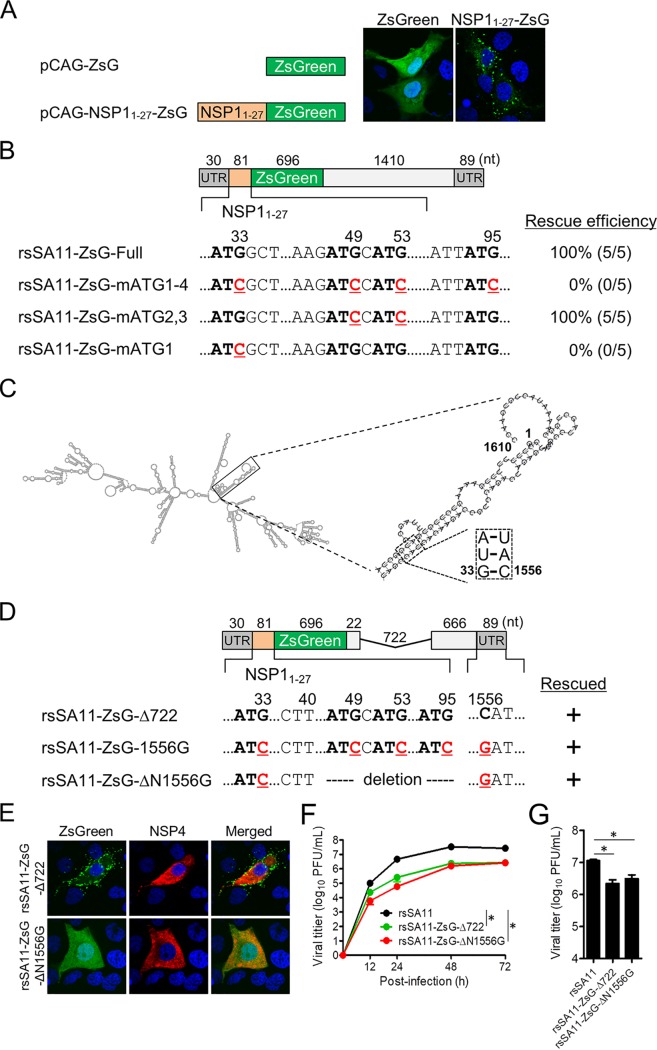
Engineered recombinant viruses expressing reporter genes have been developed for real-time monitoring of replication and for mass screening of antiviral inhibitors. Recently, we reported using a reverse genetics system to develop the first recombinant reporter rotaviruses (RVs) that expressed NanoLuc (NLuc) luciferase. Here, we describe a strategy for developing stable reporter RVs expressing luciferase and green or red fluorescent proteins. The reporter genes were inserted into the open reading frame of NSP1 and expressed as a fusion with an NSP1 peptide consisting of amino acids 1 to 27. The stability of foreign genes within the reporter RV strains harboring a shorter chimeric NSP1-reporter gene was greater than that of those in the original reporter RV strain, independent of the transgene inserted. The improved reporter RV was used to screen for neutralizing monoclonal antibodies (MAbs). Sequence analysis of escape mutants from one MAb clone (clone 29) identified an amino acid substitution (arginine to glycine) at position 441 in the VP4 protein, which resides within neutralizing epitope 5-1 in the VP5* fragment. Furthermore, to express a native reporter protein lacking NSP1 amino acids 1 to 27, the 5'- and 3'-terminal region sequences were modified to restore the predicted secondary RNA structure of the NSP1-reporter chimeric gene. These data demonstrate the utility of reporter RVs for live monitoring of RV infections and also suggest further applications (e.g., RV vaccine vectors, which can induce mucosal immunity against intestinal pathogens).IMPORTANCE Development of reporter RVs has been hampered by the lack of comprehensive reverse genetics systems. Recently, we developed a plasmid-based reverse genetics system that enables generation of reporter RVs expressing NLuc luciferase. The prototype reporter RV had some disadvantages (i.e., the transgene was unstable and was expressed as a fusion protein with a partial NSP1 peptide); however, the improved reporter RV overcomes these problems through modification of the untranslated region of the reporter-NSP1 chimeric gene. This strategy for generating stable reporter RVs could be expanded to diverse transgenes and be used to develop RV transduction vectors. Also, the data improve our understanding of the importance of 5'- and 3'-terminal sequences in terms of genome replication, assembly, and packaging.

Keywords: reporter virus; rotavirus; virus vector.


Figures

FIG 1
FIG 1
Transgene stability of reporter RV SA11 expressing NanoLuc luciferase (NLuc) or ZsGreen (ZsG). (A) Construction of the NSP1-NLuc-Full and NSP1-ZsG-Full genes. The NLuc or ZsG gene was inserted into the SA11 NSP1 gene between nucleotides 111 and 112. (B) Fluorescence imaging of rsSA11-ZsG-infected cells. MA104 cells were infected with rsSA11 or rsSA11-ZsG-Full, and ZsG expression was observed under a fluorescence microscope. As a control, NSP4 was detected in an indirect immunofluorescence assay using rabbit anti-NSP4 serum and a CF594-conjugated anti-rabbit IgG antibody. (C and D) Stability of the NLuc and ZsG genes after serial passage of reporter RVs. Electrophoresis of the dsRNA genome purified from rsSA11-NLuc-Full and rsSA11-ZsG-Full viruses, as indicated, after passages 5 and 10. Black arrow, NSP1; white arrowheads, the NSP1-NLuc gene (C) or the NSP1-ZsG gene (D); white arrows, truncated NSP1-NLuc (C) or NSP1-ZsG (D) gene.
FIG 2
FIG 2
Examination of revertant reporter viruses after 10 passages. (A and B) Schematic images of reporter genes before and after serial passage. Nucleotide sequences of dsRNA genomes purified from rsSA11-NLuc-Full (P10) and rsSA11-ZsG-Full (P10), as indicated, were analyzed. (C) Expression of NSP1 from reporter SA11 virus-infected cells (P1 or P10). MA104 cells were infected with rsSA11 or rsSA11-NLuc-Full (P1 and P10) or rsSA11-ZsG-Full (P1 and P10) at an MOI of 1 PFU/cell. NSP1 and VP7 in cell lysates were detected using a rabbit anti-NSP1 antibody and a monoclonal antibody specific for VP7, followed by appropriate HRP-conjugated secondary antibodies. An anti-β-actin antibody was used as a loading control. (D) Replication of rsSA11-NLuc and rsSA11-ZsG P1 (P5 and P10) viruses. MA104 cells were infected with each reporter virus at an MOI of 0.01 PFU per cell and incubated for 48 h. Infectious virus titers were measured in a plaque assay. Data are expressed as the means ± standard deviations (n = 3). *, P < 0.05 (Tukey’s multiple-comparison test).
FIG 3
FIG 3
Generation of reporter viruses with improved transgene stability. (A) NSP1-NLuc-Δ332, -Δ722, and -Δ1110 were generated by deleting the NSP1 gene region downstream of the NLuc ORF. (B and C) Stability of NSP1-NLuc reporter genes after serial passage. Panel B shows electropherotypes of dsRNA purified from P1, P5, and P10 reporter viruses. Black arrow, wild-type NSP1; white arrowheads, NSP1-NLuc gene. The kinetics of NLuc activity in rsSA11-NLuc viruses is shown in panel C. MA104 cells were infected with each rsSA11-NLuc virus (P1, P5, and P10) at an MOI of 0.01 PFU per cell and incubated for various times. NLuc activity in the cell lysate was measured by luminometry. Data are expressed as the means ± standard deviations (n = 3). NLuc activity values at 72 h postinfection were compared statistically. *, P < 0.05 (t test). (D and E) Replication kinetics of rsSA11-NLuc viruses. MA104 cells were infected with rsSA11 and rsSA11-NLuc-Full -Δ332, -Δ722, or -Δ1110 viruses at an MOI of 0.001 PFU per cell (D) and incubated for various times or at an MOI of 5 PFU per cell (E) and incubated for 16 h. Infectious virus titers in cell lysates were determined by plaque assay. Data are expressed as the means ± standard deviations (n = 3). Statistical significance was determined by one-way analysis of variance and Tukey’s multiple-comparison posttest. *, P < 0.05.
FIG 4
FIG 4
Generation of ZsG-expressing reporter viruses with improved transgene stability. (A) NSP1-ZsG-Δ332, -Δ722, and -Δ1110 were generated by deleting the NSP1 gene region downstream of the ZsG ORF. (B and C) Stability of NSP1-ZsG reporter genes after serial passage. (B) Electropherotypes of dsRNA purified from P1, P5, and P10 reporter viruses. Black arrow, wild-type NSP1; white arrowhead, NSP1-ZsG gene. (C) Quantitative examination of transgene stability after serial passage. MA104 cells were infected with each rsSA11-ZsG virus (P1, P5, and P10). At 24 h postinfection, the viral NSP4 antigen was detected in an indirect immunofluorescence assay using rabbit anti-NSP4 serum and a CF594-conjugated anti-rabbit IgG antibody. The number of ZsG-positive cells within the total NSP4-positive cell population was examined.
FIG 5
FIG 5
Generation of reporter RVs expressing AsRed2 (AsR). (A) Construction of the NSP1 gene containing the AsRed2 gene. The AsRed2 gene was inserted into the SA11 NSP1 gene between nucleotides 111 and 112. NSP1-AsR-Δ332 was generated by deleting the NSP1 gene fragment after the AsRed2 ORF. (B) Electropherotype of the dsRNA genome purified from rsSA11-AsR-Δ332. Viral dsRNAs were separated in 8% polyacrylamide gels and stained with ethidium bromide. (C) MA104 cells were infected with rsSA11 or rsSA11-AsR-Δ332. Expression of AsRed2 was observed under a fluorescence microscope. As a control, NSP4 was detected in an indirect immunofluorescence assay using rabbit anti-NSP4 serum and an anti-rabbit IgG antibody-CF488 conjugate. (D and E) Replication kinetics of reporter viruses. MA104 cells were infected with rsSA11-NLuc-Δ332, rsSA11-ZsG-Δ332, or rsSA11-AsR-Δ332 virus at an MOI of 0.001 PFU per cell and incubated for various times (D) or at an MOI of 5 PFU per cell and incubated for 16 h (E). Infectious virus titers in cell lysates were determined by plaque assay. Data are expressed as the means ± standard deviations (n = 3). Data were analyzed by one-way ANOVA and Tukey’s multiple-comparison posttest. A P value of <0.05 was considered statistically significant.
FIG 6
FIG 6
Screening for neutralizing monoclonal antibodies specific for SA11. (A) rsSA11-NLuc-Δ332 (1.0 × 102 PFU) was incubated at 37°C for 1 h with hybridoma culture supernatant (1:5 dilution). The virus-antibody mixture was inoculated onto MA104 cells, and NLuc titers in cell lysates were measured at 24 h postinfection. PBS was used as a negative control. Candidate NT-positive hybridoma clones are indicated by asterisks. Murine anti-SA11 serum raised after oral infection with SA11 virus was used as a positive control. (B) rsSA11-NLuc-Δ332 (1.0 × 102 PFU) was incubated with different concentrations of purified MAb clones 26, 29, 47, and 11. The virus-MAb mixture was inoculated onto MA104 cells, and NLuc titers in the cell lysates were measured at 24 h postinfection. MAb clone 11 was used as a negative control. (C) Hemagglutinin (HA)-tagged recombinant SA11 VP4 protein expressed in 293T cells was detected in an indirect immunofluorescence assay using MAb clone 29 and a rabbit anti-hemagglutinin peptide antibody, followed by anti-mouse IgG-CF594 and anti-rabbit IgG-CF488 secondary antibodies, respectively. (D) rsSA11-NLuc-Δ332, rsSA11-NLuc-esc29, and rsSA11-R441G-NLuc were incubated with various concentrations of MAb clone 29 and then inoculated onto MA104 cells. NLuc titers in the cell lysates were measured at 24 h postinfection. (E) MA104 cells were infected with rsSA11-NLuc-Δ332 or rsSA11-R441G-NLuc, and viral antigens were detected in an indirect immunofluorescence assay using a rabbit anti-NSP4 antibody and MAb clone 29, followed by an anti-rabbit IgG-CF488 conjugate or an anti-mouse IgG-CF594 conjugate, respectively. (F) Crystal structure of the trimeric VP5*CT (VP4 residues N252-L523) from a rhesus rotavirus (RRV) strain (PDB accession number 1SLQ). R441 is indicated in red. (G) Alignment of amino acid residues 428 to 455 of the VP4 proteins of RV reference strains. The P genotypes of the VP4 gene of each strain are shown. Amino acids identical to the consensus sequence (SA11) are indicated by dots. R441 is underlined.
FIG 7
FIG 7
Modification of the reporter gene. (A) Cellular localization of a recombinant ZsG protein expressed with/without the NSP11–27 peptide plasmid vector. (B) Partial nucleotide sequence of the NSP11–27-ZsG gene used for reverse genetics. Four ATG triplets (bold) and ATG-to-ATC substitutions (red, underlined) in the NSP11–27 coding sequence are shown. The results of virus rescue and the rescue efficiencies of recombinant viruses harboring each reporter gene are shown. Data represent the mean values of five independent rescue experiments. (C) Predicted secondary structure of wild-type NSP1 mRNA. Double-strands formed by the 5′- and 3′-terminal regions are shown in the insets. Base paring at positions 33 and 1556 within the stem structure are shown in the square. Nucleotide positions are numbered according to the wild-type NSP1 gene sequence. (D) Schematic showing the NSP1-ZsG-1556G and NSP1-ZsG-ΔN1556G constructs. Nucleotide positions are numbered according to the wild-type NSP1 sequence. (E) MA104 cells were infected with rsSA11-ZsG-Δ722 or rsSA11-ZsG-ΔN1556G. Viral NSP4 was detected using rabbit anti-NSP4 and an anti-rabbit-IgG-CF594 conjugate. Nuclei were stained with 4′,6-diamidino-2-phenylindole. (F and G) Replication kinetics of rsSA11-ZsG-ΔN1556G. MA104 cells were infected with rsSA11, rsSA11-NLuc-Δ722, or rsSA11-ZsG-ΔN1556G virus at MOI of 0.001 PFU per cell and incubated for various times (F) or at an MOI of 5 PFU per cell and incubated for 16 h (G). Infectious virus titers in cell lysates were determined by the plaque assay. Data are expressed as the means ± standard deviations (n = 3). Statistical significance was determined by one-way ANOVA and Tukey’s multiple-comparison posttest. *, P < 0.05.

Similar articles

Cited by

References

    1. Agapov EV, Frolov I, Lindenbach BD, Pragai BM, Schlesinger S, Rice CM. 1998. Noncytopathic Sindbis virus RNA vectors for heterologous gene expression. Proc Natl Acad Sci U S A 95:12989–12994. doi:10.1073/pnas.95.22.12989. - DOI - PMC - PubMed
    1. Tsetsarkin K, Higgs S, McGee CE, De Lamballerie X, Charrel RN, Vanlandingham DL. 2006. Infectious clones of Chikungunya virus (La Reunion isolate) for vector competence studies. Vector Borne Zoonotic Dis 6:325–337. doi:10.1089/vbz.2006.6.325. - DOI - PubMed
    1. McGee CE, Shustov AV, Tsetsarkin K, Frolov IV, Mason PW, Vanlandingham DL, Higgs S. 2010. Infection, dissemination, and transmission of a West Nile virus green fluorescent protein infectious clone by Culex pipiens quinquefasciatus mosquitoes. Vector Borne Zoonotic Dis 10:267–274. doi:10.1089/vbz.2009.0067. - DOI - PMC - PubMed
    1. Manicassamy B, Manicassamy S, Belicha-Villanueva A, Pisanelli G, Pulendran B, García-Sastre A. 2010. Analysis of in vivo dynamics of influenza virus infection in mice using a GFP reporter virus. Proc Natl Acad Sci U S A 107:11531–11536. doi:10.1073/pnas.0914994107. - DOI - PMC - PubMed
    1. Zou G, Xu HY, Qing M, Wang QY, Shi PY. 2011. Development and characterization of a stable luciferase dengue virus for high-throughput screening. Antiviral Res 91:11–19. doi:10.1016/j.antiviral.2011.05.001. - DOI - PubMed

Publication types

LinkOut - more resources