. 2019 Feb 5;93(4):e01244-18.
doi: 10.1128/JVI.01244-18. Print 2019 Feb 15.

Cellular RNA Helicase DHX9 Interacts with the Essential Epstein-Barr Virus (EBV) Protein SM and Restricts EBV Lytic Replication

Affiliations

Cellular RNA Helicase DHX9 Interacts with the Essential Epstein-Barr Virus (EBV) Protein SM and Restricts EBV Lytic Replication

Wenmin Fu et al. J Virol. .

Abstract

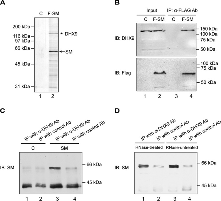
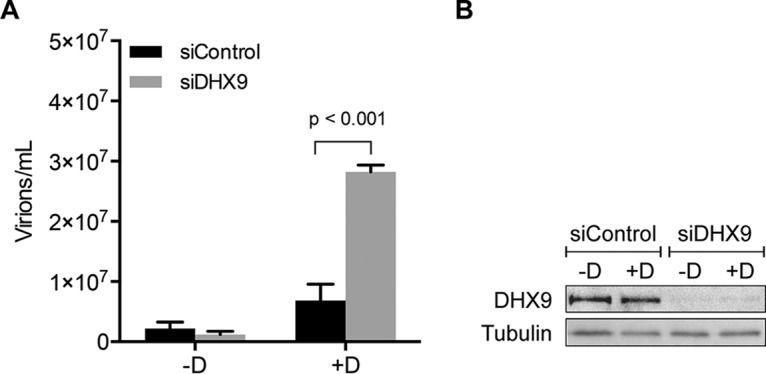
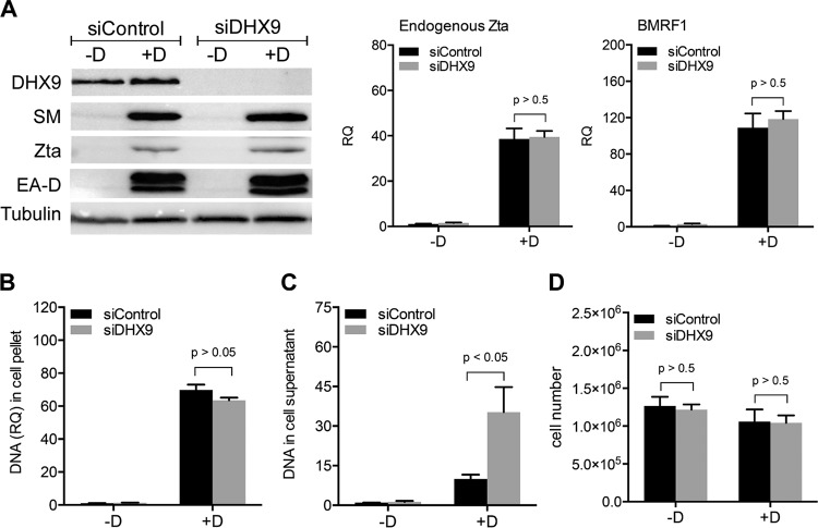
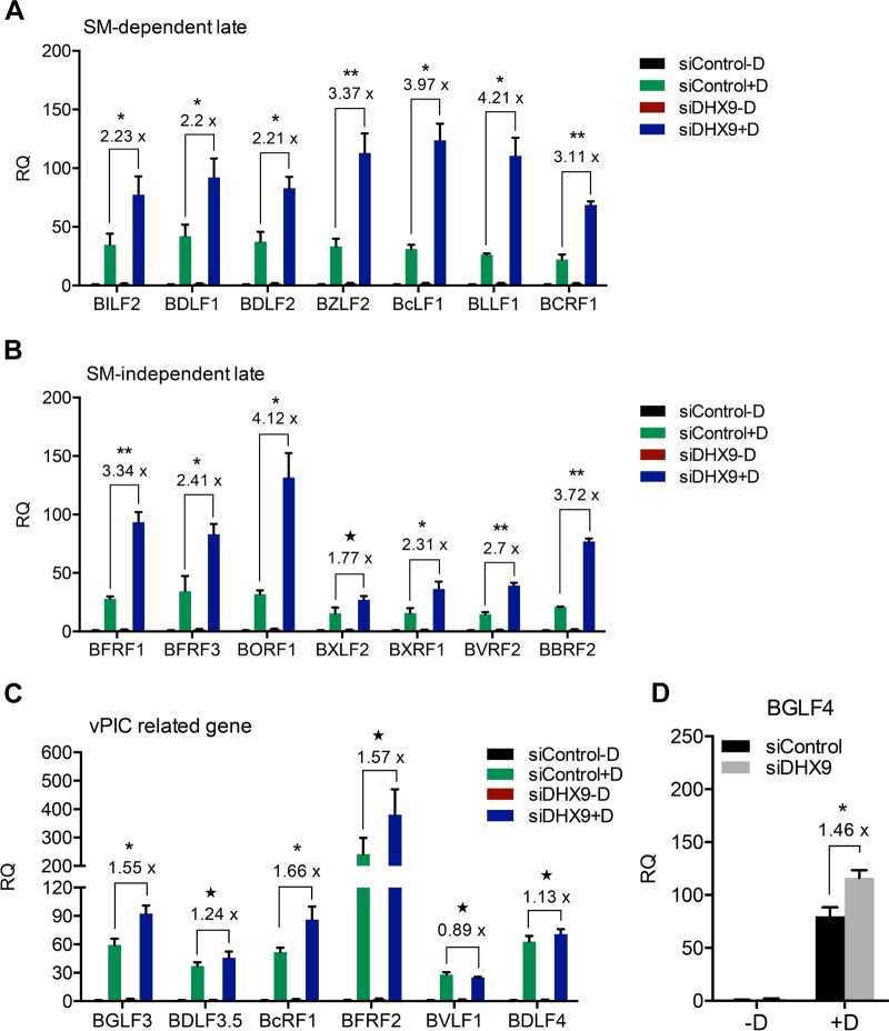
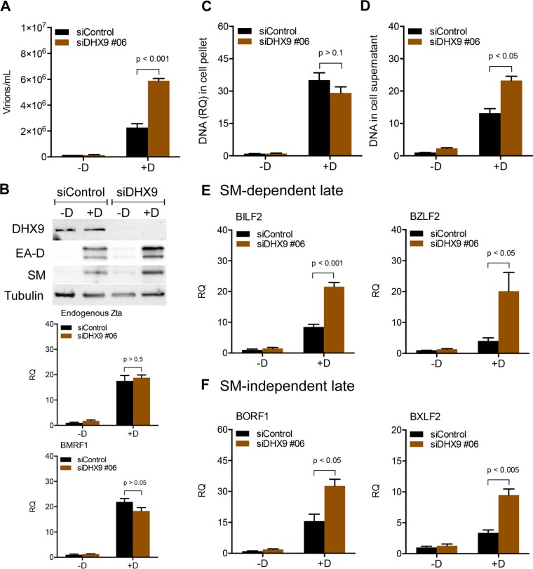
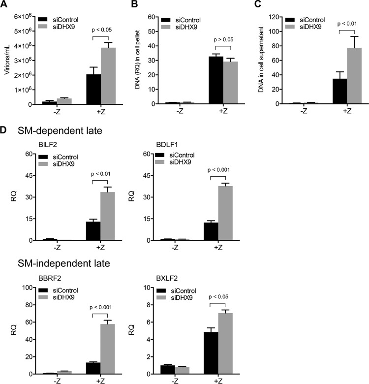
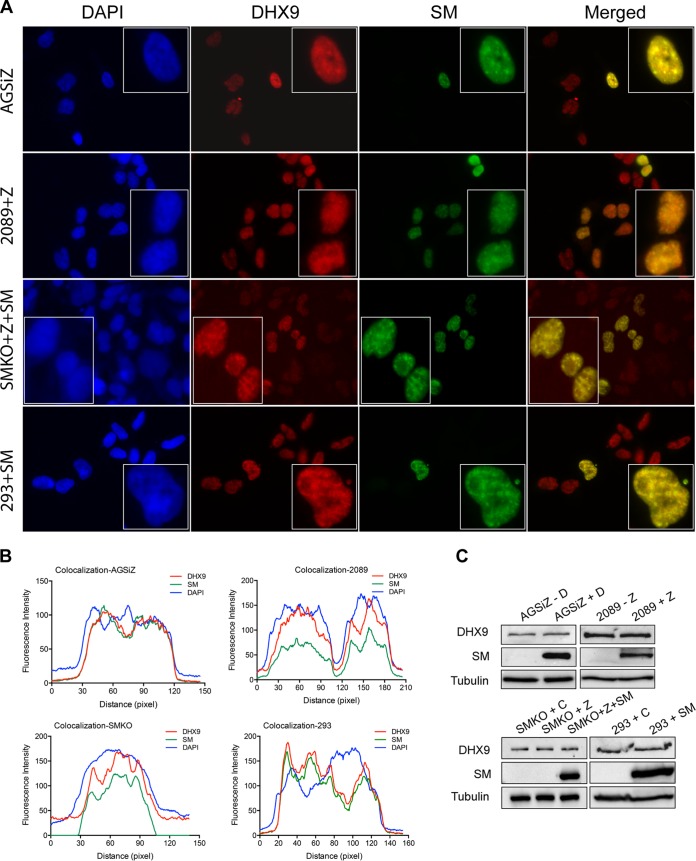
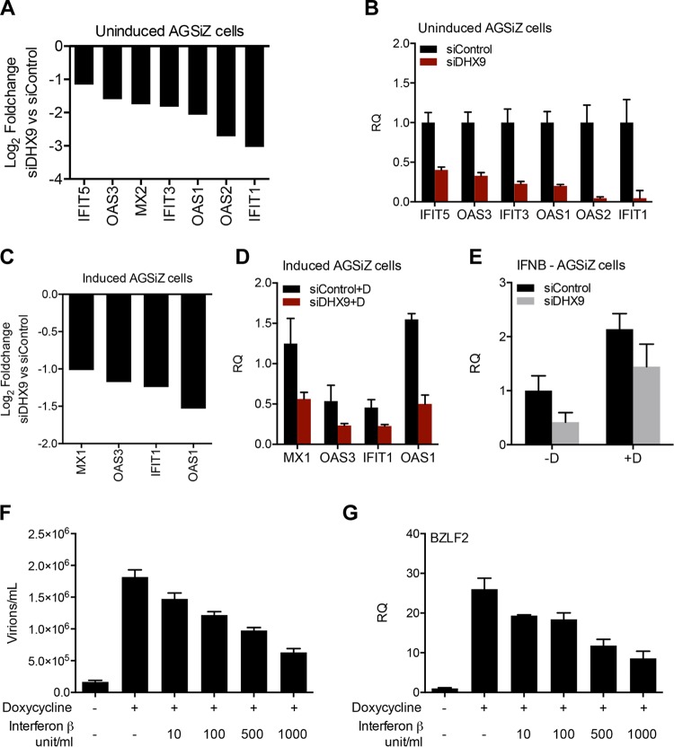
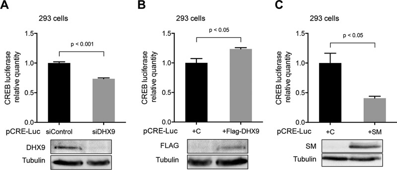
Epstein-Barr virus (EBV) SM protein is an RNA-binding protein that has multiple posttranscriptional gene regulatory functions essential for EBV lytic replication. In this study, we identified an interaction between SM and DHX9, a DExH-box helicase family member, by mass spectrometry and coimmunoprecipitation. DHX9 participates in many cellular pathways involving RNA, including transcription, processing, transport, and translation. DHX9 enhances virus production or infectivity of a wide variety of DNA and RNA viruses. Surprisingly, an increase in EBV late gene expression and virion production occurred upon knockdown of DHX9. To further characterize the SM-DHX9 interaction, we performed immunofluorescence microscopy of EBV-infected cells and found that DHX9 partially colocalized with SM in nuclear foci during EBV lytic replication. However, the positive effect of DHX9 depletion on EBV lytic gene expression was not confined to SM-dependent genes, indicating that the antiviral effect of DHX9 was not mediated through its effects on SM. DHX9 enhanced activation of innate antiviral pathways comprised of several interferon-stimulated genes that are active against EBV. SM inhibited the transcription-activating function of DHX9, which acts through cAMP response elements (CREs), suggesting that SM may also act to counteract DHX9's antiviral functions during lytic replication.IMPORTANCE This study identifies an interaction between Epstein-Barr virus (EBV) SM protein and cellular helicase DHX9, exploring the roles that this interaction plays in viral infection and host defenses. Whereas most previous studies established DHX9 as a proviral factor, we demonstrate that DHX9 may act as an inhibitor of EBV virion production. DHX9 enhanced innate antiviral pathways active against EBV and was needed for maximal expression of several interferon-induced genes. We show that SM binds to and colocalizes DHX9 and may counteract the antiviral function of DHX9. These data indicate that DHX9 possesses antiviral activity and that SM may suppress the antiviral functions of DHX9 through this association. Our study presents a novel host-pathogen interaction between EBV and the host cell.

Keywords: Epstein-Barr virus; RNA processing; helicase; innate immune response; protein-protein interactions; transcriptional activation; virus-host interactions.


Figures

FIG 1
FIG 1
DHX9 interacts with EBV SM protein in an RNase-independent manner. (A) Immunoprecipitation of SM-interacting proteins for mass spectrometry analysis (9). FLAG epitope-tagged SM (F-SM) or empty vector (C) was transfected into 293T cells, and SM was isolated by affinity purification with M2 anti-FLAG antibody-conjugated beads, followed by elution with FLAG peptide. Eluates of lysates from vector-transfected and SM-transfected cells were analyzed by SDS-PAGE and silver staining. The major SM band is shown with an arrow in lane 2. DHX9, which was confirmed by excision of the gel band and MS analysis, is shown with an asterisk. (B) Coimmunoprecipitation of SM and DHX9. 293T cells were transfected with epitope-tagged SM (F-SM) or empty vector (C), and lysates were harvested at 48 h posttransfection. The input protein samples (Input, 0.5% lysate) and samples that were immunoprecipitated with anti-FLAG antibodies (IP, 5% lysate) (lanes 3 and 4) were separated by SDS-PAGE and analyzed by immunoblotting with anti-DHX9 and anti-FLAG antibodies, respectively. (C) Reciprocal immunoprecipitation of SM by DHX9 antibody. 293T cells were transfected with untagged SM or empty vector (C) and then harvested at 48 h posttransfection. The protein samples were immunoprecipitated with anti-DHX9 antibody or control antibody as indicated and analyzed by Western blotting with anti-SM antibody. (D) RNase resistance of DHX9-SM interaction. 293T cells were transfected with untagged SM plasmid, and lysates were harvested at 48 h posttransfection. The protein samples were treated or mock treated with RNase, immunoprecipitated with anti-DHX9 antibody or control antibody, and immunoblotted for SM as in panel C. Molecular weight markers are shown in kilodaltons.
FIG 2
FIG 2
Effects of DHX9 depletion on EBV virion production. AGSiZ cells were depleted of DHX9 by siRNA transfection or mock depleted with control siRNA, followed by treatment with doxycycline (+D) to induce viral lytic replication or untreated (−D). (A) Quantification of infectious virus particles. Serial dilutions of virus-containing supernatant harvested at 5 days postinduction were used to infect 293T cells, and the number of GFP-positive cells representing infectious viral particles was measured by flow cytometry. Three independent biological experiments were performed, and each sample was measured in technical triplicates. (B) Knockdown efficiency of siDHX9. Protein cell lysates were harvested at 48 h after siRNA transfection and analyzed by Western blotting with anti-DHX9 antibody. The blot was stripped and reprobed with antitubulin antibody as a loading control.
FIG 3
FIG 3
Effects of DHX9 depletion on EBV lytic gene expression, DNA replication, and extracellular virion release. AGSiZ cells were depleted of DHX9 or mock depleted by siRNA transfections, followed by treatment with doxycycline (+D) to induce viral lytic replication or untreated (−D). (A) Expression of EBV early and immediate early gene proteins and mRNAs. Protein lysates were harvested at 48 h postinduction and immunoblotted with anti-SM, anti-Zta, or anti-EA-D to examine the expression levels of EBV lytic genes. Blots were also probed with anti-DHX9 to confirm DHX9 knockdown and with antitubulin antibodies as a loading control. Transcript levels of endogenous Zta and BMRF1 were measured by qRT-PCR. (B) Quantification of intracellular EBV DNA. DNA was isolated from cell pellets harvested at 3 days postinduction and analyzed by qPCR. BILF2 primers were used to measure viral DNA (RQ, relative quantity). (C) Quantification of viral DNA in cell supernatant. Cell supernatants from cells treated as in panels A and B were harvested at 5 days postinduction for viral DNA isolation. Viral DNA quantities were measured by qPCR. (D) Quantitation of cell numbers after DHX9 knockdown. Three independent biological experiments were performed, and cell counts in each sample were measured in technical triplicates.
FIG 4
FIG 4
Effects of DHX9 depletion on EBV late gene transcript levels. AGSiZ cells were depleted of DHX9 or mock depleted by siRNA transfections followed by treatment with doxycycline (+D) to induce viral lytic replication or untreated (−D). Specific mRNA transcripts were measured by qRT-PCR with 7 sets of SM-dependent viral late gene primers (BILF2, BDLF1, BDLF2, BZLF2, BcLF1, BLLF1, and BCRF1) (A) and 7 sets of SM-independent late gene primers (BFRF1, BFRF3, BORF1, BXLF2, BXRF1, BVRF2, and BBRF2) (B) at 48 h postinduction. Transcript levels of vPIC-related genes (BGLF3, BDLF3.5, BFRF2, BcRF1, BVLF1, and BDLF4) (C) and BGLF4 (D) were measured by qRT-PCR. Three independent biological experiments were performed, and each sample was measured in technical triplicates. RQ, relative quantity. Fold changes of lytic transcript levels after DHX9 knockdown compared to control knockdown are shown above each gene. *, P < 0.05; **, P < 0.001; ★, P > 0.05.
FIG 5
FIG 5
Effects of DHX9 depletion with second independent siRNA (siDHX9 #06) on EBV gene expression and virion production. AGSiZ cells were depleted of DHX9 or mock depleted by DHX9 siRNA transfections followed by treatment with doxycycline (+D) to induce viral lytic replication or untreated (−D). (A) Quantification of infectious virus particles. Serial dilutions of virus-containing supernatant harvested at 5 days postinduction were used to infect 293T cells, and the number of GFP-positive cells representing infectious viral particles was measured by flow cytometry. (B) Expression of EBV early and immediate early genes. (C) Quantification of intracellular EBV DNA. DNA was isolated from cell pellets harvested at 3 days postinduction and analyzed by qPCR. BILF2 primers were used to measure viral DNA (RQ, relative quantity). (D) Quantification of viral DNA in cell supernatant. Cell supernatants from cells treated as in panels A and B were harvested at 5 days postinduction for viral DNA isolation. Viral DNA quantities were measured by qPCR. (E and F) Quantification of SM-dependent (E) or SM-independent (F) EBV late gene transcripts by qRT-PCR. Three independent biological experiments were performed, and each sample was measured in technical triplicates.
FIG 6
FIG 6
Effects of DHX9 depletion on EBV gene expression and virion production in HEK293-2089 cells. HEK293-2089 cells were depleted of DHX9 or mock depleted by siRNA transfections followed by transfection with transactivator plasmid Zta (+Z) to induce viral lytic replication or transfected with empty vector (−Z). (A) Quantification of infectious virus particles. Serial dilutions of virus-containing supernatant harvested at 5 days postinduction were used to infect 293T cells, and the number of GFP-positive cells representing infectious viral particles was measured by flow cytometry. (B) Quantification of intracellular EBV DNA. DNA was isolated from cell pellets harvested at 3 days postinduction and analyzed by qPCR. BILF2 primers were used to measure viral DNA (RQ, relative quantity). (C) Quantification of viral DNA in cell supernatant. Cell supernatants from cells treated as in panels A and B were harvested at 5 days postinduction for viral DNA isolation. Viral DNA quantities were measured by qPCR. (D) Quantification of EBV SM-dependent and SM-independent late gene transcripts by qRT-PCR. Three independent biological experiments were performed, and each sample was measured in technical triplicates.
FIG 7
FIG 7
DHX9 colocalizes with SM in various cell lines. (A) Localization of DHX9 and SM in AGSiZ, HEK2089, SMKO, and HEK293 cells. AGSiZ cells were treated with doxycycline (+D) to induce viral lytic replication; 2089 cells were transfected with plasmid Zta to induce viral lytic replication; SMKO cells were cotransfected with Zta and SM to induce lytic replication; uninfected HEK293 cells were transfected with untagged SM plasmid. At 48 h postinduction, cells were costained for DHX9 (red) and SM (green) and visualized by fluorescence microcopy. The nuclei were stained with DAPI (blue). (B) Colocalization analysis with ImageJ of cells shown in the boxes as in panel A. Two-dimensional graph of the intensities of pixels along the longitudinal axis of cells in merged images. The x axis represents distance along the line, and the y axis is the pixel intensity. (C) Expression of DHX9 and SM in AGSiZ, 2089, SMKO, and 293 cells. Protein cell lysates were harvested at 48 h postinduction and analyzed by Western blotting with anti-DHX9 and anti-SM antibodies. Tubulin was probed as a loading control.
FIG 8
FIG 8
Effect of DHX9 depletion on type I interferon pathway and IFN-β expression. (A to D) Effect of DHX9 KD on ISG expression. AGSiZ cells were depleted of DHX9 or mock depleted by siRNA transfection followed by treatment with doxycycline (+D) to induce viral lytic replication or left untreated (−D). RNA was harvested at 48 h postinduction and analyzed by high-throughput sequencing and by qRT-PCR. (A) Log2 fold changes of selected interferon-stimulated genes after DHX9 depletion of uninduced AGSiZ cells derived from differential expression of RNA-seq data. (B) ISG mRNA levels of genes shown in panel A were quantitated by qRT-PCR. (C) Log2 fold changes of selected interferon-stimulated genes after DHX9 depletion of induced AGSiZ cells derived from differential expression of RNA-seq data. (D) ISG mRNA levels of genes shown in panel C were quantitated by qRT-PCR. (E) Effect of DHX9 depletion on IFN-β transcript levels. RNAs from experiments above were analyzed by qPCR with primers specific for IFN-β. (F and G) Dose-dependent effects of IFN-β treatment on EBV lytic virion production and late gene transcripts. Various concentrations of IFN as shown were added to the medium of AGSiZ cells when doxycycline was added to induce viral lytic replication. At 5 days postinduction, virus-containing supernatant was harvested and released infectious viral particles were quantitated as described above (F). Total RNA was isolated from cell pellets at 48 h postinduction, and late gene expression was measured by qRT-PCR (G). GAPDH was used as the endogenous control. RQ, relative quantity. Three independent biological experiments were performed, and each sample was measured in technical triplicates.
FIG 9
FIG 9
SM effect on transcription activation via the CRE. (A) Effect of DHX9 depletion on CRE-luciferase activity in HEK293 cells. HEK293 cells were depleted of DHX9 with siRNA or mock depleted with control siRNA. At 30 h post-siRNA transfection, cells were transfected with pCRE-luc reporter vector. At 24 h after transfection of the reporter, cells were lysed for luciferase assay and analyzed by Western blotting to examine the knockdown efficiency of DHX9. (B) Effect of DHX9 overexpression on CRE-luciferase activity. The pCRE-luc reporter vector was cotransfected with FLAG-tagged DHX9 expression plasmid or empty vector (C). At 24 h posttransfection, cells were harvested and lysed for luciferase assay. Protein lysates were analyzed by Western blotting to detect DHX9 overexpression. (C) Effect of SM expression on CRE-luciferase activity in HEK293 cells. The pCRE-luc reporter vector was cotransfected with SM plasmid or empty vector (C). At 24 h posttransfection, cells were harvested and lysates were prepared for luciferase assay. Protein lysates were analyzed by Western blotting to detect SM expression. All transfections were performed in triplicate, and technical triplicates of each sample were measured by luciferase assay.

Similar articles

Cited by

References

    1. Longnecker RM, Kieff E, Cohen JI. 2013. Epstein-Barr virus, p 1898–1959. In Knipe DM, Howley PM, Cohen JL, Griffin DE, Lamb RA, Martin MA, Racaniello VR, Roizman B (ed), Fields virology, 6th ed, vol 2 Lippincott Williams & Wilkins, Philadelphia, PA.
    1. Hutt-Fletcher LM. 2016. The long and complicated relationship between Epstein-Barr virus and epithelial cells. J Virol 91:e01677-16. doi:10.1128/JVI.01677-16. - DOI - PMC - PubMed
    1. Li D, Fu W, Swaminathan S. 2018. Continuous DNA replication is required for late gene transcription and maintenance of replication compartments in gammaherpesviruses. PLoS Pathog 14:e1007070. doi:10.1371/journal.ppat.1007070. - DOI - PMC - PubMed
    1. Li H, Liu S, Hu J, Luo X, Li N, M Bode A, Cao Y. 2016. Epstein-Barr virus lytic reactivation regulation and its pathogenic role in carcinogenesis. Int J Biol Sci 12:1309–1318. doi:10.7150/ijbs.16564. - DOI - PMC - PubMed
    1. Gruffat H, Batisse J, Pich D, Neuhierl B, Manet E, Hammerschmidt W, Sergeant A. 2002. Epstein-Barr virus mRNA export factor EB2 is essential for production of infectious virus. J Virol 76:9635–9644. - PMC - PubMed

Publication types

MeSH terms

LinkOut - more resources