. 2019 Feb 19;93(5):e02088-18.
doi: 10.1128/JVI.02088-18. Print 2019 Mar 1.

Hantavirus RdRp Requires a Host Cell Factor for Cap Snatching

Affiliations

Hantavirus RdRp Requires a Host Cell Factor for Cap Snatching

Subbiah Jeeva et al. J Virol. .

Abstract

The hantavirus RNA-dependent RNA polymerase (RdRp) snatches 5' capped mRNA fragments from the host cell transcripts and uses them as primers to initiate transcription and replication of the viral genome in the cytoplasm of infected cells. Hantavirus nucleocapsid protein (N protein) binds to the 5' caps of host cell mRNA and protects them from the attack of cellular decapping machinery. N protein rescues long capped mRNA fragments in cellular P bodies that are later processed by an unknown mechanism to generate 10- to 14-nucleotide-long capped RNA primers with a 3' G residue. Hantavirus RdRp has an N-terminal endonuclease domain and a C-terminal uncharacterized domain that harbors a binding site for the N protein. The purified endonuclease domain of RdRp nonspecifically degraded RNA in vitro It is puzzling how such nonspecific endonuclease activity generates primers of appropriate length and specificity during cap snatching. We fused the N-terminal endonuclease domain with the C-terminal uncharacterized domain of the RdRp. The resulting NC mutant, with the assistance of N protein, generated capped primers of appropriate length and specificity from a test mRNA in cells. Bacterially expressed and purified NC mutant and N protein required further incubation with the lysates of human umbilical vein endothelial cells (HUVECs) for the specific endonucleolytic cleavage of a test mRNA to generate capped primers of appropriate length and defined 3' terminus in vitro Our results suggest that an unknown host cell factor facilitates the interaction between N protein and NC mutant and brings the N protein-bound capped RNA fragments in close proximity to the endonuclease domain of the RdRp for specific cleavage at a precise length from the 5' cap. These studies provide critical insights into the cap-snatching mechanism of cytoplasmic viruses and have revealed potential new targets for their therapeutic intervention.IMPORTANCE Humans acquire hantavirus infection by the inhalation of aerosolized excreta of infected rodent hosts. Hantavirus infections cause hemorrhagic fever with renal syndrome (HFRS) and hantavirus cardiopulmonary syndrome (HCPS), with mortality rates of 15% and 50%, respectively (1). Annually 150,000 to 200,000 cases of hantavirus infections are reported worldwide, for which there is no treatment at present. Cap snatching is an early event in the initiation of virus replication in infected hosts. Interruption in cap snatching will inhibit virus replication and will likely improve the prognosis of the hantavirus disease. Our studies provide mechanistic insight into the cap-snatching mechanism and demonstrate the requirement of a host cell factor for successful cap snatching. Identification of this host cell factor will reveal a novel therapeutic target for combating this viral illness.

Keywords: cap snatching; hantavirus; negative-strand RNA virus; nucleocapsid.


Figures

FIG 1
FIG 1
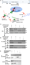
Interaction between hantavirus N protein and RdRp. (A) The domains predicted in hantavirus RdRp using sequence homology reveal the presence of an endonuclease domain from amino acids 1 to 250 and a catalytic domain from amino acids 750 to 1290 (19). The question mark represents the domains of unknown function. (B) A model showing the rescue of capped mRNA fragments (red) in cellular P-bodies by N protein (black). The interaction between the N protein and C-terminal uncharacterized domain of the RdRp (green) facilitates the specific cleavage of the capped mRNA fragment at the G residue to generate the RNA primer. Use of RNA primer (red) at the replication complex is shown. (C) HUVECs were transfected with either pHisEndo fusion plasmid, pmycSNV-NP plasmid, or both, or with pTriEx1.1 plasmid as a control. Whole-cell lysates (WCL) were examined by Western blot analysis (WB) using either anti-His tag antibody (i), anti-N protein antibody (ab20309; Abcam) (ii), or anti-β-actin antibody (Cell Signaling, 4970S) (iii). (D) Cell lysates were immunoprecipitated (IP) by anti-N protein antibody, and the immunoprecipitated material was examined by Western blot analysis using either anti-His tag antibody (A00174; GenScript) (iv) or anti-N protein antibody (v). The cell lysates were also pulled down on Ni-NTA beads, and the pulldown material was examined by Western blot analysis using either anti-N protein antibody (vi) or anti-His tag antibody (vii). (E) The experiment in this panel was carried out exactly the same way as that shown in panel D, except pmycSNV-NP plasmid was replaced with pmycHSP40 plasmid, expressing heat shock protein HSP40. Immunoprecipitation and Western blotting was carried out using anti-HSP40 antibody, as shown.
FIG 2
FIG 2
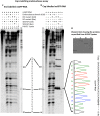
Generation of capped RNA primers in cells. (A) The pcDNAGFPnsG14 plasmid was previously constructed (4); it expresses a nonsense GFP mRNA containing a premature termination codon. The 14 nucleotides from its 5′ terminus are efficiently snatched during hantavirus cap-snatching. (B) HUVECs were cotransfected with plasmids as shown. Cell lysates were immunoprecipitated using anti-N protein antibody (Ab), and total RNA from immunoprecipitated material was reverse transcribed. The cDNA was PCR amplified using a forward primer complementary to the 5′ terminus of the nonsense GFP mRNA shown in panel A and a reverse primer complementary to the sequence 280 nucleotides downstream of the 5′ terminus. Shown are the PCR products separated on agarose gel. (C) Immunoprecipitated material from panel B (step 1) was used for the purification of shorter RNA using a mirVana miRNA isolation kit (step 2). The RNA was ligated with 5′ phosphorylated anchor primer using single-strand RNA ligase (step 3), as previously reported (19, 32), and reverse transcribed using primer P1 complementary to the anchor sequence. The cDNA was PCR amplified using primers P2 and P3 and had NcoI and NotI restriction sites. (D) The PCR product was separated on agarose gel. (E) The PCR product from panel D was gel purified and cloned between NcoI and NotI restriction sites in pTriEx1.1 vector and sequenced. The sequence of the ligated RNA segment to the anchor primer is shown.
FIG 3
FIG 3
Cap snatching in vitro. SDS-PAGE gel showing the purification of the N-terminal endonuclease domain of hantavirus RdRp (A), NC mutant (B), and N protein (C). The proteins were purified by Ni-NTA column chromatography using a native purification procedure, as mentioned in Materials and Methods. The symbols M, for marker, FT, for flowthrough, W, for wash, and E1 to E6, for eluted fractions 1 to 6, are shown on top of each gel. The digestion of nsGFP-RNA (D) by the endonuclease domain and NC mutant is shown in panel E. Briefly, the radioactively cap-labeled nsGFP-RNA was incubated with purified N protein along with purified endonuclease domain, NC mutant, or both at 37°C in endonuclease buffer for 45 min. Lanes L1, L2, and L3 show the RNA ladder generated by alkaline hydrolysis of the radioactively capped nsGFP-RNA for 10 min, 20 min, and 30 min, respectively, as previously reported (24). Lane T1 shows the RNase T1 digest of the nsGFP-RNA.
FIG 4
FIG 4
Generation of capped primers of appropriate length and defined 3′ terminus. (A) Bacterially expressed and purified N protein, NC mutant, and endonuclease domain were incubated with HUVEC lysates for 1 h at room temperature, followed by purification using Ni-NTA chromatography. Shown is the SDS-PAGE gel of repurified endonuclease domain (lane i), NC mutant (lane ii), and N protein (lane iii). The HUVEC lysates alone were also loaded on an Ni-NTA column. The eluted material from the washed column is shown in lane iv. (B) A 20% denaturing polyacrylamide urea gel showing the separation of radioactively capped nsGFP-RNA that was incubated with the N protein along with either endonuclease domain or NC mutant under different experimental conditions (lanes 1 to 8). Lane T1 shows the RNase T1 digest of the nsGFP-RNA, and lane 9 shows the RNA sample that was incubated with the eluted material from lane iv of panel A. SNV-NP (Bac) indicates the bacterially expressed and purified Sin Nombre hantavirus N protein. SNV-NP (Cell) indicates the bacterially expressed N protein that was incubated with HUVEC lysates for 1 h at room temperature and then repurified by native purification procedure using Ni-NTA chromatography. The same applies to endonuclease (Bac), endonuclease (Cell), NC mutant (Bac), and NC mutant (Cell). (C) The experiment was performed as for panel B, except the nsGFP-RNA was uncapped and end labeled with (γ-32P)GTP.
FIG 5
FIG 5
Effect of RdRp fusion mutant on hantavirus replication. HUVECs in 6-well plates were transfected with a plasmid expressing either hantavirus N protein (green) or NC mutant (red) or both (purple) or transfected with empty vector (blue). Twenty-four hours posttransfection, cells were infected with Sin Nombre hantavirus at an MOI of 1.0. Cells were lysed at increasing time points postinfection, and virus replication was monitored by quantitative estimation of viral S-segment RNA by real-time PCR, as previously reported (36). The significance of the differences was calculated by the t test.
FIG 6
FIG 6
Model showing the role of N protein and an unknown host cell factor in hantavirus cap snatching. N protein (black) binds to mRNA caps and protects capped mRNA fragments (red line) up to 180 nucleotides in length in P-bodies. An unknown host cell factor (HCF) binds to N protein and transports it to the replication complex along with bound capped mRNA fragment. The simultaneous binding of HCF to both the N protein and RdRp (green) brings the capped mRNA fragment close to the endonuclease domain (scissors) of RdRp for specific endonucleolytic cleavage at the G residue located 14 nucleotides downstream of the 5′ cap to generate the RNA primer. The HCF is then released, which starts another round of shuttling the N protein-associated capped mRNA fragments to the replication complex. N protein may remain associated with the 5′ cap of the newly synthesized mRNA and facilitate its translation, consistent with our previously reported observations (28, 34).

References

    1. Jonsson CB, Figueiredo LT, Vapalahti O. 2010. A global perspective on hantavirus ecology, epidemiology, and disease. Clin Microbiol Rev 23:412–441. doi:10.1128/CMR.00062-09. - DOI - PMC - PubMed
    1. Llah ST, Mir S, Sharif S, Khan S, Mir MA. 2018. Hantavirus induced cardiopulmonary syndrome: a public health concern. J Med Virol 90:1003–1009. doi:10.1002/jmv.25054. - DOI - PubMed
    1. Schountz T, Prescott J. 2014. Hantavirus immunology of rodent reservoirs: current status and future directions. Viruses 6:1317–1335. doi:10.3390/v6031317. - DOI - PMC - PubMed
    1. Cheng E, Mir MA. 2012. Signatures of host mRNA 5' terminus for efficient hantavirus cap snatching. J Virol 86:10173–10185. doi:10.1128/JVI.05560-11. - DOI - PMC - PubMed
    1. Garcin D, Lezzi M, Dobbs M, Elliott RM, Schmaljohn C, Kang CY, Kolakofsky D. 1995. The 5' ends of Hantaan virus (Bunyaviridae) RNAs suggest a prime-and-realign mechanism for the initiation of RNA synthesis. J Virol 69:5754–5762. - PMC - PubMed

Publication types