. 2019 Feb 19;93(5):e01679-18.
doi: 10.1128/JVI.01679-18. Print 2019 Mar 1.

Strawberry Mottle Virus (Family Secoviridae, Order Picornavirales) Encodes a Novel Glutamic Protease To Process the RNA2 Polyprotein at Two Cleavage Sites

Affiliations

Strawberry Mottle Virus (Family Secoviridae, Order Picornavirales) Encodes a Novel Glutamic Protease To Process the RNA2 Polyprotein at Two Cleavage Sites

Krin S Mann et al. J Virol. .

Abstract

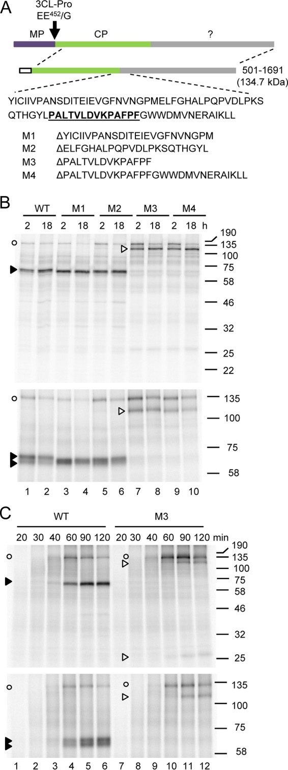
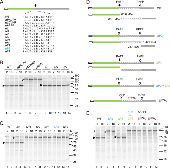
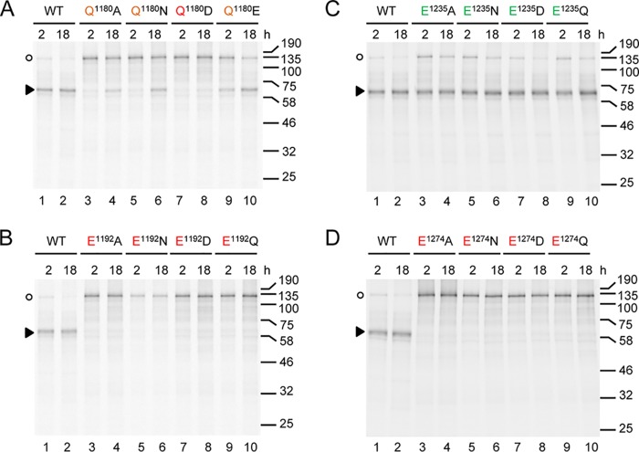
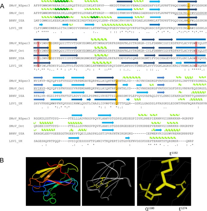
Strawberry mottle virus (SMoV) belongs to the family Secoviridae (order Picornavirales) and has a bipartite genome with each RNA encoding one polyprotein. All characterized secovirids encode a single protease related to the picornavirus 3C protease. The SMoV 3C-like protease was previously shown to cut the RNA2 polyprotein (P2) at a single site between the predicted movement protein and coat protein (CP) domains. However, the SMoV P2 polyprotein includes an extended C-terminal region with a coding capacity of up to 70 kDa downstream of the presumed CP domain, an unusual characteristic for this family. In this study, we identified a novel cleavage event at a P↓AFP sequence immediately downstream of the CP domain. Following deletion of the PAFP sequence, the polyprotein was processed at or near a related PKFP sequence 40 kDa further downstream, defining two protein domains in the C-terminal region of the P2 polyprotein. Both processing events were dependent on a novel protease domain located between the two cleavage sites. Mutagenesis of amino acids that are conserved among isolates of SMoV and of the related Black raspberry necrosis virus did not identify essential cysteine, serine, or histidine residues, suggesting that the RNA2-encoded SMoV protease is not related to serine or cysteine proteases of other picorna-like viruses. Rather, two highly conserved glutamic acid residues spaced by 82 residues were found to be strictly required for protease activity. We conclude that the processing of SMoV polyproteins requires two viral proteases, the RNA1-encoded 3C-like protease and a novel glutamic protease encoded by RNA2.IMPORTANCE Many viruses encode proteases to release mature proteins and intermediate polyproteins from viral polyproteins. Polyprotein processing allows regulation of the accumulation and activity of viral proteins. Many viral proteases also cleave host factors to facilitate virus infection. Thus, viral proteases are key virulence factors. To date, viruses with a positive-strand RNA genome are only known to encode cysteine or serine proteases, most of which are related to the cellular papain, trypsin, or chymotrypsin proteases. Here, we characterize the first glutamic protease encoded by a plant virus or by a positive-strand RNA virus. The novel glutamic protease is unique to a few members of the family Secoviridae, suggesting that it is a recent acquisition in the evolution of this family. The protease does not resemble known cellular proteases. Rather, it is predicted to share structural similarities with a family of fungal and bacterial glutamic proteases that adopt a lectin fold.

Keywords: Picornavirales; Secoviridae; plant viruses; proteases.


Figures

FIG 1
FIG 1
In vitro cis-processing of the partial P2 polyprotein precursor clone 501-1691 delineates the CP-X cleavage site. (A) Schematic representation of the wild-type 501-1691 polyprotein precursor. The entire P2 polyprotein is shown at the top of the figure, and the upper dashed lines represent the sequence corresponding to the 501-1691 construct. The amino acid sequence of the polyprotein at the approximate location of the cleavage site is shown below the diagram along with the specific amino acid sequences that were deleted for each individual construct. Deduced functional domains are shown as follows: MP, purple; CP-like domain, green;? (uncharacterized C-terminal region of the polyprotein), gray. The white box represents the 15-amino-acid S tag (derived from the N terminus of RNase A), which is present in the pCITE4a(+) vector and is fused in frame with the N terminus of the polyprotein. (B) In vitro processing reactions of the wild-type 501-1691 polyprotein (lanes 1 and 2) or the deletion mutants (lanes 3 to 10). Translation reactions were performed at 23°C for 2 h and were arrested immediately after translation (t = 2 h) or after an additional 16 h of incubation at 16C in the presence of protease reaction buffer (t = 18 h). The precursor polyprotein and potential cleavage products were separated by 11% (top) or 8% (bottom) SDS-PAGE. Migration of the unprocessed polyprotein is shown with the white circles on the left. The migrations of the predominant putative cleavage product and of the alternative secondary cleavage product are indicated with the black and white arrowheads, respectively. The migration positions of molecular mass markers (kDa) are indicated on the right. (C) Detailed time course during the translation phase of the reaction. Translation reactions were performed at 23°C as above but were arrested at the indicated time points (indicated in minutes) by addition of 2 × SDS-PAGE protein loading buffer. Translation products were separated by 11% (top) or 8% (bottom) SDS-PAGE.
FIG 2
FIG 2
In vitro processing of the partial P2 polyprotein precursor clone 501-1691 identifies two putative cleavage sites recognized by the RNA2-encoded protease. (A) Schematic representation of the wild-type 501-1691 precursor polyprotein is shown as in Fig. 1 along with the specific amino acid sequences that were deleted for each individual construct. (B and C) In vitro processing reactions of the wild-type 501-1691 polyprotein and deletion mutants were processed as in Fig. 1 and separated by 11% SDS-PAGE. (D) Schematic representation of the wild-type and mutated polyprotein precursors along with the detected cleavage products. The calculated molecular masses of these cleavage products are based on putative cleavage at the PAFP and PKFP sequences. The approximate location of the E1274A mutation is also shown. (E) In vitro processing of the single and double mutants or wild-type precursors. The migration positions of molecular mass markers (in kilodaltons) are indicated on the right of each gel.
FIG 3
FIG 3
Activity of the SMoV RNA2 protease in planta and in E. coli and sequencing of the cleavage product using N-terminal Edman degradation. (A) Schematic representation of the GFP-tagged wild-type 921-1691 and 921-1485 precursor polyproteins with the calculated molecular masses of each region of the polyproteins. (B) Nicotiana benthamiana leaves were coagroinfiltrated with tomato bushy stunt virus p19 RNA silencing suppressor protein and wild-type (WT) or E1274A mutant derivative GFP-tagged polyproteins. Total protein extracts from infiltrated patches were analyzed by Western blot at 4 days postinfection (dpi) using a commercial GFP antibody. The migration positions of molecular mass markers (kDa) are indicated on the left. (C) Analysis of cytoplasmic (S30) or membrane-enriched (P30) subcellular fractions derived from infiltrated patches expressing the 921-1485 GFP-tagged polyprotein. Western blots were processed as in panel B. (D) Schematic representation of the GST-tagged wild-type 921-1485 precursor polyprotein with the calculated molecular mass of each region of the polyprotein. (E) Expression of the wild-type (WT) or E1274A mutant derivative GST-tagged polyproteins in E. coli after induction with IPTG. Fourteen microliters of the soluble (S) and insoluble (I) fractions was separated by 12% SDS-PAGE and analyzed by immunoblot using a commercial GST antibody. (F) Purification of the cleavage product derived from the soluble fraction obtained from E. coli expressing the wild-type GST-tagged polyprotein. The equivalent of 1 μl of the total extract (T), soluble fraction (S), and unbound material (U), 3 μl of the first wash (W), and 10 μl of eluted fractions 1 to 5 were separated by 12% SDS-PAGE. Western blot was processed as in panel E to detect the GST-fused cleaved product (indicated by the arrow). The PVDF membrane was subsequently stripped and stained with Coomassie blue to detect all proteins present in the different fractions. The cleaved product was eluted from the PVDF membrane for five cycles of Edman degradation sequencing. Please note that the cleaved product is not readily detectable in the total and soluble fractions. This is because smaller amounts of material loaded were on the gel compared to that in panel E and because the E. coli growth conditions were modified to limit overexpression and prevent aggregation (see Materials and Methods). (G) Results of the Edman degradation of the purified cleaved products. The determined first five amino acids of the cleaved product are underlined. The deduced position of the cleavage site is shown with the arrow.
FIG 4
FIG 4
Sequence alignment of the putative RNA2-encoded protease domain for selected members of the family Secoviridae. The deduced amino acid sequences of the P2 polyproteins from SMoV isolates NSper3 (accession number AMR36337.1) (14), Ontario (AMS36887.1) (14), and 1134 (NP_599087.1) (16), from BRNV isolates USA (YP_654556.1) (17) and Alyth (CBM42549.1) (54), and from lettuce secovirus 1 (LSV1; ATA66954.1) (21) were used for the alignments. Alignments were produced with Clustal Omega. The positions of the determined P↓AFP and putative P↓KFP cleavage sites are shown above the SMoV NSper3 sequence by the black and open arrows, respectively. Amino acids highlighted in yellow represent mutations that impacted recognition of the proximal cleavage site but did not otherwise inhibit protease activity. Amino acids highlighted in green were those for which replacement by an alanine had little or no effect on protease activity. Amino acids highlighted in orange or red were those for which replacement by an alanine reduced the efficiency of cleavage (orange) or eliminated cleavage (red) (see Fig. 6 and 7). The number of each amino acid is indicated above the sequence. Numbers that are underlined represent those that underwent more detailed mutagenesis analysis as shown in Fig. 7. The green, orange, and red arrows show the positions of the C-terminal deletion mutants tested in Fig. 5 and their impact on cleavage efficiency (same color coding as above).
FIG 5
FIG 5
C-terminal truncation of the polyprotein precursor 501-1691 delineates the minimal region required for protease activity. (A) A schematic representation of the wild-type and truncated 501-1691 polyprotein precursors along with the detected cleavage products. The exact endpoint of each truncation can be found in Fig. 4. (B) In vitro processing reactions of the wild-type 501-1691 polyprotein and deletion mutants. Migration of the unprocessed polyprotein (white circles) and cleavage products (black arrowheads) on 11% SDS-PAGE is shown. Molecular mass markers are indicated on the right.
FIG 6
FIG 6
Point mutation analysis of conserved residues indicates that the RNA2-encoded protease does not belong to the family of serine, cysteine, or aspartic acid proteases. (A to D) In vitro processing reactions of the wild-type 501-1691 polyprotein and mutants were separated by 11% SDS-PAGE as in Fig. 1. In each case, the indicated amino acid was mutated to alanine. The effect of each mutation is indicated with the color coding (green, little or no effect on cleavage efficiency; orange, significant reduction of cleavage efficiency). The exact position of each amino acid mutation can be found on the alignment shown in Fig. 4.
FIG 7
FIG 7
Point mutations of selected glutamine and glutamic acid residues demonstrate the absolute requirement for E1192 and E1274 for the activity of the RNA2-encoded protease. (A to D) In vitro processing reactions of the wild-type 501-1691 polyprotein or mutants were separated by 11% SDS-PAGE as in Fig. 1. Color coding indicate mutations that had little or no effect on cleavage efficiency (green), a significant reduction in cleavage efficiency (orange), or the complete inhibition of cleavage (red). The exact position of each amino acid mutation can be found on the alignment shown in Fig. 4.
FIG 8
FIG 8
Predicted secondary structure of the Pro2-Glu domain and putative tertiary structure model for the catalytic region. (A) Amino acid alignments were produced using Clustal Omega. Secondary structures were predicted for each isolate using Phyre2 and are depicted above the alignments. Blue arrows represent beta-sheets and green helices represent alpha-helices. Darker colors indicate higher degrees of confidence in the prediction. Amino acids highlighted in orange or red are those that were shown to reduce or eliminate, respectively, the activity of the SMoV NSper3 protease (Fig. 6 and 7). (B) A putative tertiary structure model for the SMoV Ontario isolate catalytic region of the protease. Amino acids considered in the model are underlined in panel A. The homology-based model had a 75.9% degree of confidence with the concanavalin A-like lectin/glucanase fold of galectin, an animal S-lectin. Amino acids E1192 and E1274, which were shown to be essential for protease activity (Fig. 7), are in close proximity in this model and are predicted to form a catalytic pocket. Amino acid Q1180 which also contributes to protease activity (Fig. 7) is also in close proximity to amino acids E1192 and E1274. The overall model is shown on the left with a rainbow color model (N terminus of the considered region of the protein in red and C terminus shown in blue). Amino acids Q1180, E1192, and E1274 are highlighted in yellow in the model (middle), with a closeup shown on the right. Residue D1198, which is less well conserved in the alignment in panel A, was not positioned in proximity to the other residues and is not shown in the model. The deduced amino acid sequences of the P2 polyproteins from SMoV isolates NSper3 (AMR36337.1) (14) and Ontario (AMS36887.1) (14), BRNV isolate USA (YP_654556.1) (17), and lettuce secovirus 1 (LSV1; accession number ATA66954.1) (21) were used for the alignments.
FIG 9
FIG 9
Amino acid alignment and predicted secondary structures of a putative Pro2-Glu-like domain in the central region of the CPm of crini- and velariviruses. (A) The deduced amino acid sequences corresponding to the catalytic region of the Pro2-Glu domain of SMoV (isolate NSper3, AMR36337.1) (14) and BRNV (isolate USA, YP_654556.1) (17) were aligned with a putative Pro2-Glu domain found in the central region of the CPm of Tomato infectious chlorosis virus (TICV; crinivirus, accession number CDG34552) (59), beet pseudoyellows virus (BPYV; crinivirus, NP_940793) (60), strawberry pallidosis-associated virus (SpaV; crinivirus, YP_025090) (61), cordyline virus 1 (CDV1; velarivirus, ADU03661) (62), and little cherry virus 1 (LCV1; AGB06247) (63). Note that we only show the putative catalytic region of the Pro2-Glu like domains (see Fig. S2 in the supplemental material for an alignment of the full-length secovirid Pro2-Glu domain with the full-length CPm domains of additional criniviruses and velariviruses). Secondary structures were predicted using Phyre2 and are depicted above the alignments. Blue arrows represent beta-sheets and green helices represent alpha-helices. Darker colors indicate higher degrees of confidence in the prediction. Amino acids highlighted in orange or red are those that were shown to reduce or eliminate, respectively, the activity of the SMoV NSper3 protease (Fig. 6 and 7). (B) Genomic organization of the RNA2 of Tomato infectious chlorosis virus (TICV), a crinivirus (29). The CPm domain is shown in the lower part of the figure with the CP-like domain highlighted in orange and the Pro2-Glu-like domain highlighted with the gray shading.
FIG 10
FIG 10
Updated processing map of the SMoV P2 polyprotein and comparison with related viruses. A schematic representation of SMoV isolates NSper3 and 1134 P2 polyproteins is shown at the top. Putative cleavage sites identified in this study and previously (MP-CP cleavage site) (18) are depicted. The predicted genomic organization of the closely related species Black raspberry necrosis virus (BRNV) is also shown. Red arrows represent characterized and putative cleavage sites recognized by the RNA2-encoded glutamic protease (Pro2-Glu). Black arrows represent characterized and putative cleavage sites recognized in trans by the RNA1-encoded 3CL-Pro protease. The deduced genome organization of other viruses discussed in the text is shown (see Discussion), including Chocolate lily virus A (CLVA) and Dioscorea mosaic associated virus (DMaV), both currently assigned to the family Secoviridae but not to a specific genus, Satsuma dwarf virus (SDV; genus Sadwavirus), Grapevine fanleaf virus (GFLV; genus Nepovirus, subgroup A) and Tomato ringspot virus (ToRSV; genus Nepovirus, subgroup C). The genomic organizations of GFLV and ToRSV have been characterized and 3CL-Pro cleavage sites have been identified (64–66). For SDV, the cleavage sites defining the CP domains are known (67), but the organization of the N-terminal region of the polyprotein has not been elucidated. The DMaV polyprotein is short and likely only includes the MP and CP domains (68). In the case of CLVA, although the P2 polyprotein extends beyond the presumed CP domain, the C-terminal region of the polyprotein does not share significant sequence identity with the SMoV Pro2-Glu (69). For CLVA, DMaV, and BRNV, proposed cleavage sites were based on alignments with the corresponding SMoV cleavage sites and the number of CP subunits has not been experimentally confirmed ( and this study).

Similar articles

Cited by

References

    1. Tautz N, Tews BA, Meyers G. 2015. The molecular biology of pestiviruses. Adv Virus Res 93:47–160. doi:10.1016/bs.aivir.2015.03.002. - DOI - PubMed
    1. Rodamilans B, Shan H, Pasin F, Garcia JA. 2018. Plant viral proteases: beyond the role of peptide cutters. Front Plant Sci 9:666. doi:10.3389/fpls.2018.00666. - DOI - PMC - PubMed
    1. Laitinen OH, Svedin E, Kapell S, Nurminen A, Hytonen VP, Flodstrom-Tullberg M. 2016. Enteroviral proteases: structure, host interactions and pathogenicity. Rev Med Virol 26:251–267. doi:10.1002/rmv.1883. - DOI - PMC - PubMed
    1. Jensen LM, Walker EJ, Jans DA, Ghildyal R. 2015. Proteases of human rhinovirus: role in infection. Methods Mol Biol 1221:129–141. doi:10.1007/978-1-4939-1571-2_10. - DOI - PubMed
    1. Sun D, Chen S, Cheng A, Wang M. 2016. Roles of the picornaviral 3C proteinase in the viral life cycle and host cells. Viruses 8:82. doi:10.3390/v8030082. - DOI - PMC - PubMed

Publication types

LinkOut - more resources