. 2019 Feb 19;93(5):e02026-18.
doi: 10.1128/JVI.02026-18. Print 2019 Mar 1.

Insulin-Like Growth Factor 2 Receptor Expression Is Promoted by Human Herpesvirus 8-Encoded Interleukin-6 and Contributes to Viral Latency and Productive Replication

Affiliations

Insulin-Like Growth Factor 2 Receptor Expression Is Promoted by Human Herpesvirus 8-Encoded Interleukin-6 and Contributes to Viral Latency and Productive Replication

Qian Li et al. J Virol. .

Abstract

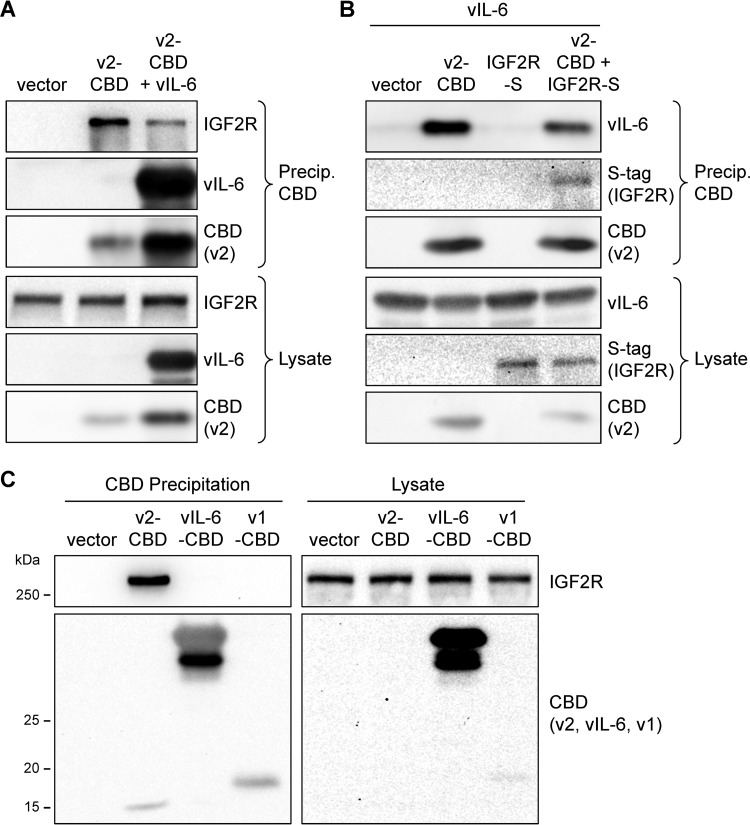
Human herpesvirus 8 (HHV-8) viral interleukin-6 (vIL-6) localizes largely to the endoplasmic reticulum (ER) and here associates functionally with both the gp130 signal transducer and the novel ER membrane protein vitamin K epoxide reductase complex subunit 1 variant-2 (VKORC1v2). The latter interaction contributes to the viability of latently infected primary effusion lymphoma (PEL) cells and to HHV-8 productive replication, in part via promotion of ER-associated degradation (ERAD) of nascent pro-cathepsin D (pCatD) and consequent suppression of lysosome-localized proapoptotic mature CatD. Here we report that VKORC1v2 associates with insulin-like growth factor 2 receptor (IGF2R), also known as cation-independent mannose-6-phosphate receptor, which is involved in trafficking of mannose-6-phosphate-conjugated glycoproteins to lysosomes. VKORC1v2 effected reduced IGF2R expression in a manner dependent on VKORC1v2-IGF2R interaction, while vIL-6, which could inhibit VKORC1v2-IGF2R interaction, effected increased expression of IGF2R. These effects were independent of changes in IGF2R mRNA levels, indicating likely posttranslational mechanisms. In kinetic analyses involving labeling of either newly synthesized or preexisting IGF2R, vIL-6 promoted accumulation of the former while having no detectable effect on the latter. Furthermore, vIL-6 led to decreased K48-linked ubiquitination of IGF2R and suppression of ERAD proteins effected increased IGF2R expression and loss of IGF2R regulation by vIL-6. Depletion-based experiments identified IGF2R as a promoter of PEL cell viability and virus yields from lytically reactivated cultures. Our findings identify ER-transiting nascent IGF2R as an interaction partner of VKORC1v2 and target of vIL-6 regulation and IGF2R as a positive contributor to HHV-8 biology, thereby extending understanding of the mechanisms of VKORC1v2-associated vIL-6 function.IMPORTANCE HHV-8 vIL-6 promotes productive replication in the context of reactivated lytic replication in primary effusion lymphoma (PEL) and endothelial cells and sustains latently infected PEL cell viability. Viral IL-6 is also considered to contribute significantly to HHV-8-associated pathogenesis, since vIL-6 can promote cell proliferation, cell survival, and angiogenesis that are characteristic of HHV-8-associated Kaposi's sarcoma, PEL and multicentric Castleman's disease (MCD), in addition to proinflammatory activities observed in MCD-like "Kaposi's sarcoma-associated herpesvirus-induced cytokine syndrome." We show in the present study that vIL-6 can promote productive replication and latent PEL cell viability through upregulation of the mannose-6-phosphate- and peptide hormone-interacting receptor IGF2R, which is a positive factor in HHV-8 biology via these activities. VKORC1v2-enhanced ER-associated degradation of IGF2R and vIL-6 promotion of IGF2R expression through prevention of its interaction with VKORC1v2 and consequent rescue from degradation represent newly recognized activities of VKOCR1v2 and vIL-6.

Keywords: ER-associated degradation; cation-independent mannose-6-phosphate receptor; endoplasmic reticulum; human herpesvirus 8; insulin-like growth factor 2 receptor; latency; replication; viral interleukin-6; vitamin K epoxide reductase complex subunit 1 variant-2.


Figures

FIG 1
FIG 1
Interaction between VKORC1v2 and IGF2R. (A) Coprecipitation assays were carried out using transfected HEK293T cell lysates as a source for affinity-tagged VKORC1v2 (CBD-fused, v2-CBD), or negative-control VKORC1v1-CBD (v1-CBD), and IGF2R (S-peptide-linked, IGF2R-S), which were precipitated with chitin and S-protein beads, respectively. Coprecipitated IGF2R-S and VKORC1-CBD proteins were identified by immunoblotting of the respective sedimented material. Appropriate protein expression in vector-transfected cells was confirmed by immunoblotting of cell lysates. (B) CBD-tagged VKORC1v2 variants (Δ1 to Δ5) containing various deletions in the ER-lumenal portion of the receptor, C-terminal to the transmembrane (TM) domain, were used in similar coprecipitation assays. Endogenous IGF2R coprecipitated with chitin bead-bound VKORC1v2-CBD was detected by immunoblotting. IGF2R expression in cell lysates was also checked, along with β-actin, used as a protein loading and membrane-transfer control. The arrowhead indicates the position of the VKORC1v2Δ1 band, expressed at much lower levels than the other VKORC1v2 proteins.
FIG 2
FIG 2
Competitive vIL-6 and IGF2R interactions with VKORC1v2. (A) VKORC1v2-CBD (v2-CBD)-based coprecipitation assays were carried out in the absence or presence of vIL-6 coexpression to assess the influence of vIL-6 on VKORC1v2-IGF2R interaction. Chitin-bead precipitates and cell lysates were analyzed by immunoblotting for detection of IGF2R (endogenous), VKORC1v2-CBD, and vIL-6. (B) A similar experiment was undertaken to explore the effect of overexpressed IGF2R (S-tagged, IGF2R-S) on vIL-6 interaction with VKORC1v2 in appropriately transfected cells. Chitin-bead precipitates and cell lysates were immunoblotted for detection of CBD (VKORC1v2), vIL-6, and S-tag (IGF2R). (C) Chitin bead-based precipitation analyses of (endogenous) IGF2R interactions with VKORC1v2-CBD (v2-CBD) and vIL-6-CBD; VKORC1v1-CBD (v1-CBD) and empty vector were transfected into parallel cultures to provide negative controls.
FIG 3
FIG 3
Regulation of IGF2R expression by VKORC1v2. (A) Expression of IGF2R in HEK293T cell was monitored as a function of the dose of transfected VKORC1v2-CBD (v2-CBD) expression plasmid (0.4, 0.8, and 1.2 μg) and encoded protein. IGF2R and VKORC1v2-CBD levels were assessed by immunoblotting of cell extracts; probing for β-actin provided a loading control. Quantified levels of IGF2R, relative to the level in empty vector (vec)-transfected cells (set at 1), are shown below the IGF2R blot. (B) RT-qPCR analysis of IGF2R mRNA levels in parallel cultures transfected with 1.2 μg of either empty vector (vec) or VKORC1v2-CBD (v2) expression plasmid. RNA samples were derived from duplicate cultures and each reverse-transcribed sample was analyzed in duplicate by qPCR; averages of the mean qPCR values for each biological replicate were calculated, along with the standard deviations from these average values. The average GAPDH mRNA-normalized IGF2R mRNA level in the VKORC1v2 vector-transfected cultures is shown relative to that in empty vector-transfected cells (set at 1). (C) Wild-type (WT) and vBD-mutated (ΔvBD, lacking residues 31 to 39 [1]) VKORC1v2 proteins were expressed in vector-transfected cells to determine the requirement for (vBD-dependent) VKORC1v2-IGF2R interaction for VKORC1v2 suppression of IGF2R. Representative immunoblots from one of five experiments are shown, together with quantified data from all experiments (chart). IGF2R levels were normalized to β-actin and expressed relative to levels (set at 1) in empty vector-transfected cultures. Standard deviations from average values are indicated along with Student t test P value for VKORC1v2 suppression of IGF2R.
FIG 4
FIG 4
Regulation of IGF2R expression by vIL-6. (A) Expression of endogenous IGF2R was measured in response to increasing levels of vIL-6 (expressed from 0.1 to 0.5 μg of vector per transfected culture) relative to empty vector-transfected cells (vec, 0.5 μg). Cell extracts from cultures harvested 30 h after transfection were analyzed by immunoblotting for detection of IGF2R, vIL-6, and β-actin (loading control). Levels of IGF2R (normalized to β-actin) in vIL-6-expressing cells are shown relative to the level (set at 1.0) in empty vector-transfected cells. (B) IGF2R mRNA levels from parallel cultures transfected with 0.5 μg of empty vector or vIL-6 expression plasmid were determined by RT-qPCR, as outlined in the legend to Fig. 3B. (C) Similar analyses of IGF2R protein expression were undertaken in transfected native and VKORC1v2-null (5) HEK293T cells. The levels of IGF2R in vIL-6-expressing cells relative to levels in empty vector (vec)-transfected wild-type and VKORC1 gene-mutated cells (each set at 1.0) are shown below the IGF2R blot. Relative transfection efficiencies in WT cells (50%) and VKORC1v2-null cells (70%) were determined by cotransfection of a GFP expression plasmid. (D) VKORC1v2 (StrepII+Flag-tagged, SF) was introduced exogenously into VKORC1v2-deficient HEK293T cells by transfection of a VKORC1v2 expression plasmid (altered, codon-synonymously, in gRNA target sequences and therefore Cas9-resistant [“CR”]), along with either empty vector (vec) or vIL-6 expression plasmid to verify the role of VKORC1v2 in vIL-6-mediated augmentation of IGF2R expression. IGF2R expression levels, normalized to β-actin and expressed relative to the IGF2R levels (set at 1) in empty vector-transfected cultures, are shown below the IGF2R blot.
FIG 5
FIG 5
vIL-6 regulation of nascent IGF2R. (A) Halo-tagged IGF2R was introduced into HEK292T cells via expression vector transfection, along with empty vector or vIL-6 expression plasmid. Pulse-labeling of IGF2R-Halo was achieved by addition of fluorescent Halo-ligand probe TMR to cultures 24 h posttransfection, replacing blocking ligand, HaloTag succinimidyl ester O4 (“Halo-O4”), added immediately after transfection. Parallel cultures were harvested at different times post-TMR addition, extracts were run on a polyacrylamide gel, and TMR-labeled IGF2R-Halo was visualized under UV illumination. Proteins were then transferred to nitrocellulose membrane for immunoblotting for vIL-6 (to check expression) and β-actin (normalization control). Digitally quantified levels of IGF2R-Halo/TMR fluorescence, normalized to β-actin, are shown as fold increases above background levels (at the zero time points). (B) A similar experiment was undertaken, but here TMR was added for 15 min to label all IGF2R-Halo in “unblocked” cultures, and then was removed and replaced with blocking agent (Halo-O4) to prevent any labeling of subsequently produced IGF2R-Halo. Replicate cultures were harvested at different times postlabeling to track IGF2R-Halo decay as a function of vIL-6 expression. Analyses were as outlined for panel A. The levels of TMR-labeled IGF2R, normalized to β-actin, are shown relative to levels (set at 1) expressed at 0 h in both the absence and presence of vIL-6 (ND, not determined; specific signal versus background uncertain). (C) Testing of ER-restricted, KDEL motif-tagged vIL-6 (vIL-6.K), relative to native vIL-6, for enhancement of IGF2R expression. Equal amounts of the respective vIL-6 vectors and empty vector (vec) were transfected separately into parallel HEK293T cell cultures and extracts, made 24 to 48 h posttransfection (depending on the particular experiment), and coharvested media were analyzed by immunoblotting. Proteins from media were first precipitated with trichloroacetic acid to enable concentration. Immunoblot data from one of five experiments are shown; β-actin-normalized levels of IGF2R in vIL-6- and vIL-6-KDEL-expressing cells relative to those in empty vector-transfected cells are shown in the chart, using the same presentation and analysis outlined for Fig. 3C. (D) To test exogenously added vIL-6 for potential IGF2R-regulatory activity, media from vIL-6 vector-transfected HEK293T cells or from empty vector (vec)-transfected cells were applied to naive HEK293T cultures for 24 h prior to preparation of cell extracts. Relative levels of IGF2R (shown normalized to β-actin) were determined by immunoblotting. Immunodetection of active phospho-STAT3 (pSTAT3, Y705-phosphorylated) and total STAT3 (normalization control) confirmed vIL-6-induced signaling and therefore functional levels of vIL-6. A representative of three experiments is shown.
FIG 6
FIG 6
IGF2R regulation by vIL-6 via ERAD. (A) Influence of vIL-6 on IGF2R ubiquitination. HEK293T cultures were transfected with either empty vector (vec, negative control) or vIL-6 expression plasmid along with vectors expressing Flag-tagged ubiquitin proteins (wild-type [Flag-Ub], K48-only [Flag-K48], or K-null [Flag-K0]). Cells were harvested 48 h after transfection, Flag-tagged ubiquitin was immunoprecipitated from cell lysates, and precipitated, urea-washed material was immunoblotted for detection of IGF2R. Equivalent Flag-Ub precipitation between paired samples was verified via Flag immunoblotting of immunoprecipitates. Relative amounts (rel. amt.) of ubiquitinated IGF2R precipitated from lysates of empty vector-transfected cells (set at 1) and vIL-6-expressing cells are shown below the IGF2R blot. Cell lysates were probed for vIL-6 and β-actin to verify appropriate vIL-6 expression. (B) Plasmid vector-expressed shRNAs directed to mRNA of either HRD1 or gp78 ERAD E3 ligases were tested relative to control (NS) shRNA for their efficacies in HEK293T cells cotransfected with vectors expressing S-epitope-tagged HRD1 or gp78. Cell extracts were made 48 h posttransfection and analyzed by immunoblotting. (C) The involvement of ERAD in vIL-6 regulation of IGF2R was tested directly by comparing vIL-6 activity in response to shRNA-mediated depletion of HRD1 and gp78. Cell lysates from test (HRD1 and gp78) and control (NS) shRNA (sh) vector-transfected cultures cotransfected with either empty vector (vec) or vIL-6 expression plasmid were analyzed for IGF2R expression 48 h posttransfection. The relative levels of IGF2R, normalized to β-actin, are shown below the IGF2R blot.
FIG 7
FIG 7
Regulation of IGF2R by vIL-6 and VKORC1v2 in the context of infection. (A) IGF2R expression as a function of lentiviral vector-mediated vIL-6 overexpression in BCBL-1 (TRExBCBL1-RTA) and JSC-1 PEL cells was determined by immunoblotting of cell extracts made 48 h after lentiviral vector transduction. Lentivirus with no inserted ORF (vec) was used for transduction of parallel cultures to allow comparisons. Levels of IGF2R expression relative to those in empty vector-transduced cells (set at 1) and normalized to β-actin are shown. (B) Similar experiments were carried out to investigate the effect of vIL-6 depletion on IGF2R levels in the PEL cell lines. vIL-6 mRNA-directed shRNA or nonsilencing (NS) control shRNA was transduced via lentiviral vector infection. The relative levels of IGF2R expression, calculated as outlined in panel A, are shown. (C) The same approach was used in TRExBCBL1-RTA cells to investigate the effect of vIL-6 depletion on IGF2R expression in the context of lytic replication, induced by doxycycline (Dox) treatment (+Dox). Cells were harvested 48 h postinduction (lytic) or mock treatment (latent). Latent and lytic cell extracts were run on the same gel; the dotted lines indicate deletion of a blank lane. Levels of IGF2R expression in vIL-6-depleted cells relative to those (set at 1) in NS shRNA-transduced cells are shown (values are β-actin-normalized). (D) Expression of IGF2R in latently infected and lytically reactivated (doxycycline- and sodium butyrate-treated) iSLK cells infected with wild-type (WT) or vIL-6-null (vIL-6X) BAC16-derived HHV-8. Cells were harvested 48 h postinduction for preparation of cell extracts for immunoblotting. The LANA immunoblot confirms equivalent latent viral loads in cultures infected with identical infectious doses of the wild-type and mutated viruses. Relative IGF2R expression levels, normalized to β-actin, between cells infected with wild-type virus (set at 1) and vIL-6-null virus are shown below the IGF2R blot. (E) The role of endogenously expressed VKORC1v2 on IGF2R expression was tested in BCBL-1 (TRExBCBL1-RTA) and JSC-1 cells using lentivirally delivered VKORC1v2 (v2) mRNA-targeting shRNA or control NS shRNA. Cells were harvested 4 days (BCBL-1) or 5 days (JSC-1) postransduction for immunoblot analysis of IGF2R expression; the values shown represent β-actin-normalized IGF2R expression levels in VKORC1v2-depleted cells relative to controls (set at 1) for each cell type. RT-PCR was applied to mRNA extracted from samples of the same cultures to verify shRNA-mediated depletion of VKORC1v2 mRNA; VKORC1v1 (v1) mRNA (lacking the shRNA target site) was unaffected. RT-PCR for detection of GAPDH mRNA provided a normalization control.
FIG 8
FIG 8
Role of IGF2R in HHV-8 biology. (A) and (B) Effect of IGF2R on latent PEL cell growth. BCBL-1 (TRExBCBL1-RTA) (A) and JSC-1 (B) cultures were transduced at high efficiency (>90%) with (GFP+) lentiviral vectors expressing either of two shRNAs (designated sh3 and sh4) directed to IGF2R mRNA or nonsilencing (NS) shRNA (control). After 3 days, trypan blue-excluding, viable-cell densities determined by hemocytometric counting were normalized (day 0) and then monitored for a further 3 days. On day 3, culture samples were analyzed by annexin V-Cy3 staining and nuclear counterstaining with Hoechst 33342 to determine rates of apoptosis. Data were collected from multiple, random fields from each culture (>400 cells viewed per culture). Average values from triplicate cultures are shown for the growth and apoptosis analyses (top and bottom charts, respectively); error bars show standard deviations from these averages. P values from Student t test (paired, two-tailed) are indicated. The immunoblots (bottom) show effective depletion of IGF2R by each of the IGF2R-specific shRNAs relative to the NS shRNA control; samples were derived from pooled terminal BCBL-1 and JSC-1 cultures in each treatment group. (C) Effect of IGFR2 depletion on HHV-8 productive replication. TRExBCBL1-RTA cells were induced into lytic replication by treatment with doxycycline (see Materials and Methods) and after 4 days, culture media were harvested for assessment of released viral titers, as determined by latency-associated nuclear antigen (LANA) immunofluorescence staining of inoculated iSLK cells (infectious assay) or qPCR-measured copy numbers of DNase I-resistant (encapsidated) viral genomes (virion DNA assay), using established methods (3, 28). Data were derived from duplicate cultures; error bars show standard deviations from the plotted mean values. Student t test-derived P values are indicated. Cell lysates from terminal cultures were immunoblotted to confirm IGF2R depletion.

Similar articles

Cited by

References

    1. Chen D, Cousins E, Sandford G, Nicholas J. 2012. Human herpesvirus 8 viral interleukin-6 interacts with splice variant 2 of vitamin K epoxide reductase complex subunit 1. J Virol 86:1577–1588. doi:10.1128/JVI.05782-11. - DOI - PMC - PubMed
    1. Cousins E, Nicholas J. 2013. Role of human herpesvirus 8 interleukin-6-activated gp130 signal transducer in primary effusion lymphoma cell growth and viability. J Virol 87:10816–10827. doi:10.1128/JVI.02047-13. - DOI - PMC - PubMed
    1. Cousins E, Gao Y, Sandford G, Nicholas J. 2014. Human herpesvirus 8 viral interleukin-6 signaling through gp130 promotes virus replication in primary effusion lymphoma and endothelial cells. J Virol 88:12167–12172. doi:10.1128/JVI.01751-14. - DOI - PMC - PubMed
    1. Chen D, Nicholas J. 2015. Promotion of endoplasmic reticulum-associated degradation of procathepsin D by human herpesvirus 8-encoded viral interleukin-6. J Virol 89:7979–7990. doi:10.1128/JVI.00375-15. - DOI - PMC - PubMed
    1. Chen D, Xiang Q, Nicholas J. 2017. Human herpesvirus 8 interleukin-6 interacts with calnexin cycle components and promotes protein folding. J Virol 91:e00965-17. doi:10.1128/JVI.00965-17. - DOI - PMC - PubMed

Publication types

MeSH terms