. 2019 Feb 19;93(5):e01635-18.
doi: 10.1128/JVI.01635-18. Print 2019 Mar 1.

The Potato Virus X TGBp2 Protein Plays Dual Functional Roles in Viral Replication and Movement

Affiliations

The Potato Virus X TGBp2 Protein Plays Dual Functional Roles in Viral Replication and Movement

Xiaoyun Wu et al. J Virol. .

Abstract

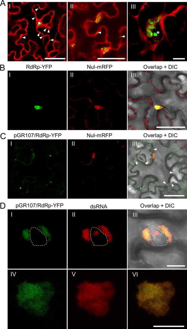
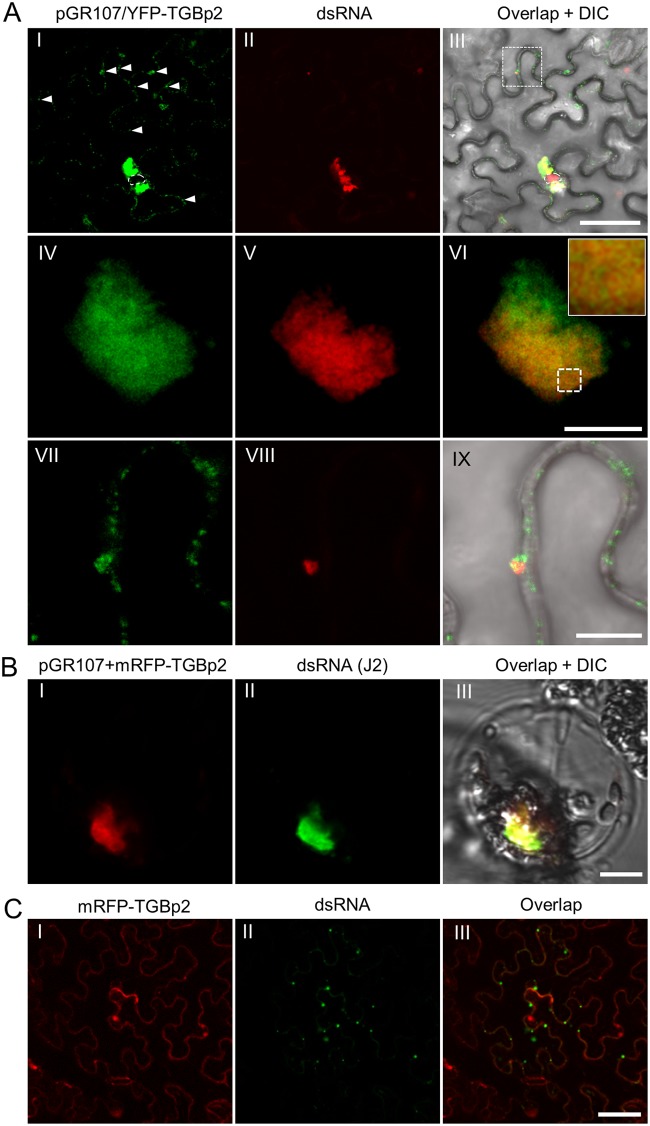
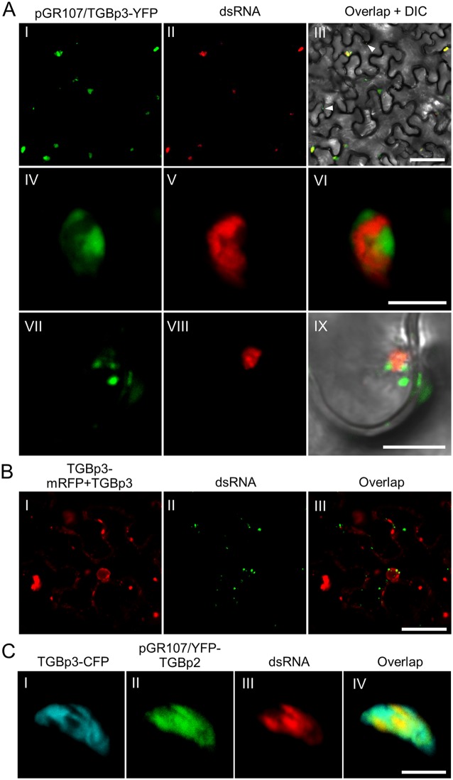
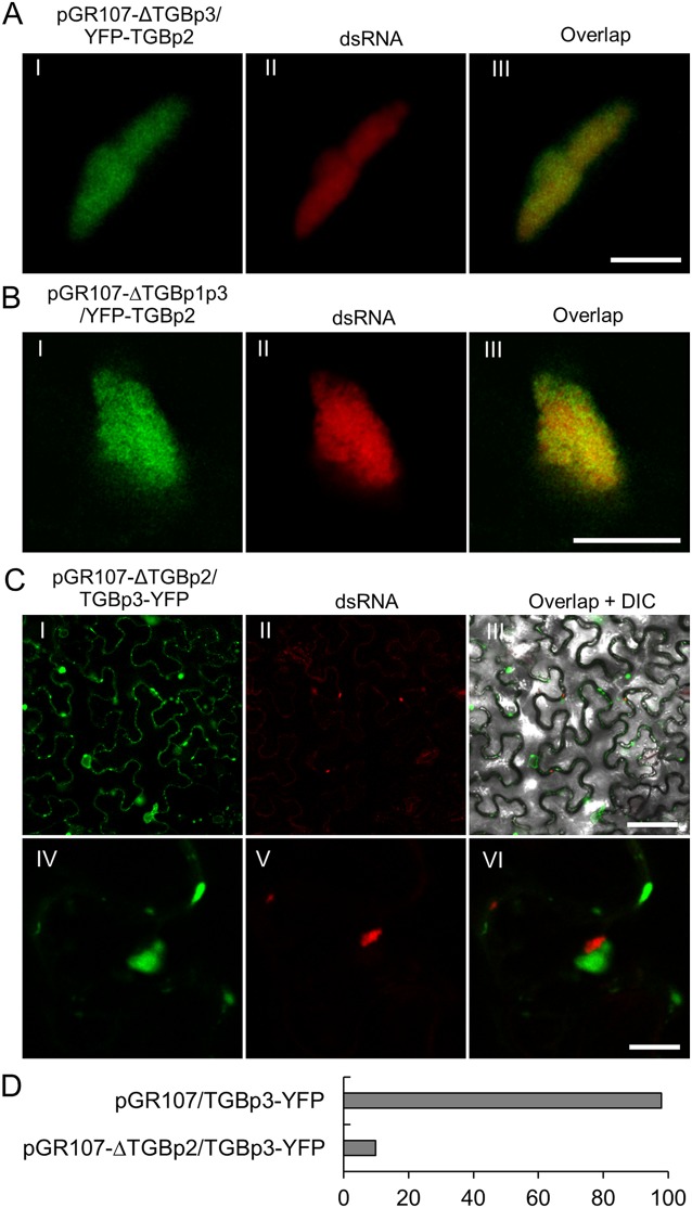
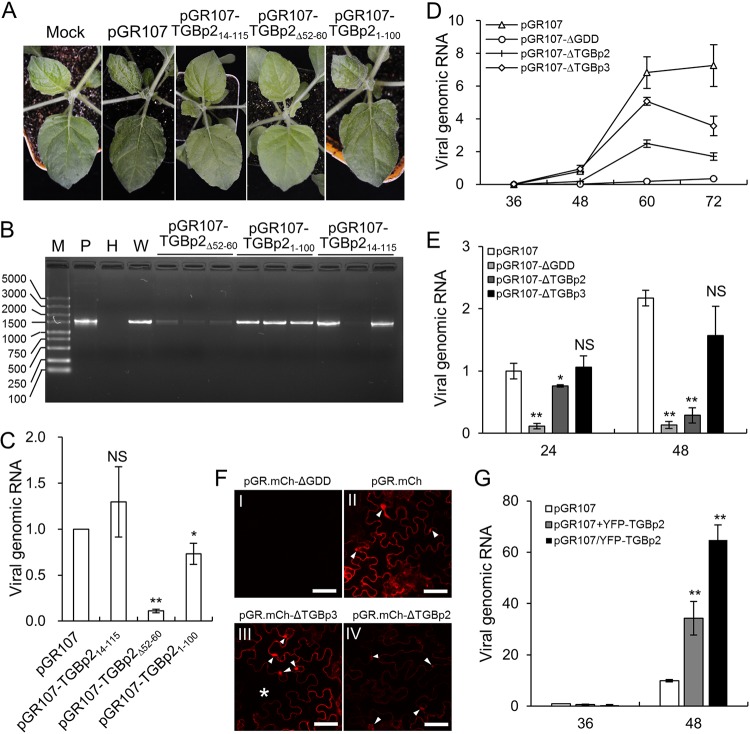
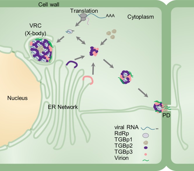
Plant viruses usually encode one or more movement proteins (MP) to accomplish their intercellular movement. A group of positive-strand RNA plant viruses requires three viral proteins (TGBp1, TGBp2, and TGBp3) that are encoded by an evolutionarily conserved genetic module of three partially overlapping open reading frames (ORFs), termed the triple gene block (TGB). However, how these three viral movement proteins function cooperatively in viral intercellular movement is still elusive. Using a novel in vivo double-stranded RNA (dsRNA) labeling system, we showed that the dsRNAs generated by potato virus X (PVX) RNA-dependent RNA polymerase (RdRp) are colocalized with viral RdRp, which are further tightly covered by "chain mail"-like TGBp2 aggregates and localizes alongside TGBp3 aggregates. We also discovered that TGBp2 interacts with the C-terminal domain of PVX RdRp, and this interaction is required for the localization of TGBp3 and itself to the RdRp/dsRNA bodies. Moreover, we reveal that the central and C-terminal hydrophilic domains of TGBp2 are required to interact with viral RdRp. Finally, we demonstrate that knockout of the entire TGBp2 or the domain involved in interacting with viral RdRp attenuates both PVX replication and movement. Collectively, these findings suggest that TGBp2 plays dual functional roles in PVX replication and intercellular movement.IMPORTANCE Many plant viruses contain three partially overlapping open reading frames (ORFs), termed the triple gene block (TGB), for intercellular movement. However, how the corresponding three proteins coordinate their functions remains obscure. In the present study, we provided multiple lines of evidence supporting the notion that PVX TGBp2 functions as the molecular adaptor bridging the interaction between the RdRp/dsRNA body and TGBp3 by forming "chain mail"-like structures in the RdRp/dsRNA body, which can also enhance viral replication. Taken together, our results provide new insights into the replication and movement of PVX and possibly also other TGB-containing plant viruses.

Keywords: Potato virus X; TGB; TGBp2; movement; replication.


Figures

FIG 1
FIG 1
In vivo visualization of dsRNA and RdRp in PVX infected N. benthamiana epidermal cells. (A) Visualization of the dsRNA during PVX infection at 36 (I), 48 (II), and 60 (III) hpi. The fluorescence from the dRBFC assay is shown in green, whereas the fluorescence from pGR.mCh is shown in red. The nucleus is indicated by a white asterisk and the peripheral dsRNA fluorescent foci are indicated by white arrowheads. Scale bar = 50 μm (I and II) or 10 μm (III). Note that the dRBFC signal in the nucleus represents endogenous d-bodies (28). (B and C) Subcellular localization of transiently expressed RdRp-YFP (green) in N. benthamiana epidermal cells in the absence of (B) or during (C) PVX infection at 48 hpi. The RdRp-YFP in panel C was expressed from pGR107/RdRp-YFP to ensure infection of PVX in the same cell. The cytoplasmic green fluorescent foci from RdRp-YFP are indicated by white arrowheads. The nuclei were labeled by a nuclear localization signal peptide (NLS)-tagged mRFP (Nul-mRFP). Scale bars = 50 μm. (D) Subcellular localization of perinuclear (I to III) or cytoplasmic (IV to VI) RdRp and dsRNA during PVX infection at 60 hpi; the RdRp-YFP (green) was expressed from pGR107/RdRp-YFP, and dsRNAs (red) were labeled by mRFP-based dRBFC plasmids. The nucleus is indicated by a white dashed line. All scale bars = 10 μm.
FIG 2
FIG 2
Subcellular localization of TGBp2 and dsRNA during PVX infection. (A) Confocal micrographs of YFP-TGBp2 (green) and dsRNA (red) during PVX infection in N. benthamiana epidermal cells at 48 hpi at lower (I to III) and higher (IV to IX) magnifications. YFP-TGBp2 was expressed from pGR107/YFP-TGBp2 and dsRNA was labeled using the mRFP-based dRBFC assay. The white arrowheads in frame I indicates the TGBp2-induced small granules. The nuclei are indicated by white dashed lines. The inset in frame VI is an enlargement of the dashed area to show the typical chain mail-like structure of TGBp2 on dsRNA. Scale bar = 50 μm (I to III) or = 10 μm (IV to IX). (B) Subcellular localization of dsRNA (green) and mRFP-TGBp2 (red) in a typical globular-like X-body in the N. benthamiana protoplast. The dsRNA was detected using J2 monoclonal antibodies. Scale bar = 10 μm. (C) Subcellular localization of dsRNA (green) and mRFP-TGBp2 (red) in the absence of PVX infection. Scale bar = 50 μm.
FIG 3
FIG 3
Subcellular localization of TGBp3 and dsRNA in PVX infected N. benthamiana cells. (A) Confocal micrographs of dsRNA (red) and TGBp3-YFP (green) in N. benthamiana leaf tissue during PVX infection at 48 hpi. TGBp3-YFP was expressed from pGR107/TGBp3-YFP, and the dsRNA was labeled using the mRFP-based dRBFC assay. The white arrowheads indicate TGBp3 foci at the cell periphery. Scale bar = 50 μm (I to III) or 10 μm (IV to IX). (B) TGBp3-mRFP is not associated with dsRNA in the absence of PVX infection. Scale bar = 10 μm. (C) Confocal micrographs of TGBp3-CFP (cyan), YFP-TGBp2 (green), and dsRNA (red) in N. benthamiana epidermal cells during PVX infection at 48 hpi. YFP-TGBp2 was expressed from pGR107/YFP-TGBp2. Scar bar = 10 μm.
FIG 4
FIG 4
Recruitment of TGBp2 to dsRNA occurs independently of TGBp1 and TGBp3. (A and B) Confocal micrographs of YFP-TGBp2 (green) and dsRNA (red) in N. benthamiana epidermal cells during infection by pGR107-ΔTGBp3/YFP-TGBp2 (A) or pGR107-ΔTGBp1p3/YFP-TGBp2 (B) at 48 hpi. The dsRNAs were labeled using the mRFP-based dRBFC assay. Scar bar = 10 μm. (C) Subcellular localization of TGBp3-YFP and dsRNA in N. benthamiana cells infected with a TGBp2-defective PVX infectious clone (pGR107-ΔTGBp2/TGBp3-YFP) at 48 hpi. Scar bar = 50 μm (I to III) or = 10 μm (IV to VI). A differential interference contrast (DIC) channel was included in the overlap micrograph to illustrate the outline of the cell. (D) Percentage of RdRp/dsRNA bodies associated with TGBp3 in N. benthamiana epidermal cells infected with pGR107/TGBp3-YFP or pGR107-ΔTGBp2/TGBp3-YFP.
FIG 5
FIG 5
TGBp2 interacts with PVX RdRp. (A) Schematic representations of the PVX RdRp domains. MET, methyltransferase domain; UNL, unstructured loop domain; HEL, helicase domain; REP, replicase domain; RepN, N terminus of the REP domain; RepC, C terminus of the REP domain. The numbers represent the amino acid positions of the domain boundaries. (B) Subcellular localization of MET-YFP, UNL-YFP, HEL-YFP, and REP-YFP in N. benthamiana epidermal cells at 48 hpi. The insets show the typical cytoplasmic granules of MET-YFP and HEL-YFP, as well as the nuclear signal of UNL-YFP and REP-YFP. (C) BiFC for protein-protein interactions between TGBp2 and RdRp domains in N. benthamiana epidermal cells at 48 hpi. Micrographs were obtained using the same settings. Scale bars = 50 μm. (D) Subcellular localization of RepN-YFP and RepC-YFP in N. benthamiana epidermal cells at 48 hpi. The insets show the typical nuclear signal of RepN-YFP and cytoplasmic vesicles of RepC-YFP. The nuclei are indicated by white asterisks, and the typical vesicle-like structures are indicated by white arrowheads. Scale bars = 50 μm. (E) BiFC for protein-protein interactions between TGBp2 and RepN or RepC. Micrographs were obtained using the same parameters. Scale bars = 50 μm. (F) MYTH for protein-protein interaction between TGBp2 and RdRp domains. (G) Influence of TGBp2 on the subcellular localization of RdRp and REP domain in N. benthamiana epidermal cells at 48 dpi. RdRp-YFP and REP-YFP are shown in green, and mRFP-TGBp2 is shown in red. Scale bars = 50 μm.
FIG 6
FIG 6
The central and C-terminal hydrophilic domains of TGBp2 are required for interactions with viral RdRp. (A) Illustration of TGBp2 mutants. The two transmembrane motifs (TM1 and TM2) are shown in black. (B) BiFC for protein-protein interactions between RepN and TGBp2 mutants in N. benthamiana epidermal cells at 48 hpi. Micrographs were obtained using the same parameters. Scale bars = 50 μm. (C) MYTH for protein-protein interactions between RepN and TGBp2 mutants. (D) Subcellular localization of TGBp2 mutants. The insets show the typical morphology of ER-derived vesicles induced by TGBp2 or its mutants. Scare bar = 50 μm. (E) Subcellular localization of dsRNA and TGBp2 mutants during PVX infection. The nuclei are indicated by white asterisks. Scale bar = 50 μm.
FIG 7
FIG 7
TGBp2 affects PVX replication. (A) Phenotype of wild-type PVX and its mutants containing various deletions in TGBp2 on N. benthamiana seedlings at 8 dpi. (B) RT-PCR detection of PVX genomic RNA on N. benthamiana systemic leaves at 10 dpi. M, DL5000 DNA marker (Vazyme Biotech Co., Ltd., Nanjing, China); P, positive plasmid; H, healthy N. benthamiana leaf; W, N. benthamiana leaf infected by wild-type PVX. (C) Quantitative RT-PCR analysis of the genomic RNA of pGR107, pGR107-TGBp2ΔND, pGR107-TGBp2Δ52-60, and pGR107-TGBp2ΔCD in N. benthamiana leaves at 48 hpi. The viral RNA level of wild-type PVX was normalized to 1. Bars represent SDs from three biological repeats. *, **, and NS, respectively, indicate P values of <0.01, <0.001, and >0.1 compared with the amount of wild-type PVX genomic RNA at the same time point by the Student t test. (D) Time course study of the replication of pGR107, pGR107-ΔGDD, and TGBp2 or TGBp3 knockout PVX infectious clones in N. benthamiana leaves. Bars represent the SEs from 5 biological repeats. (E) Accumulation of wild-type and TGBp2-mutated PVX in N. benthamiana protoplasts at 24 and 48 h posttransformation (hpt) The viral genomic RNA level of the wild type at 24 hpt was normalized to 1. Bars represent the SDs from three biological repeats. *, **, and NS, respectively, indicate P values of <0.01, <0.001, or >0.1 compared with amount of wild-type PVX genomic RNA at the same time point by the Student t test, respectively. (F) Comparison of the fluorescence of wild-type, replication-defective (ΔGDD), and TGBp2 or TGBp3 knockout PVX infectious clones in N. benthamiana leaves at 48 hpi. All micrographs were obtained using the same parameters. The typical irregularly shaped X-bodies are indicated by white arrowheads; the uninfected cell in image III is indicated by an asterisk. Scale bars= 50 μm. (G) Replication of wild-type PVX under the condition of overexpression of TGBp2. The viral genomic RNA level of wild-type at 36 hpt was normalized to 1. Bars represent SDs from three biological repeats.
FIG 8
FIG 8
Schematic model of the PVX X-body. RdRp/dsRNA bodies, TGBp1, TGBp2, TGBp3, viral RNA, and virions are represented by gray spherules, brown helices, purple spherules, roseous spherules, cyan strings, and green streaks, respectively. The ribosome, nucleus, cytoplasm, cell wall, ER network, and PD are also indicated.

Similar articles

Cited by

References

    1. Sieczkarski SB, Whittaker GR. 2002. Dissecting virus entry via endocytosis. J Gen Virol 83:1535–1545. doi:10.1099/0022-1317-83-7-1535. - DOI - PubMed
    1. Schoelz JE, Harries PA, Nelson RS. 2011. Intracellular transport of plant viruses: finding the door out of the cell. Mol Plant 4:813–831. doi:10.1093/mp/ssr070. - DOI - PMC - PubMed
    1. Morozov SY, Solovyev AG. 2003. Triple gene block: modular design of a multifunctional machine for plant virus movement. J Gen Virol 84:1351–1366. doi:10.1099/vir.0.18922-0. - DOI - PubMed
    1. Verchot-Lubicz J, Torrance L, Solovyev AG, Morozov SY, Jackson AO, Gilmer D. 2010. Varied movement strategies employed by triple gene block–encoding viruses. Mol Plant Microbe Interact 23:1231–1247. doi:10.1094/MPMI-04-10-0086. - DOI - PubMed
    1. Beck DL, Guilford PJ, Voot DM, Andersen MT, Forster RL. 1991. Triple gene block proteins of white clover mosaic potexvirus are required for transport. Virology 183:695–702. doi:10.1016/0042-6822(91)90998-Q. - DOI - PubMed

Publication types

MeSH terms