. 2019 Feb 19;93(5):e01125-18.
doi: 10.1128/JVI.01125-18. Print 2019 Mar 1.

Identification of the Receptor Used by the Ecotropic Mouse GLN Endogenous Retrovirus

Affiliations

Identification of the Receptor Used by the Ecotropic Mouse GLN Endogenous Retrovirus

Jhen Tsang et al. J Virol. .

Abstract

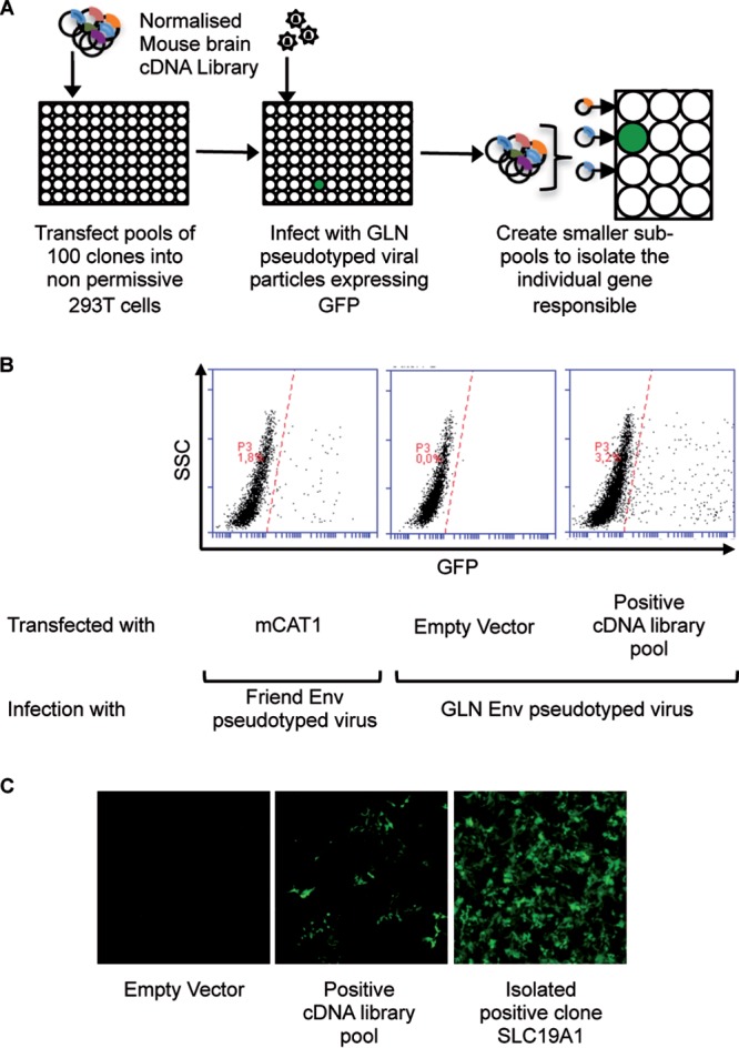
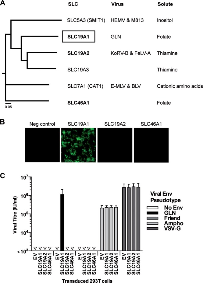
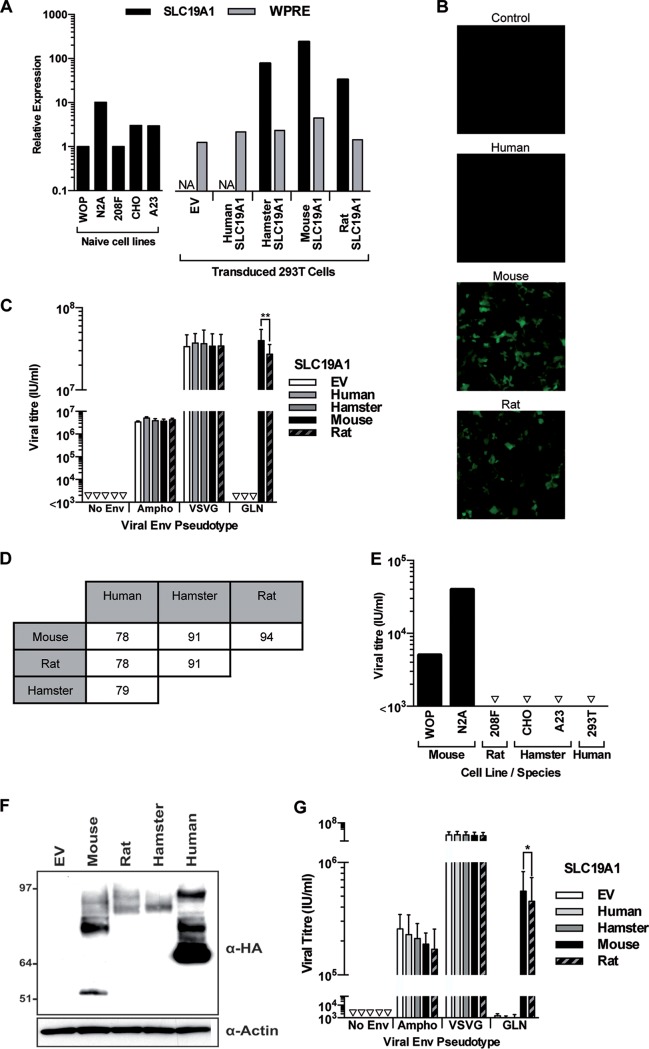
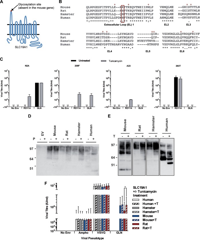
Approximately 10% of the mouse genome is composed of endogenous retroviruses belonging to different families. In contrast to the situation in the human genome, several of these families correspond to recent, still-infectious elements capable of encoding complete viral particles. The mouse GLN endogenous retrovirus is one of these active families. We previously identified one fully functional provirus from the sequenced genome of the C57BL/6 mouse strain. The GLN envelope protein gives the infectious viral particles an ecotropic host range, and we had demonstrated that the receptor was neither CAT1 nor SMIT1, the two previously identified receptors for mouse ecotropic retroviral envelope proteins. In this study, we have identified SLC19A1, the reduced folate carrier, as the cellular protein used as a receptor by the GLN retrovirus. The ecotropic tropism exhibited by this envelope is due to the presence or absence of an N-linked glycosylation site in the first extracellular loop as well as the specific amino acid sequence of the extracellular domains of the receptor. Like all the other retroviral envelope proteins from the gammaretrovirus genus whose receptors have been identified, the GLN envelope protein uses a member of the solute carrier superfamily as a receptor.IMPORTANCE Endogenous retroviruses are genomic traces of past infections present in all vertebrates. Most of these elements degenerate over time and become nonfunctional, but the mouse genome still contains several families with full infection abilities. The GLN retrovirus is one of them, and its members encode particles that are able to infect only mouse cells. Here, we identified the cellular protein used as a receptor by GLN for cell entry. It is SLC19A1, the reduced folate carrier. We show that GLN infection is limited to mouse cells due to both a mutation in the mouse gene preventing the glycosylation of SLC19A1 and also other residues conserved within the rat but not in the hamster and human proteins. Like all other gammaretroviruses whose receptors have been identified, GLN uses a member of the solute carrier superfamily for cell entry, highlighting the role of these proteins for retroviral infection in mammals.

Keywords: ERV; Env; GLN; endogenous retrovirus; gammaretrovirus; receptor.


Figures

FIG 1
FIG 1
cDNA library screen to identify the GLN cellular receptor. (A) Schematic representation of the screening method. Nonpermissive 293T cells were transfected with pools of 100 cDNAs from a normalized mouse brain library and then exposed to GFP-expressing GLN-pseudotyped virions 48 h later. Seventy-two hours after viral exposure, the 293T cells were analyzed for GFP expression by fluorescence microscopy and flow cytometry. Positive pools were retransformed into competent bacteria and used to generate subpools until the unique positive clone was identified. (B) Flow cytometric analysis of the cDNA screen showing the cDNA pool containing the GLN receptor. Cells transfected with mouse CAT-1 (mCAT1) and infected with Friend Env-pseudotyped viruses were used as positive controls for the assay. SSC, side scatter. (C) Fluorescence microscopy images of GFP-positive 293T cells after infection with GLN-pseudotyped viruses when transfected with the positive cDNA pool and the isolated cDNA (SLC19A1) responsible for enabling infection.
FIG 2
FIG 2
Specificity of SLC19A1 as the receptor for GLN. (A) Neighbor-joining phylogenetic tree without distance corrections showing the GLN receptor SLC19A1 and its similarities to other SLC proteins at the amino acid level. A closely related SLC19 family member, SLC19A2, and the other folate transporter, SLC46A1 (in boldface type), were tested for their ability to render 293T cells permissive to infection by GLN. HEMV, hortulanus endogenous murine leukemia virus; BLV, bovine leukemia virus. (B) 293T cells were transduced to express SLC19A1, SLC19A2, SLC46A1, or an empty vector (EV) control. After selection with hygromycin, the cell populations were exposed to GLN-pseudotyped viruses, and fluorescence was analyzed 72 h later by microscopy. Images are from one infection assay representative of results from 3 independent experiments. (C) Viral titers of different pseudotyped viruses on the same transduced cells were measured using flow cytometry (n = 3; the standard deviations [SD] are represented by the error bars).
FIG 3
FIG 3
Relative expression levels of SLC19A1 in different mouse tissues. RT-qPCR was used to measure the expression of SLC19A1 transcripts from pooled RNA obtained from multiple organs. SLC19A1 transcript levels were normalized to the level of the housekeeping gene RPLPO and are shown relative to levels for the embryo body. The lowest expression level of SLC19A1 is in the testis (threshold cycle [CT], 27) compared to the housekeeping gene RPLP0 (CT, 20). This measurement was performed once. Bars represent average values from 2 technical replicates.
FIG 4
FIG 4
Specificity of SLC19A1 as the cellular receptor for GLN. 293T cells were transduced to express SLC19A1 from different species before being exposed to GFP-expressing pseudotyped viruses. (A) RT-qPCR was used to measure the relative expression of SLC19A1 from different rodent cell lines and the exogenous expression of the SLC19A1 transgenes in stably transduced 293T cells. NA, not applicable, as the SLC19A1 qPCR oligonucleotides are able to detect only rodent versions of the transcript (100% homology with mouse, rat, and hamster sequences) and were not used to analyze empty vector- and human SLC19A1-transduced cells. Therefore, relative expression of the transgene was also assessed by detection of the 3′ untranslated region (UTR) (WPRE) present in all lentiviral constructs. Levels of both transcripts were normalized to the level of the housekeeping gene RPLPO. Expression levels of SLC19A1 and WPRE are shown relative to those in WOP and empty vector-transduced 293T cells, respectively. This measurement was performed once using one set of cells that were used for the experiment in panel B. Bars represent average values from 2 technical replicates. (B and C) Infection by GLN was assessed by fluorescence (B), and the titers of viruses pseudotyped with Ampho, VSV-G, GLN Envs, or a no-Env control were calculated by flow cytometry (C) (**, P < 0.01 by a paired t test; n = 3 except for GLN infections [n = 6]; the SD are represented by the error bars). (D) Amino acid similarity of the SLC19A1 sequences between different species. (E) Demonstration of the species tropism of GLN by infection of cell lines of different rodent species as well as of the human 293T cell line after transduction with mouse SLC19A1. Note that the differences in the titers observed between the mouse WOP and N2A cell lines is likely due to the difference in SLC19A1 expression levels (panel A), since we ensured by sequencing that both cell lines express identical versions of the SLC19A1 gene. Shown are data from one representative experiment out of three. (F) 293T cells were transduced with 3′ HA-tagged versions of the SLC19A1 genes. Western blotting was used to detect expression of the protein using an anti-HA antibody. The image shown is representative of results from 3 independent experiments. (G) 293T cells expressing the HA-tagged versions of the SLC19A1 constructs were exposed to GFP-expressing pseudotyped viruses. (*, P < 0.05 by a paired t test; n = 4).
FIG 5
FIG 5
Effects of glycosylation on GLN tropism. (A) Schematic of SLC9A1 showing the 12 transmembrane domains and the 6 extracellular loops. A putative N-linked glycosylation site is present in the first extracellular loop of all species except for mouse. (B) Clustal analysis of the amino acid sequences of SLC19A1 showing the amino acid similarities of the six extracellular loops between mouse, rat, hamster, and human proteins. The glycosylation site at N56 (boxed in red) is absent in the mouse protein. The red + symbols indicate residues shared by mouse and rat proteins but not human and hamster proteins. (C) Titers of viruses pseudotyped with RD114 or GLN envelopes were measured on different cell lines with or without 16 h of tunicamycin (0.2 μg/ml) pretreatment to remove N-linked glycosylation sites (n = 3; the SD are represented by the error bars). (D) 293T cells were transduced to express HA-tagged SLC19A1 from different species. Cell lysates were treated with PNGase F (shortened to P) to remove N-linked glycans before probing for SLC19A1 by Western blotting using an anti-HA antibody. (E) Cells were treated with tunicamycin (shortened to T) before collection of cell lysates, and SLC19A1 proteins were detected by Western blotting using an anti-HA antibody. (F) The titers of viruses pseudotyped with different Envs were measured on 293T cells expressing HA-tagged SLC19A1 from different species with and without tunicamycin pretreatment.

Similar articles

Cited by

References

    1. Finkelshtein D, Werman A, Novick D, Barak S, Rubinstein M. 2013. LDL receptor and its family members serve as the cellular receptors for vesicular stomatitis virus. Proc Natl Acad Sci U S A 110:7306–7311. doi:10.1073/pnas.1214441110. - DOI - PMC - PubMed
    1. Albritton LM, Tseng L, Scadden D, Cunningham JM. 1989. A putative murine ecotropic retrovirus receptor gene encodes a multiple membrane-spanning protein and confers susceptibility to virus infection. Cell 57:659–666. doi:10.1016/0092-8674(89)90134-7. - DOI - PubMed
    1. Miller DG, Miller AD. 1992. Tunicamycin treatment of CHO cells abrogates multiple blocks to retrovirus infection, one of which is due to a secreted inhibitor. J Virol 66:78–84. - PMC - PubMed
    1. Wang H, Klamo E, Kuhmann SE, Kozak SL, Kavanaugh MP, Kabat D. 1996. Modulation of ecotropic murine retroviruses by N-linked glycosylation of the cell surface receptor/amino acid transporter. J Virol 70:6884–6891. - PMC - PubMed
    1. Eiden MV, Farrell K, Wilson CA. 1994. Glycosylation-dependent inactivation of the ecotropic murine leukemia virus receptor. J Virol 68:626–631. - PMC - PubMed

Publication types

MeSH terms

LinkOut - more resources