. 2019 Feb 19;93(5):e01736-18.
doi: 10.1128/JVI.01736-18. Print 2019 Mar 1.

Porcine Hemagglutinating Encephalomyelitis Virus Activation of the Integrin α5β1-FAK-Cofilin Pathway Causes Cytoskeletal Rearrangement To Promote Its Invasion of N2a Cells

Affiliations

Porcine Hemagglutinating Encephalomyelitis Virus Activation of the Integrin α5β1-FAK-Cofilin Pathway Causes Cytoskeletal Rearrangement To Promote Its Invasion of N2a Cells

Xiaoling Lv et al. J Virol. .

Abstract

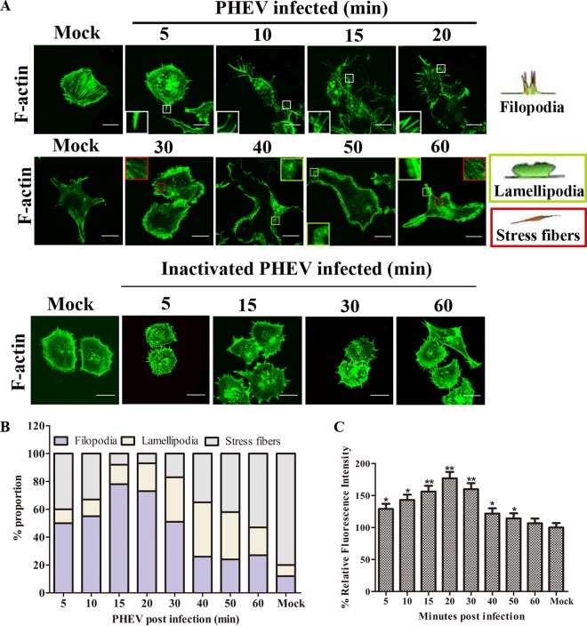
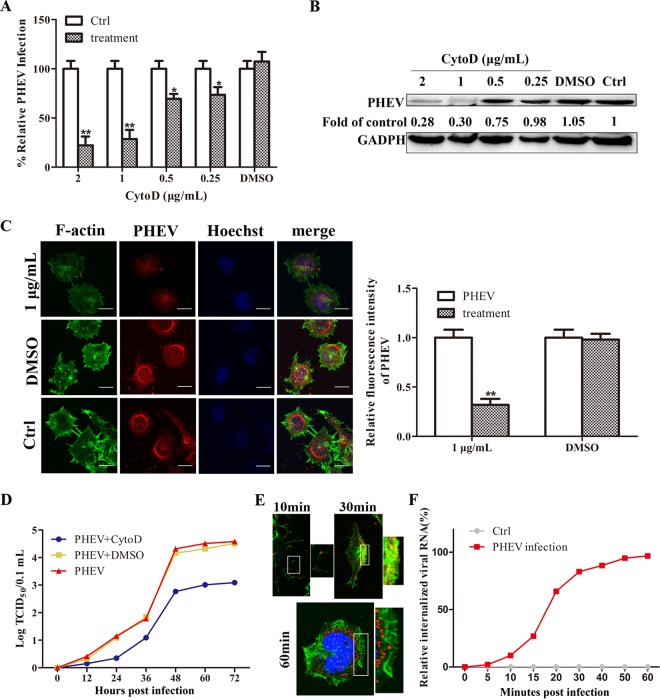
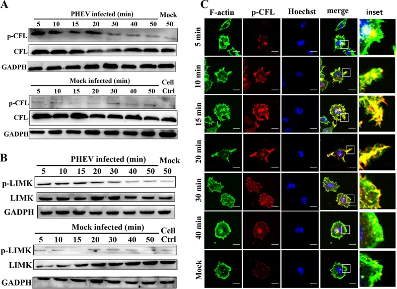
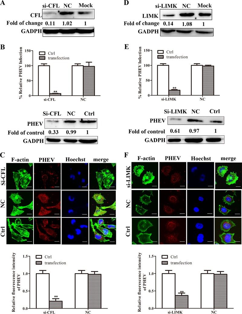
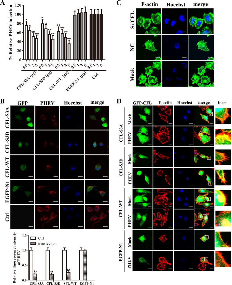
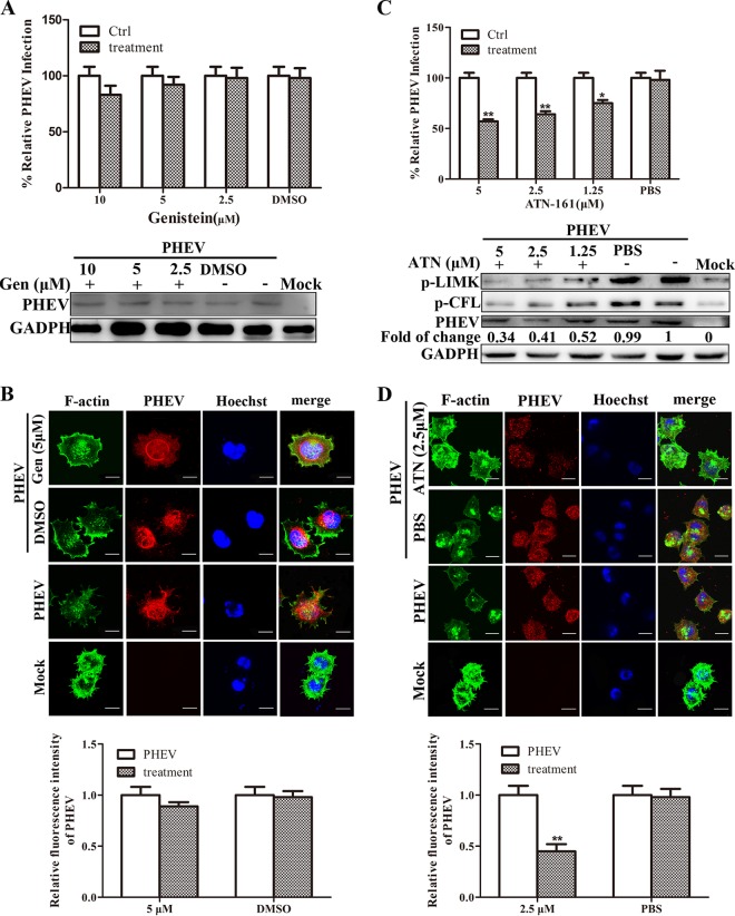
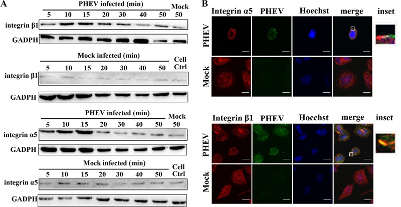
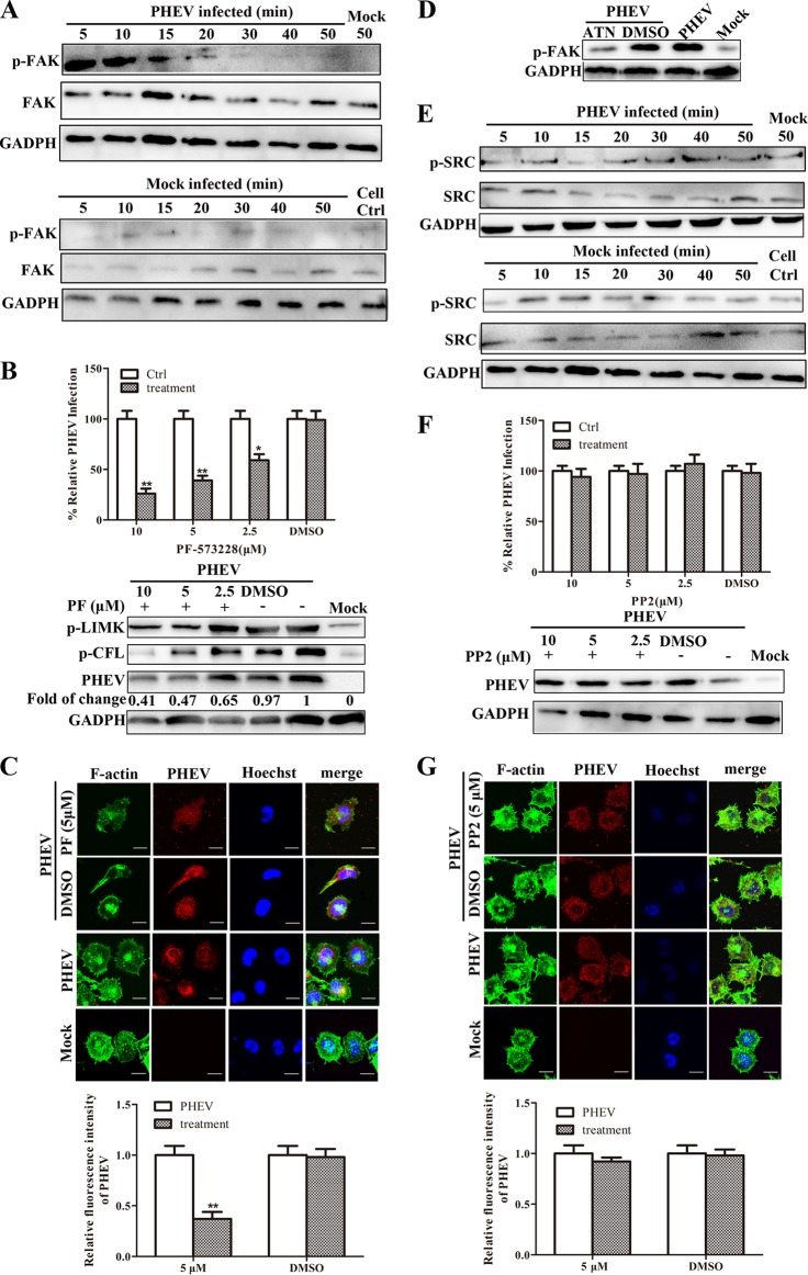
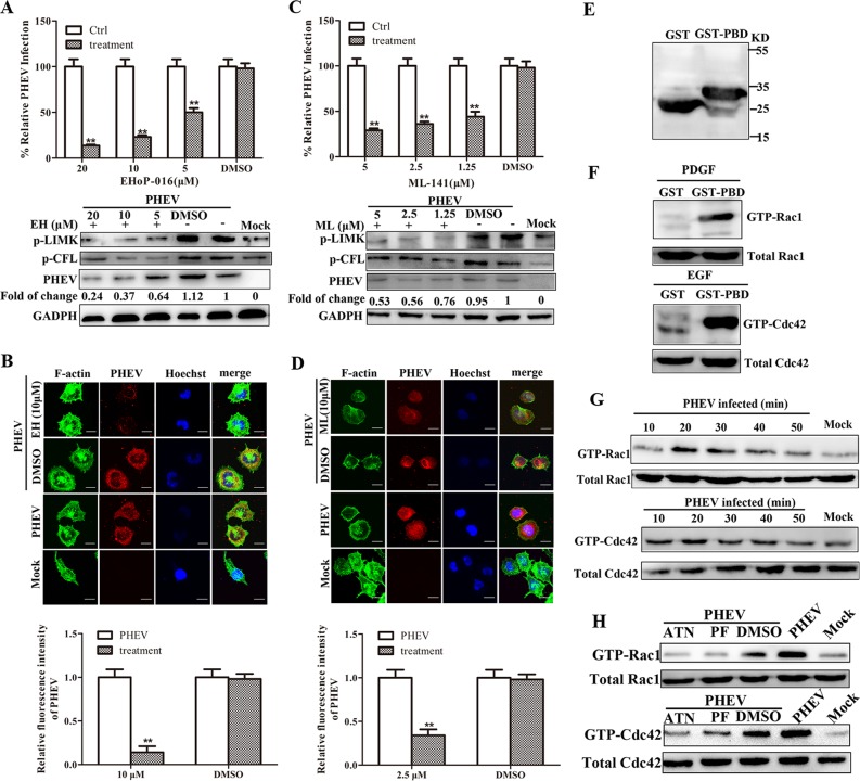
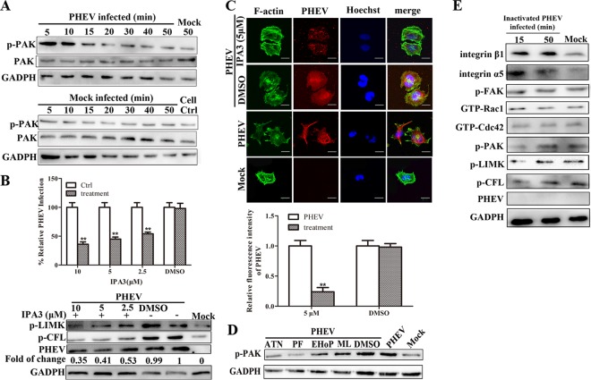
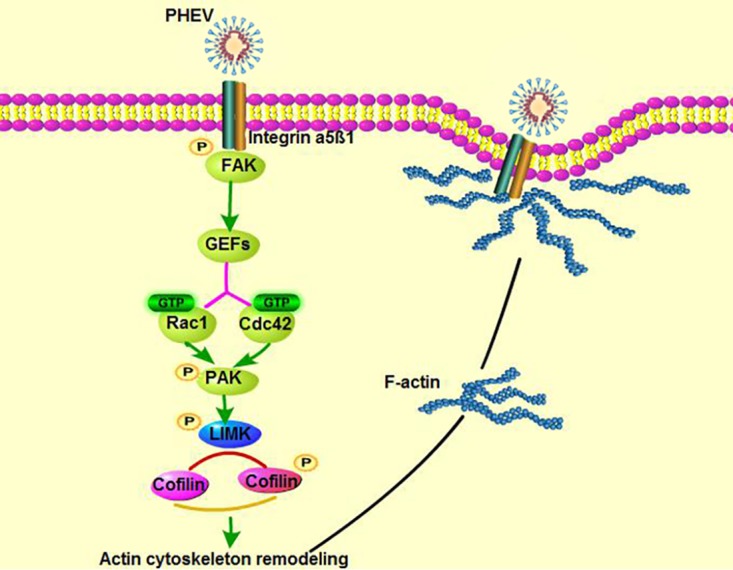
Porcine hemagglutinating encephalomyelitis virus (PHEV) is a highly neurotropic virus that causes diffuse neuronal infection with neurological damage and high mortality. Virus-induced cytoskeletal dynamics are thought to be closely related to this type of nerve damage. Currently, the regulation pattern of the actin cytoskeleton and its molecular mechanism remain unclear when PHEV enters the host cells. Here, we demonstrate that entry of PHEV into N2a cells induces a biphasic remodeling of the actin cytoskeleton and a dynamic change in cofilin activity. Viral entry is affected by the disruption of actin kinetics or alteration of cofilin activity. PHEV binds to integrin α5β1 and then initiates the integrin α5β1-FAK signaling pathway, leading to virus-induced early cofilin phosphorylation and F-actin polymerization. Additionally, Ras-related C3 botulinum toxin substrate 1 (Rac1), cell division cycle 42 (Cdc42), and downstream regulatory gene p21-activated protein kinases (PAKs) are recruited as downstream mediators of PHEV-induced dynamic changes of the cofilin activity pathway. In conclusion, we demonstrate that PHEV utilizes the integrin α5β1-FAK-Rac1/Cdc42-PAK-LIMK-cofilin pathway to cause an actin cytoskeletal rearrangement to promote its own invasion, providing theoretical support for the development of PHEV pathogenic mechanisms and new antiviral targets.IMPORTANCE PHEV, a member of the Coronaviridae family, is a typical neurotropic virus that primarily affects the nervous system of piglets to produce typical neurological symptoms. However, the mechanism of nerve damage caused by the virus has not been fully elucidated. Actin is an important component of the cytoskeleton of eukaryotic cells and serves as the first obstacle to the entry of pathogens into host cells. Additionally, the morphological structure and function of nerve cells depend on the dynamic regulation of the actin skeleton. Therefore, exploring the mechanism of neuronal injury induced by PHEV from the perspective of the actin cytoskeleton not only helps elucidate the pathogenesis of PHEV but also provides a theoretical basis for the search for new antiviral targets. This is the first report to define a mechanistic link between alterations in signaling from cytoskeleton pathways and the mechanism of PHEV invading nerve cells.

Keywords: cofilin; coronavirus; cytoskeletal rearrangement; integrin α5β1; neurotropic virus; porcine hemagglutinating encephalomyelitis virus.


Figures

FIG 1
FIG 1
PHEV infection induces actin cytoskeletal remodeling. (A) PHEV infection induces cell protrusion formation. N2a cells were incubated with PHEV or inactivated PHEV (MOI of 50) for 1 h at 4°C and then moved to 37°C and fixed at the indicated time points. The untreated cells were used as a control (Mock). F-actin was stained with FITC-phalloidin (green) and observed by confocal microscopy. Scale bar, 10 μm. (B) Quantitative detection of cell ruffle changes at different time points after infection. Cells with F-actin stress fibers, lamellipodia, or filopodia were designated positive cells; each value represents the average value from 60 to 80 cells from at least 5 regions of 3 representative experiments. (C) PHEV treatment causes cell actin polymerization and depolymerization. F-actin was stained with FITC-phalloidin and analyzed by flow cytometry.
FIG 2
FIG 2
Inhibitors of F-actin inhibit virus entry. (A to C) CytoD inhibits the entry of PHEV in a concentration-dependent manner. Cells were pretreated with different concentrations of CytoD, and the internalization of PHEV was assessed using real-time PCR, Western blotting, and indirect immunofluorescence (see Materials and Methods). PHEV-infected cells were used as controls (Ctrl). DMSO, dimethylsulfoxide. (D) Growth analysis of PHEV. N2a cells were infected with PHEV after treatment with CytoD (1 μg/ml), and samples were collected at the indicated time points. Titers are expressed as TCID50/0.1 ml. (E) Kinetics of PHEV entry into cells. Cells treated with PHEV were labeled with anti-S protein monoclonal antibody (red), FITC-phalloidin (green), and Hoechst (blue) at different time points. The figure shows the quantification of viral particle locations at different times. At least 30 cells from five representative fields were counted in each experiment. Scale bar, 10 μm. (F) PHEV bound to N2a cells at 4°C for 1 h were transferred to 37°C; at the indicated times, bound viral particles that did not enter the cells were removed, and internalized viral RNA was extracted and assayed by real-time PCR. All results were considered statistically significant at a P value of <0.05.
FIG 3
FIG 3
PHEV infection induces cofilin biphasic activation. (A) Cofilin activity test. Detection of cofilin phosphorylation with Western blotting at the designated times after infection. In all of the mock-infected experimental groups, the untreated cells were used as a control. In the PHEV-infected group, the cells that were incubated at 4°C for 1 h and then transferred to 37°C for the indicated times without PHEV infection to mimic infection were used as the control (Mock). (B) LIMK activity test. Detection of LIMK phosphorylation with Western blotting at the designated times after infection. (C) The level of p-cofilin increases early in PHEV infection. Cells treated with PHEV were labeled with anti-p-cofilin monoclonal antibody (red), FITC-phalloidin, and Hoechst at different time points. At least 30 cells from five representative fields were counted in each experiment. Scale bar, 10 μm.
FIG 4
FIG 4
Cofilin and LIMK siRNAs affect viral entry. (A) Cofilin protein levels in N2a cells 24 h posttransfection with cofilin siRNA and siR-cofilin negative control (NC). (B and C) PHEV entry is inhibited when cofilin is knocked down. Cells were transfected with siRNA. Twenty-four hours after transfection, cells were infected with PHEV for 2 h, and PHEV entry was assessed by real-time PCR, Western blotting, and indirect immunofluorescence. Scale bar, 10 μm. (D) LIMK protein levels in N2a cells 24 h posttransfection with LIMK siRNA and siR-LIMK negative control (NC). (E and F) PHEV entry is inhibited when LIMK is knocked down. The same detection method was used as that for panel B. All results were considered statistically significant at a P value of <0.05.
FIG 5
FIG 5
Cofilin activity influences virus entry and participates in virus-induced cell membrane ruffle formation. (A) Overexpression of wild-type cofilin or mutants (S3A and S3D) inhibits PHEV entry. The cells were transfected with plasmids (at different concentrations) and infected with PHEV for 2 h, and real-time PCR was used to detect PHEV entry. (B) Confocal microscopy showed overexpression of cofilin inhibited viral entry. Cells were transfected with green fluorescent protein (GFP)-tagged plasmids (2 μg) and then infected with PHEV for 2 h, fixed, and stained with anti-PHEV-S (red). In each independent experiment, at least 30 cells from five representative fields were counted. Scale bar, 10 μm. (C) Effects of cofilin siRNA on the actin cytoskeleton. Cells were fixed at 24 h after transfection with siRNA, F-actin was labeled with FITC-phalloidin, and nuclei were labeled with Hoechst, followed by observation with confocal microscopy. Scale bar, 10 μm. (D) Cofilin is involved in virus-induced cell protrusion formation. Cells were infected for 30 min, and then F-actin (red) or cofilin (green) was stained and examined by laser confocal microscopy. Scale bar, 10 μm. All results were considered statistically significant at a P value of <0.05.
FIG 6
FIG 6
Integrins are involved in cofilin phosphorylation and viral entry. (A and B) RTKs are not involved in cofilin phosphorylation and virus entry. Cells were treated with different doses of genistein, an RTK specific inhibitor, at 37°C for 1 h and then infected with PHEV for 2 h. The internalization of PHEV was assessed by real-time PCR, Western blotting, and indirect immunofluorescence. (C and D) Integrin inhibitors inhibit viral entry and affect cofilin phosphorylation. Different doses of integrin α5β1-specific inhibitor ATN-161 were used to treat cells at 37°C for 1 h. Cofilin and LIMK phosphorylation were detected by Western blotting, and the internalization of PHEV was assessed by real-time PCR, Western blotting, and indirect immunofluorescence. All results were considered statistically significant at a P value of <0.05.
FIG 7
FIG 7
Integrin α5β1 affects viral entry. Integrin α5β1 expression is upregulated during PHEV entry. N2a cells were incubated with PHEV (MOI of 50) for 1 h at 4°C and then moved to 37°C and fixed at the indicated time points. Integrin α5β1 was probed with integrin α5 and integrin β1 antibodies. (B) Correlation of viral particles with integrin α5β1 in infected cells. Cells were infected with PHEV for 15 min and then fixed, and indirect immunofluorescence assays were performed with anti-integrin α5/β1 and anti-PHEV-S antibodies. Scale bar, 10 μm.
FIG 8
FIG 8
FAK is involved in cofilin phosphorylation and viral entry. (A) Activation of FAK during PHEV entry. PHEV-infected cell lysates were collected at different time points. Active FAK was probed with a p-FAK antibody. (B and C) FAK inhibitors inhibit viral entry and affect cofilin phosphorylation. Different doses of the FAK-specific inhibitor PF-573228 were used to treat cells at 37°C for 1 h and then infected with PHEV for 2 h. Cofilin and LIMK phosphorylation were detected by Western blotting, and the internalization of PHEV was assessed by real-time PCR, Western blotting, and indirect immunofluorescence. (D) Integrins are upstream proteins that regulate FAK activation. Cells were pretreated with ATN-161 (2.5 μM) for 1 h before infection. Ten minutes after PHEV infection, FAK phosphorylation levels of cell lysates were analyzed. (E to G) SRC is not involved in PHEV entry. Cells were treated with different doses of SRC-specific inhibitor PP2 at 37°C for 1 h and then infected with PHEV for 2 h. The internalization of PHEV was assessed by real-time PCR, Western blotting, and indirect immunofluorescence. All results were considered statistically significant at a P value of <0.05.
FIG 9
FIG 9
Rac1 GTPase and Cdc42 GTPase are involved in early cofilin phosphorylation and promote PHEV entry. (B) Rac GTPase specific inhibitor EHoP-016 (EH) affects viral entry and cofilin phosphorylation. Different doses of EHoP-016 were used to treat cells for 1 h, and then cofilin and LIMK phosphorylation were detected by Western blotting and the internalization of PHEV was assessed by real-time PCR, Western blotting, and indirect immunofluorescence. (C and D) ML-141 (ML), which is a Cdc42 GTPase-specific inhibitor, affects viral entry and cofilin phosphorylation. The same detection method was used as that for panels A and B. (E) GST-PBD protein purification. KD, molecular mass in kilodaltons. (F) GST-PBD specifically binds GTP-Rac1 and GTP-Cdc42. Lysates of cells that were treated with PDGF (20 ng/ml) or EGF (50 ng/ml) were incubated with GST-PBD. GST-PBD binds GTP-bound Rac1 and GTP-bound Cdc42 (pulldown), and total Rac1 and Cdc42 proteins in lysates were analyzed by Western blotting. (G) Rac1 and Cdc42 activation assay. Cells were harvested and lysed at different time points after PHEV treatment, and pulldown assays were performed as described above. (H) ATN-161 (2.5 μM) and PF-573228 (5 μM) affect Rac1 and Cdc42 activity. Cells were harvested and lysed after inhibitor treatment and virus inoculation for 20 min, and pulldown assays were performed as described above. All results were considered statistically significant at a P value of <0.05.
FIG 10
FIG 10
PAK is a downstream effector of PHEV-induced integrin α5β1-FAK-Rac1/Cdc42 pathway in early cofilin phosphorylation. PAK is activated during PHEV entry. Cells were infected with PHEV, and cell lysates were analyzed with anti-PAK and anti-p-PAK antibodies. (B and C) PAK-specific inhibitor IPA3 affects viral entry and cofilin phosphorylation. Different doses of IPA3 were used to treat cells for 1 h, and then cofilin and LIMK phosphorylation were detected. (D) Inhibitors block the activation of PAK. Cells were pretreated with ATN-161 (2.5 μM), PF-573228 (5 μM), EHoP-016 (10 μM), or ML-141 (2.5 μM) for 1 h before infection. Ten minutes after PHEV infection, PAK phosphorylation levels of cell lysates were analyzed. (E) Integrin pathway was not activated in inactivated PHEV-infected N2a cells. N2a cells were incubated with inactivated PHEV (MOI of 50) for 1 h at 4°C and then moved to 37°C, and cells were harvested at the indicated time points. Detection of integrin pathway activation by Western blotting is shown. All results were considered statistically significant at a P value of <0.05.
FIG 11
FIG 11
Model of cofilin regulating the actin cytoskeleton and initiating PHEV entry. In early stages of infection, PHEV stimulates the dynamic phosphorylation of cofilin and polymerizes F-actin via the integrin α5β1-FAK-Rac1/Cdc42-PAK-LIMK signaling pathway.

Similar articles

Cited by

References

    1. Spear M, Wu Y. 2014. Viral exploitation of actin: force-generation and scaffolding functions in viral infection. Virol Sin 29:139–147. doi:10.1007/s12250-014-3476-0. - DOI - PMC - PubMed
    1. MacLeod IJ, Minson T. 2010. Binding of herpes simplex virus type-1 virions leads to the induction of intracellular signalling in the absence of virus entry. PLoS One 5:e9560. doi:10.1371/journal.pone.0009560. - DOI - PMC - PubMed
    1. Salameh S, Sheth U, Shukla D. 2012. Early events in herpes simplex virus lifecycle with implications for an infection of lifetime. Open Virol J 6:1–6. doi:10.2174/1874357901206010001. - DOI - PMC - PubMed
    1. Wurth MA, Schowalter RM, Smith EC, Moncman CL, Dutch RE, McCann RO. 2010. The actin cytoskeleton inhibits pore expansion during PIV5 fusion protein-promoted cell-cell fusion. Virology 404:117–126. doi:10.1016/j.virol.2010.04.024. - DOI - PMC - PubMed
    1. Foo KY, Chee HY. 2015. Interaction between flavivirus and cytoskeleton during virus replication. Biomed Res Int 2015:427814. doi:10.1155/2015/427814. - DOI - PMC - PubMed

Publication types

MeSH terms