. 2019 Feb 19;93(5):e01739-18.
doi: 10.1128/JVI.01739-18. Print 2019 Mar 1.

Rotavirus Infection Alters Splicing of the Stress-Related Transcription Factor XBP1

Affiliations

Rotavirus Infection Alters Splicing of the Stress-Related Transcription Factor XBP1

Mariela Duarte et al. J Virol. .

Abstract

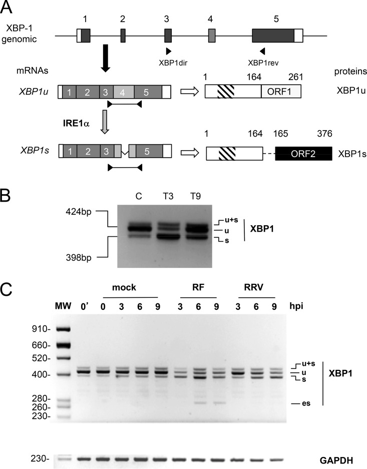
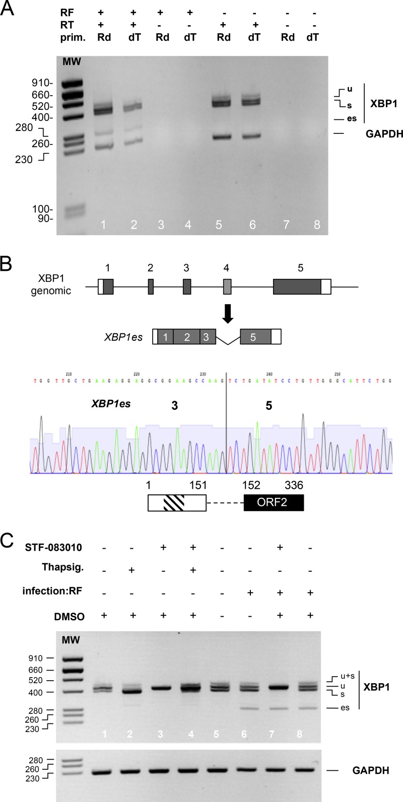
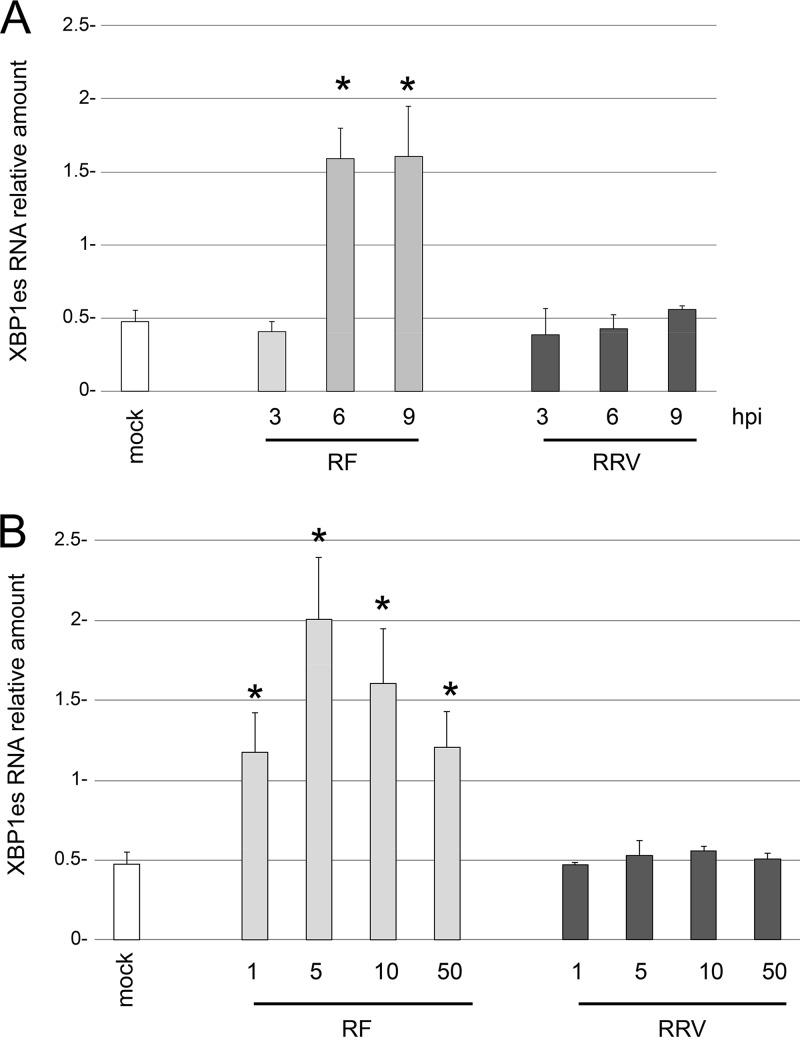
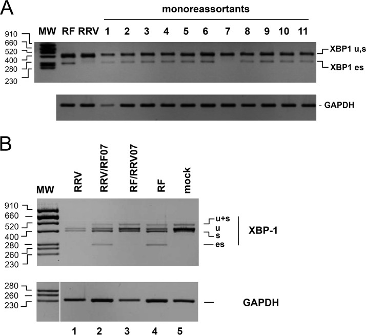
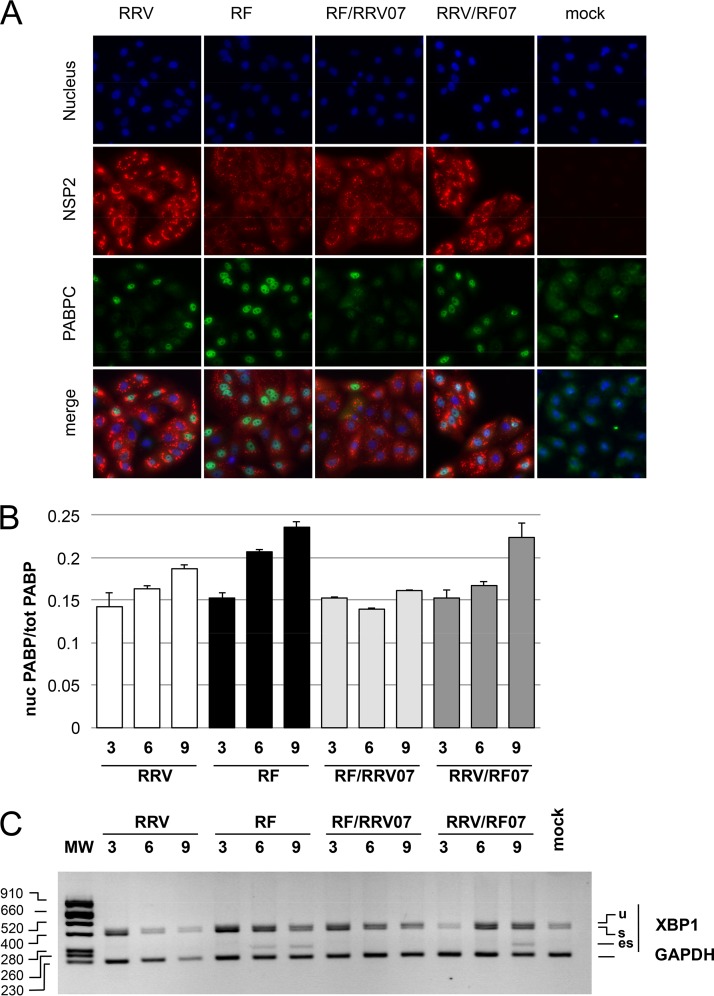
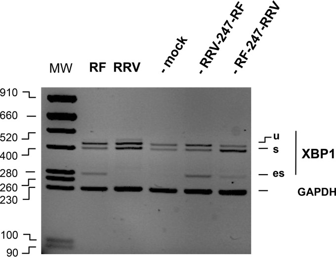
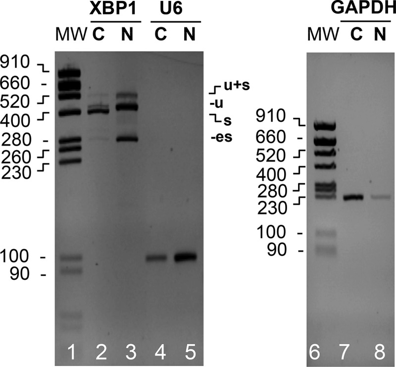
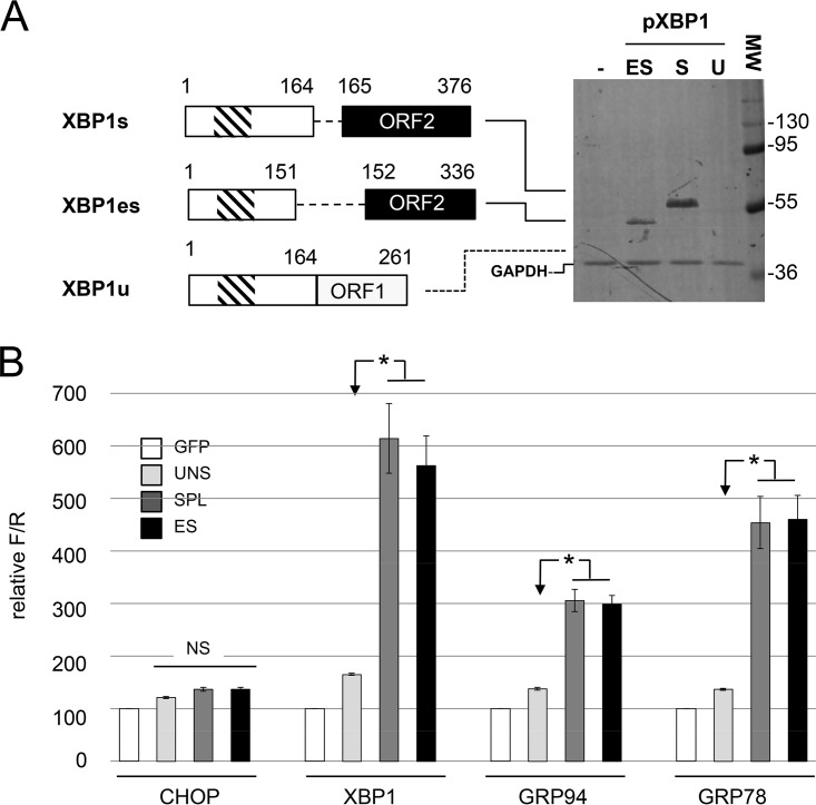
XBP1 is a stress-regulated transcription factor also involved in mammalian host defenses and innate immune response. Our investigation of XBP1 RNA splicing during rotavirus infection revealed that an additional XBP1 RNA (XBP1es) that corresponded to exon skipping in the XBP1 pre-RNA is induced depending on the rotavirus strain used. We show that the translation product of XBP1es (XBP1es) has trans-activation properties similar to those of XBP1 on ER stress response element (ERSE) containing promoters. Using monoreassortant between ES+ ("skipping") and ES- ("nonskipping") strains of rotavirus, we show that gene 7 encoding the viral translation enhancer NSP3 is involved in this phenomenon and that exon skipping parallels the nuclear relocalization of cytoplasmic PABP. We further show, using recombinant rotaviruses carrying chimeric gene 7, that the ES+ phenotype is linked to the eIF4G-binding domain of NSP3. Because the XBP1 transcription factor is involved in stress and immunological responses, our results suggest an alternative way to activate XBP1 upon viral infection or nuclear localization of PABP.IMPORTANCE Rotavirus is one of the most important pathogens causing severe gastroenteritis in young children worldwide. Here we show that infection with several rotavirus strains induces an alternative splicing of the RNA encoding the stressed-induced transcription factor XBP1. The genetic determinant of XBP1 splicing is the viral RNA translation enhancer NSP3. Since XBP1 is involved in cellular stress and immune responses and since the XBP1 protein made from the alternatively spliced RNA is an active transcription factor, our observations raise the question of whether alternative splicing is a cellular response to rotavirus infection.

Keywords: NSP3; PABP; XBP1; immune response; nuclear transport; rotavirus; splicing; stress response.


Figures

FIG 1
FIG 1
Rotavirus infection induces cytoplasmic splicing of XBP1. (A) Schematic structures of the XBP1u and XBP1s mRNAs. The general organization of the XBP1 gene is indicated with exons as gray boxes, noncoding regions are indicated as white boxes, and introns are indicated as lines (not to scale). The mRNA encoding XBP1u is produced by canonical nuclear splicing and then exported to the cytoplasm (black arrow) to be translated (white arrow). The XBP1u protein translated from XBP1u mRNA is 261 amino acids long and contains a DNA-binding domain (hatched box). The mRNA encoding XBP1s is produced by an unconventional cytoplasmic splicing (gray arrow) of XBP1u mRNA at exon 4 (light gray) catalyzed by the IRE1α endoribonuclease. The XBP1s protein is 376 amino acids long and contains DNA-binding and transactivating (black box) domains. (B) Detection of XBP1u splicing by IRE1α. The positions of the primers XBP1dir and XBP1rev on the XBP1 gene and mRNAs are indicated. The RT-PCR products (424 and 398 bp) obtained using these primers on RNA purified from unstressed MA104 cells (lane C) or MA104 cells treated with thapsigargin (400 nM) for 3 and 9 h (T3, T9) are illustrated. (C) The DNA products obtained by RT-PCR from RNA extracted from mock-infected cells or from cells infected (MOI of 10) with rotavirus RF or RRV for the indicated time (in hours) were analyzed by agarose gel electrophoresis. The top panel shows the XBP1 RT-PCR products, and the bottom panel shows the GAPDH RT-PCR products used as a loading control. The 0′ lane corresponds to untreated cells, and the 0 lane corresponds to mock-infected cells. The sizes of the molecular weight markers (MW) are indicated in base pairs on the left side.
FIG 2
FIG 2
XBP1es is a poly(A) RNA that results from exon skipping, not cytoplasmic splicing. (A) RNA purified from mock (RF)- or rotavirus RF (RF+)-infected cells was used as the template for a reverse transcription reaction with (RT+) or without (RT) reverse transcriptase and using oligo(dT) (dT) or random hexanucleotides (Rd) as primers. DNA products obtained by the PCRs using either XBPdir and XBPrev primers or GAPDH primers were analyzed by electrophoresis on the same agarose gel. The sizes of the molecular weight markers (MW) are indicated (in base pairs) on the left side. (B) A chromatogram from Sanger sequencing of the XBP1es RT-PCR DNA product is shown below a schematic representation of the organization of the XBP1 gene and that of XBP1es RNA resulting from exon 4 skipping. The vertical line marks the junction of the exon 3 and 5 sequences. A schematic representation of the putative translation product of XBP1es RNA is shown with the DNA-binding (hatched box) and transactivating domains (black box). The numbers indicate amino acid positions. (C) RNA purified from MA104 cells infected (for 9 h) or not by the RF strain of rotavirus and treated with the IRE1α inhibitor STF-083010 in DMSO (60 μM), with the IRE1α activator thapsigargin (400 nM) or with DMSO (used as vehicle for STF-083010), was subjected to RT-PCR with XBP1 primers and GAPDH primers. The PCR DNA products were analyzed by electrophoresis on two agarose gels. The sizes of the molecular weight markers (MW) are indicated (in base pairs) on the left.
FIG 3
FIG 3
XBP1 exon skipping depends on the rotavirus strain but not on the multiplicity of infection. The amount of XPB1es RNA was quantified by RT-qPCR using primers specific for XBP1es (Table 1) and RNA extracted from mock-infected cells or from cells infected with the rotavirus RF or RRV for the indicated time (in hours postpostinfection) (A) or at the indicated MOI (B). The relative amount of XBP1es RNA is presented. The means ± the standard errors of the mean (SEM) for three independent experiments performed in triplicate are shown. Asterisks indicate significant differences from the mock-infected control (P < 0.05) as determined by a two-tailed Student t test.
FIG 4
FIG 4
Rotavirus gene 7 determines the ES phenotype. (A) RNA purified from MA104 cells infected (9 h, MOI of 10) with the different monoreassortants and parental strains was subjected to RT-PCR with XBP1 and GAPDH primers. The PCR DNA products were analyzed by electrophoresis and ethidium bromide staining on two agarose gels (middle and lower panels). The sizes of the molecular weight markers (MW) in base pairs are indicated on the left. (B) RNA purified from mock-infected MA104 cells (lane 5) or cells infected with either an RRV monoreassortant carrying gene 7 from RF (RRV/RF07 lane 2), an RF monoreassortant carrying gene 7 from RRV (RF/RRV07 lane 3), or the RRV (lane 1) or RF (lane 4) parental strains was subjected to RT-PCR with XBP1 and GAPDH primers. The PCR DNA products were analyzed by electrophoresis and ethidium bromide staining on two agarose gels. The sizes of the molecular weight markers (MW) in base pairs are indicated on the left.
FIG 5
FIG 5
Nuclear translocation of PABPC and XBP1 exon skipping in parental RF and RRV and gene 7 monoreassortants. (A) Localization of PABPC in rotavirus-infected cells. MA104 cells infected (or mock infected) with bovine RF, rhesus RRV, or gene 7 monoreassortants for 9 h were fixed and incubated with NSP2- and PABPC-specific antibodies. Secondary antibodies coupled to the Alexa fluorophore stains NSP2 (red) and PABPC (green). Nuclei were stained blue with DAPI. (B) Quantification of nuclear PABC1 in rotavirus-infected cells. Images such as those shown in panel A were taken at 3, 6, and 9 h after infection (hpi) with the indicated virus and analyzed. The ratio of nuclear to total green (PABPC) fluorescence (corrected for background) is reported. The results are mean values ± the SEM of three fields with >50 cells. (C) Kinetics of XBP1 splicing. The XBP1 DNA products obtained by RT-PCR of RNA extracted from mock-infected cells or from cells infected (MOI of 10) for 3, 6, and 9 h with the indicated parental or monoreassortant virus were analyzed by agarose gel electrophoresis, together with the GAPDH PCR control. The sizes of the molecular weight markers (MW) are indicated in base pairs on the left.
FIG 6
FIG 6
ES phenotypes of recombinant viruses bearing NSP3 chimeras The DNA products obtained by RT-PCR (with XBP1 or GAPDH primers) of RNA extracted from mock-infected cells or from cells infected (MOI of 10 [9 hpi]) with RF or RRV or a recombinant virus (SA11 background, bearing RRV-247-RF or RF-247-RRV gene 7 chimeras) were analyzed by agarose gel electrophoresis and ethidium bromide staining, together with the GAPDH PCR control.
FIG 7
FIG 7
Alternative splicing of selected genes in ES or ES+ rotavirus-infected cells. RNA purified from mock-infected MA104 cells (lanes “M.”) or from MA104 cells infected with the RF (ES+) or RRV (ES) strain of rotavirus was used for RT-PCR with primers specific for the cellular gene indicated on the top of the lanes. On the left, the sizes of the molecular weight markers (MW) are indicated in base pairs. The expected sizes (in base pairs) of the RT-PCR products are indicated on the right.
FIG 8
FIG 8
Localization of XBP1es RNA. RNA purified from the cytoplasmic (C) or nuclear (N) fractions of MA104 cells infected with the RF strain of rotavirus (MOI of 10, 9 h postinfection) was subjected to RT-PCR using XBP1, U6, or GAPDH primers. The PCR DNA products were analyzed by electrophoresis and ethidium bromide staining on two agarose gels. The positions of the XBP1u, XBP1s, and XBP1es PCR DNA products and of the molecular weight markers (MW; sizes are denoted in base pairs) are indicated.
FIG 9
FIG 9
XBP1es and XBP1s proteins have similar transactivating properties. (A) Expression of XBP1u, XBP1s, and XBP1es in HEK293 cells. On the left, schematic representations of the XBP1u, XBP1s and XBP1es cDNA constructs used are shown together with schematic representations of the proteins they encode (numbers indicate amino acid positions). The functional DNA-binding (hatched box) and transcription-activating domains (black box) are indicated. On the right, lysates of HEK293 cells transfected with XBP1 cDNA constructs were resolved by SDS-PAGE and probed with an antiserum directed against XBP1. The numbers indicate the positions and molecular weights (in kDa) of the markers. The dotted line indicates the expected position of XBP1u. (B) Transactivating properties of XBP1u, XBP1s, and XBP1es. MA104 cells were cotransfected with a combination of three expression plasmids: (i) either one of the XBP1 cDNA constructs described in panel A or a control plasmid encoding eGFP; (ii) a reporter plasmid carrying the firefly luciferase gene under the control of the CHOP, XBP1, GRP94, or GRP78 promoter; and (iii) a normalization plasmid encoding the Renilla luciferase gene under the control of the CMV promoter. Luciferase activities were measured 48 h after transfection and reported as the firefly/Renilla ratio with the GFP control set to 100. The mean values ± the SEM for three independent experiments performed in triplicate are shown. Bars: XBP1u (UNSPL), light gray; XBP1s (SPL), dark gray; XBP1es (ES), black. Asterisks indicate significant differences compared to the XBP1u-transfected sample (P < 0.05) as determined by a two-tailed Student t test. NS, not significant.

Similar articles

Cited by

References

    1. Hetz C, Martinon F, Rodriguez D, Glimcher LH. 2011. The unfolded protein response: integrating stress signals through the stress sensor IRE1α. Physiol Rev 91:1219–1243. doi:10.1152/physrev.00001.2011. - DOI - PubMed
    1. Walter P, Ron D. 2011. The unfolded protein response: from stress pathway to homeostatic regulation. Science 334:1081–1086. doi:10.1126/science.1209038. - DOI - PubMed
    1. Yoshida H, Matsui T, Yamamoto A, Okada T, Mori K. 2001. XBP1 mRNA is induced by ATF6 and spliced by IRE1 in response to ER stress to produce a highly active transcription factor. Cell 107:881–891. doi:10.1016/S0092-8674(01)00611-0. - DOI - PubMed
    1. Calfon M, Zeng H, Urano F, Till JH, Hubbard SR, Harding HP, Clark SG, Ron D. 2002. IRE1 couples endoplasmic reticulum load to secretory capacity by processing the XBP-1 mRNA. Nature 415:92–96. doi:10.1038/415092a. - DOI - PubMed
    1. Jurkin J, Henkel T, Nielsen AF, Minnich M, Popow J, Kaufmann T, Heindl K, Hoffmann T, Busslinger M, Martinez J. 2014. The mammalian tRNA ligase complex mediates splicing of XBP1 mRNA and controls antibody secretion in plasma cells. EMBO J 33:2922–2936. doi:10.15252/embj.201490332. - DOI - PMC - PubMed

Publication types

LinkOut - more resources