. 2019 Jan 15;202(2):591-597.
doi: 10.4049/jimmunol.1801196. Epub 2018 Dec 12.

Extent of MHC Clustering Regulates Selectivity and Effectiveness of T Cell Responses

Affiliations

Extent of MHC Clustering Regulates Selectivity and Effectiveness of T Cell Responses

Nadia Anikeeva et al. J Immunol. .

Abstract

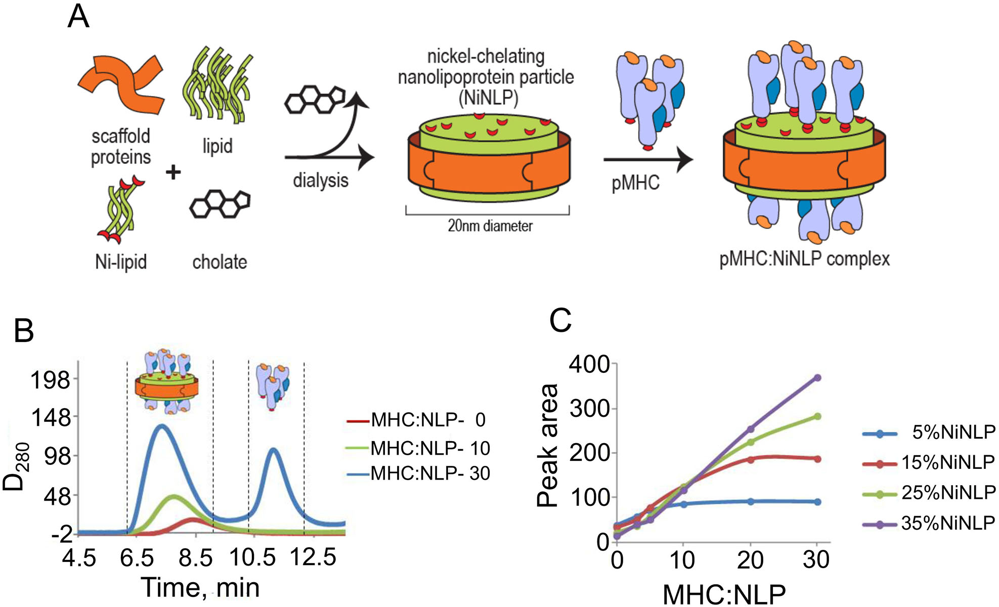
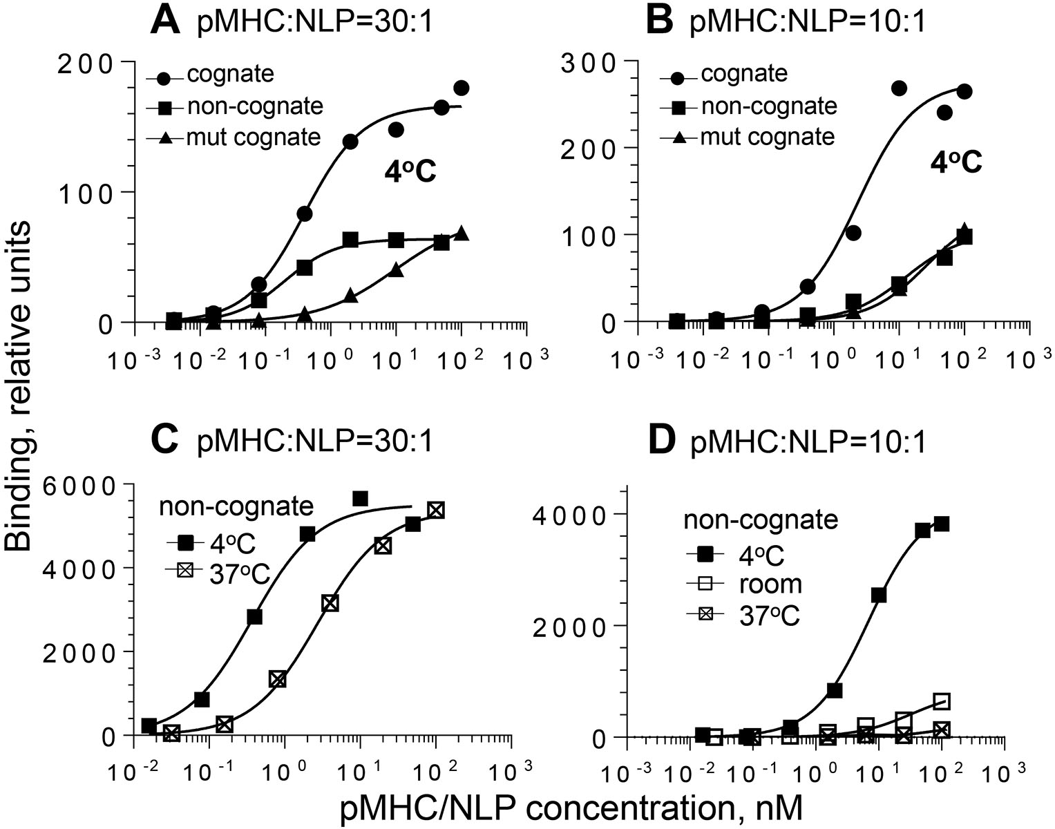
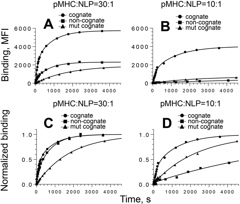
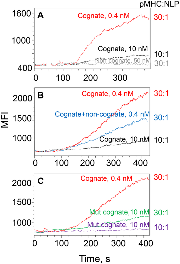
MHC proteins that present peptide ligands for recognition by TCR form nanoscale clusters on the cell membrane of APCs. How the extent of MHC clustering controls productive TCR engagement and TCR-mediated signaling has not been systematically studied. To evaluate the role of MHC clustering, we exploited nanoscale discoidal membrane mimetics (nanolipoprotein particles) to capture and present peptide-MHC (pMHC) ligands at various densities. We examined the binding of these model membrane clusters to the surface of live human CD8+ T cells and the subsequent triggering of intracellular signaling. The data demonstrate that the proximity of pMHC ligands, high association rate of CD8-MHC interactions, and relatively long lifetime of cognate TCR-pMHC complexes emerge as essential parameters, explaining the significance of MHC clustering. Rapid rebinding of CD8 to MHC suggests a dual role of CD8 in facilitating the T cells' hunt for a rare foreign pMHC ligand and the induction of rapid T cell response. Thus, our findings provide a new understanding of how MHC clustering influences multivalent interactions of pMHC ligands with CD8 and TCR on live T cells that regulate Ag recognition, kinetics of intracellular signaling, and the selectivity and efficiency of T cell responses.


Figures

Figure 1.
Figure 1.. NiNLP assembley and non-covalent conjugation of pMHC to NiNLP platform
A. NiNLPs are assembled with a functional nickel-chelating lipid, which allows for surface conjugation of His6-tagged proteins. B and C. Analytical size exclusion analysis (aSEC) of pMHC binding to NiNLPs. NiNLPs were incubated with pMHC at increasing pMHC:NiNLP molar ratios. An increase in molar ratio resulted in a gradual increase in the area under the main pMHC/NiNLP peak, which then plateaued at higher ratios concomitant with the appearance of a peak corresponding to free, unbound protein (B). The integrated area under the pMHC/NiNLP peak was analyzed as a function of the pHLA-A2:NiNLP ratio for NiNLP containing various concentrations of nickel-chelating lipids (C). Molar ratios for 5% and 25% of NiNLPs were found to be 10:1 and 30:1, respectively.
Figure 2.
Figure 2.. Equilibrium binding of pMHC/NiNLPs to the surface of live CTL.
68A62 CD8+ T cells were mixed with increasing amounts of preformed pMHC/NiNLP conjugates at a pMHC-to-NLP ratio of 30:1 (A and C) or 10:1 (B and D) at indicated temperature. The data are representative graphs for binding of strong agonist IV9-HLA-A2/NiNLP at 4oC (filled circle), non-cognate Tax-HLA-A2/NiNLP at 4oC (filled square), cognate IV9-HLA-A2mut/NiNLP at 4oC (filled triangle), non-cognate Tax-HLA-A2/NiNLP at room temperature (open square), and non-cognate Tax-HLA-A2/NiNLP at 37oC (cross-hatched square).
Figure 3.
Figure 3.. Association kinetics for the binding of pMHC/NiNLPs to the surface of live CTL.
CER-43 CD8+ T cells were mixed with 5 nM pMHC/NiNLP conjugates at 18°C and binding was measured as a function of time by flow cytometry. The experimental data and best fit are shown for strong agonist GL9-HLA-A2/NiNLP (circle), non-cognate Tax-HLA-A2/NiNLP (square) and cognate GL9-HLA-A2mut/NiNLP (triangle) conjugates at pMHC to NiNLP ratios of 30:1 (A and C) and 10:1 (B and D). For comparison, normalized data and their fits (C and D) are presented. Results of representative experiment (N=3) are shown.
Figure 4.
Figure 4.. Variations in the kinetics of intracellular Ca2+ accumulation in CTL induced by pMHC/NiNLP conjugates with different pMHC density.
Fluo-3 labelled 68A62 CD8+ T cells were mixed with cognate pMHC/NiNLP conjugates at indicated concentrations, and changes in intracellular fluorescent intensity were recorded by flow cytometry. NiNLPs were loaded with: (A) 30 (red) and 10 (black) cognate IV9-HLA-A2 molecules; (B) 30 (red) or 10 (black) cognate IV9-HLA-A2 molecules or mixture of 10 cognate IV9-HLA-A2 molecules and 20 non-cognate Tax-HLA-A2 molecules (blue); (C) 30 cognate IV9-HLA-A2 molecules (red), 30 cognate IV9-HLA-A2mut molecules (green) and 10 IV9-HLA-A2mut (violet). Data shown are representative from 2 to 4 independent experiments.

References

    1. Lebedeva T, Anikeeva N, Kalams SA, Walker BD, Gaidarov I, Keen JH, and Sykulev Y. 2004. Major histocompatibility complex class I-intercellular adhesion molecule-1 association on the surface of target cells: implications for antigen presentation to cytotoxic T lymphocytes. Immunology 113: 460–471. - PMC - PubMed
    1. Lebedeva T, Dustin ML, and Sykulev Y. 2005. ICAM-1 co-stimulates target cells to facilitate antigen presentation. Curr Opin Immunol 17: 251–258. - PubMed
    1. Ferez M, Castro M, Alarcon B, and van Santen HM. 2014. Cognate peptide-MHC complexes are expressed as tightly apposed nanoclusters in virus-infected cells to allow TCR crosslinking. J Immunol 192: 52–58. - PubMed
    1. Abbas AK, Dorf ME, Karnovsky MJ, and Unanue ER. 1976. The distribution of Ia antigens on the surfaces of lymphocytes. J Immunol 116: 371–378. - PubMed
    1. Hwang J, Gheber LA, Margolis L, and Edidin M. 1998. Domains in cell plasma membranes investigated by near-field scanning optical microscopy. Biophys J 74: 2184–2190. - PMC - PubMed

Publication types

Substances