. 2019 Jan 15;202(2):539-549.
doi: 10.4049/jimmunol.1800982. Epub 2018 Dec 12.

Microglia Regulate Neuroglia Remodeling in Various Ocular and Retinal Injuries

Affiliations

Microglia Regulate Neuroglia Remodeling in Various Ocular and Retinal Injuries

Eleftherios I Paschalis et al. J Immunol. .

Abstract

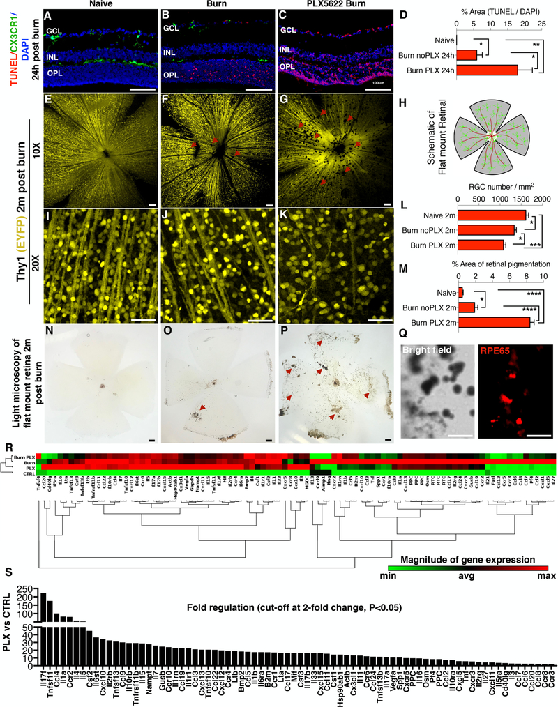
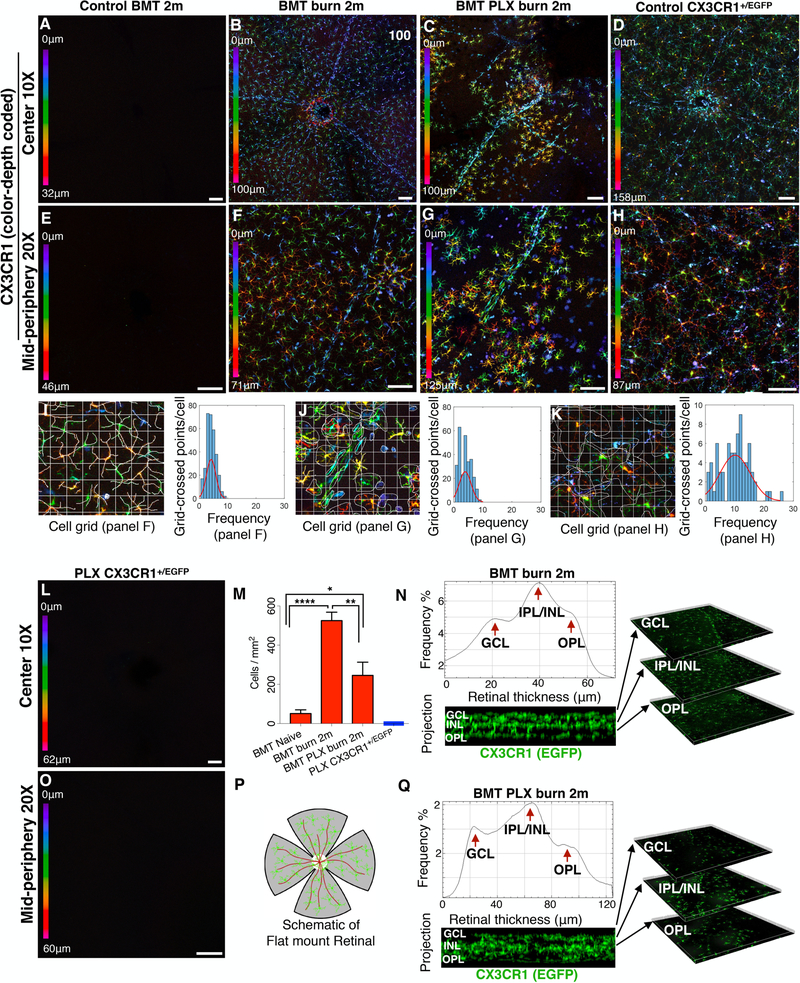
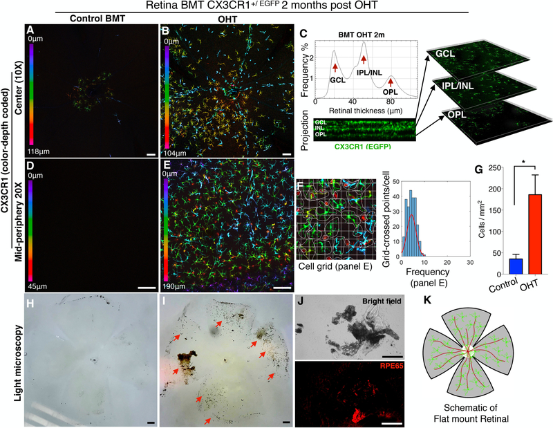
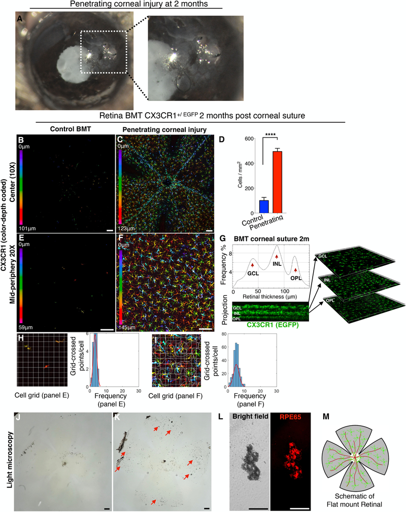
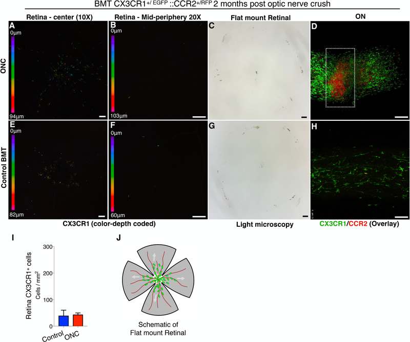
Reactive microglia and infiltrating peripheral monocytes have been implicated in many neurodegenerative diseases of the retina and CNS. However, their specific contribution in retinal degeneration remains unclear. We recently showed that peripheral monocytes that infiltrate the retina after ocular injury in mice become permanently engrafted into the tissue, establishing a proinflammatory phenotype that promotes neurodegeneration. In this study, we show that microglia regulate the process of neuroglia remodeling during ocular injury, and their depletion results in marked upregulation of inflammatory markers, such as Il17f, Tnfsf11, Ccl4, Il1a, Ccr2, Il4, Il5, and Csf2 in the retina, and abnormal engraftment of peripheral CCR2+ CX3CR1+ monocytes into the retina, which is associated with increased retinal ganglion cell loss, retinal nerve fiber layer thinning, and pigmentation onto the retinal surface. Furthermore, we show that other types of ocular injuries, such as penetrating corneal trauma and ocular hypertension also cause similar changes. However, optic nerve crush injury-mediated retinal ganglion cell loss evokes neither peripheral monocyte response in the retina nor pigmentation, although peripheral CX3CR1+ and CCR2+ monocytes infiltrate the optic nerve injury site and remain present for months. Our study suggests that microglia are key regulators of peripheral monocyte infiltration and retinal pigment epithelium migration, and their depletion results in abnormal neuroglia remodeling that exacerbates neuroretinal tissue damage. This mechanism of retinal damage through neuroglia remodeling may be clinically important for the treatment of patients with ocular injuries, including surgical traumas.


Figures

Figure 1.
Figure 1.. Microglia depletion exacerbates neuroretinal tissue damage after corneal chemical injury.
(A) TUNEL signal in naive CX3CR1+/EGFP mouse retina. CX3CR1+/EGFP cells are present in the ganglion cell (GCL), inner plexiform/inner nuclear (IPL/INL), and outer plexiform (OPL) layers but TUNEL signal is not present. (B) Increase in TUNEL signal in the retina 24 hours after corneal chemical injury. CX3CR1+/EGFP cells are present in all three retinal microglia strata. (C) TUNEL signal is significantly elevated 24 hours after ocular injury in microglia depleted mice. (D) Quantification of TUNEL signal in the retina 24 hours after the injury. (E-G) Images of Thy1 EYFP mice show reduction in retinal nerve fiber layer (RNFL) (red arrows) and (I-K) retinal ganglion cells (RGC) 2 months after the ocular injury. However, microglia depleted mice exhibit significantly more RNFL (red arrows) and RGC loss compared to injured mice with intact microglia. (L) Quantification of retinal ganglion cell number 2 months after the injury. (H) Schematic representation of the flat mount retina. (M) Quantification of the pigmented area shows marked increase in microglia depleted mice. (N) Light microscopy of naive flat mount, (O) 2 months after ocular injury, and (P) 2 months after injury in microglia depleted mice. (N-P) Ocular injury leads to pigmentation of the inner retina (P) (red arrows) which is exacerbated in microglia depleted mice (red arrows). (Q) Pigmented cells RPE65+ as shown with immunolocalization of flat mount retinas. (R) Gene expression analysis of 84 major genes involved in inflammation using RT2 Profiler PCR Array of cytokines and receptors in retinas of Naïve C57BL/6 (CTRL), microglia depleted (PLX), corneal burned (Burn), and corneal burned with microglia depletion mice (Burn PLX). Non-supervised hierarchical clustering of the entire dataset with heat map and dendrograms indicating co-regulated genes. Elevation of various inflammatory genes as compared to CTRL. (S) Microglia depletion leads to significant upregulation of a wide-range of inflammatory genes. Cut-off at 2-fold, P<0.05, Single experiment with 3 retinas per group. (A-Q) Five independent experiments with 1 mouse per group per experiment. GCL: ganglion cell layer, IPL/INL: inner plexiform/inner nuclear layer, OPL: outer plexiform layer. (A-C, N-P) Scale bar: 100μm, (E-G, I-K, Q) Scale bar: 50μm. Multiple comparisons using Tukey’s method *P<0.05, **P<0.01, ***P<0.001, ****P<0.0001.
Figure 2.
Figure 2.. Microglia depletion in injured eyes causes abnormal infiltration and engraftment of peripheral CX3CR1+ monocytes into the retina.
(A, E) Peripheral monocytes do not infiltrate into the retina after busulfan myelodepletion and bone marrow transfer of CX3CR1+/EGFP cells. (B, F) Ocular injury causes infiltration of peripheral monocytes in the retina and subsequent engraftment. These cells migrate homogeneously in the retina tissue at 2 months. (C, G) Same type of injury in microglia depleted mice results in repopulation of the retina by peripheral CX3CR1+ cells at 2 months. Repopulating CX3CR1+ cells do not form a homogeneous distribution of cells, but rather form aggregates of cells with semi-ramified morphology. (D, H) Normal distribution of CX3CR1+ cell in the retina of naive CX3CR1+/EGFP mouse. (I-K) Quantification of cell ramification using grid analysis. Representation of the data using frequency plot. Cell ramification in the BMT burn group was similar to BMT PLX burn group (P= 0.464) but significantly reduced compared to control CX3CR1+/EGFP group (P< 0.0001). (L, O) Confocal microscopy of retinal flat mounts of CX3CR1+/EGFP mice after 3 weeks of continuous PLX5622 treatment showing complete depletion of CX3CR1+ cells from the (L) central and (O) peripheral retina. (M) PLX5622 treatment leads to significant reduction of the number of peripheral CX3CR1+ monocytes that engraft into the retina 2 months after the injury. (N, Q) Engrafted peripheral monocytes migrate in all 3 distinct microglia strata (GCL: ganglion cell layer, IPL/INL: inner plexiform/inner nuclear layer, OPL: outer plexiform layer) 2 months after ocular injury in mice with intact microglia system as well as in mice with depleted microglia. (P) Schematic representation of flat mount retina used for confocal microscopy. Five independent experiments with 1 mouse per group per experiment. GCL: ganglion cell layer, IPL/INL: inner plexiform/inner nuclear layer, OPL: outer plexiform layer, BMT: bone marrow transfer. (A-D, L) Scale bar: 100μm, (E-H, O) Scale bar: 50μm. Multiple comparisons using Tukey’s method *P<0.05, **P<0.01, ****P<0.0001.
Figure 3.
Figure 3.. Acute ocular hypertension causes peripheral CX3CR1+ infiltration and engraftment into the retina.
Ocular hypertension (OHT) in BMT CX3CR1+/EGFP mice using anterior chamber cannulation (intraocular pressure elevation to 100 mmHg for 45 minutes). (A, D) In control mice CX3CR1+ cells are present in the retina 2 months after BMT. (B, G, E) OHT causes significant infiltration (P<0.05) of peripheral CX3CR1+ monocytes into the retina and subsequent differentiation to ramified cells within 2 months. (C) Infiltrated peripheral CX3CR1+ monocytes migrate in all three distinct microglia strata (ganglion cell, inner nuclear cell, and outer plexiform cell layers) 2 months after the injury and engraft into the tissue. (F) Peripheral monocytes that infiltrate the retina after OHT injury adopt a ramified morphology but remain less ramified (P<0.0001) compared to resident (native) microglia. (H) In control eyes, the inner retina shows no evidence of secondary pigmentation. (I, J) Two months after the injury, the inner retina becomes significantly pigmented (red arrows) with RPE65+ cells. (K) Schematic representation of flat mount retina used for confocal microscopy. BMT: bone marrow transfer, OHT: ocular hypertension. Three independent experiments with 1 mouse per group per experiment. (A, B, D, E, H, I, J) Scale bar: 100μm. Student t-test *P<0.05.
Figure 4.
Figure 4.. Penetrating corneal injury leads to peripheral CX3CR1+ infiltration and engraftment into the retina.
(A) Penetrating cornea injury performed by full-thickness placement of 11–0 vycril suture in the cornea. (B, E) CX3CR1+/EGFP bone marrow chimera model shows no peripheral monocyte infiltration in the absence of a suture. (C, D, F) Penetrating corneal injury causes engraftment of peripheral CX3CR1+ monocytes into the retina with a ramified appearance at 2 months. (G) Engrafted peripheral CX3CR1+ monocytes migrate into the three distinct microglia strata (ganglion cell, inner nuclear cell, and outer plexiform cell layers) 2 months after the injury. (H, I) Peripheral monocytes that infiltrate the retina after penetrating ocular injury have more ramified morphology compared to control BMT mice (P<0.0001) but appear less ramified compared to resident (native) microglia (P<0.0001). (J-L) Penetrating corneal injury causes pigmentation of the inner retina (red arrows) by RPE65+ cells. (M) Schematic representation of flat mount retina used for confocal microscopy. BMT: bone marrow transfer, OHT: ocular hypertension. Three independent experiments with 1 mouse per group per experiment. (B, C, E, F, J, K) Scale bar: 100μm. (L) Scale bar: 50μm. Student t-test ****P<0.0001.
Figure 5.
Figure 5.. Optic nerve crush does not cause peripheral CX3CR1+ infiltration and engraftment into the retinal.
Optic nerve crush (ONC) injury in CX3CR1+/EGFP::CCR2+/RFP bone marrow chimeras. (A, B, E, F) ONC does not cause peripheral CX3CR1+ CCR2+ cell infiltration or engraftment into the retina, as assessed at 2 months or inner retina pigmentation (C, G). (D, H) Flat mount confocal microscopy of the optic nerve shows a significant increase in peripheral CCR2+ CX3CR1−negative cells at the site of the ONC, and CCR2−negative CX3CR1+ cells adjacent to the ONC, in the uninjured tissue. (I) No engraftment of peripheral CX3CR+ cells in the retina is seen 2 months after ONC injury. (J) Schematic representation of flat mount retina used for confocal microscopy. BMT: bone marrow transfer, ONC: optic nerve crush, Six independent experiments with 1 mouse per group per experiment. (A-H) Scale bar: 100μm. Student t-test ****P<0.0001.

References

    1. Ginhoux F, Greter M, Leboeuf M, Nandi S, See P, Gokhan S, Mehler MF, Conway SJ, Ng LG, Stanley ER, Samokhvalov IM, and Merad M. 2010. Fate mapping analysis reveals that adult microglia derive from primitive macrophages. Science 330: 841–845. - PMC - PubMed
    1. Kierdorf K, Erny D, Goldmann T, Sander V, Schulz C, Perdiguero EG, Wieghofer P, Heinrich A, Riemke P, Holscher C, Muller DN, Luckow B, Brocker T, Debowski K, Fritz G, Opdenakker G, Diefenbach A, Biber K, Heikenwalder M, Geissmann F, Rosenbauer F, and Prinz M. 2013. Microglia emerge from erythromyeloid precursors via Pu.1- and Irf8-dependent pathways. Nat Neurosci 16: 273–280. - PubMed
    1. Crotti A, and Ransohoff RM. 2016. Microglial Physiology and Pathophysiology: Insights from Genome-wide Transcriptional Profiling. Immunity 44: 505–515. - PubMed
    1. Streit WJ 2002. Microglia as neuroprotective, immunocompetent cells of the CNS. Glia 40: 133–139. - PubMed
    1. Ginhoux F, Greter M, Leboeuf M, Nandi S, See P, Gokhan S, Mehler MF, Conway SJ, Ng LG, Stanley ER, Samokhvalov IM, and Merad M. 2010. Fate mapping analysis reveals that adult microglia derive from primitive macrophages. In Science. 841–845. - PMC - PubMed

Publication types

MeSH terms

Substances