

Bronchial epithelial cells of young and old mice directly regulate the differentiation of Th2 and Th17
- PMID: 30541898
- PMCID: PMC6356035
- DOI: 10.1042/BSR20181948
Bronchial epithelial cells of young and old mice directly regulate the differentiation of Th2 and Th17
Abstract
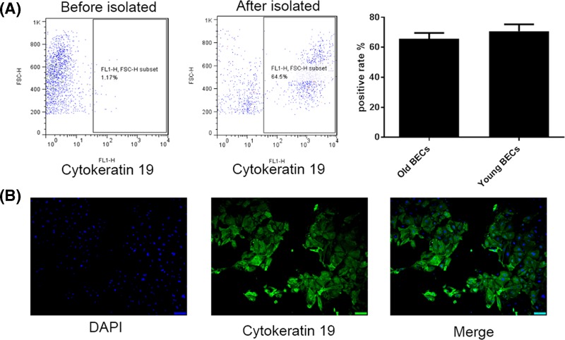
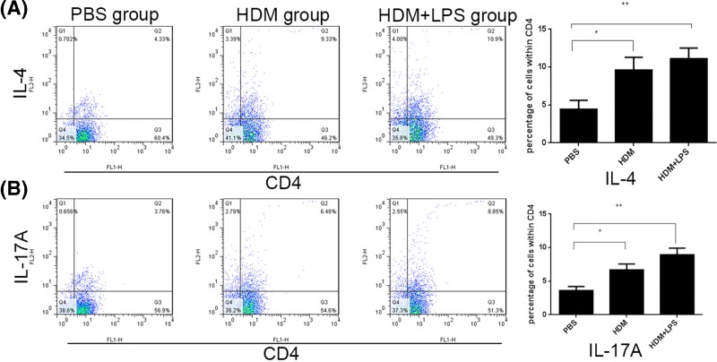
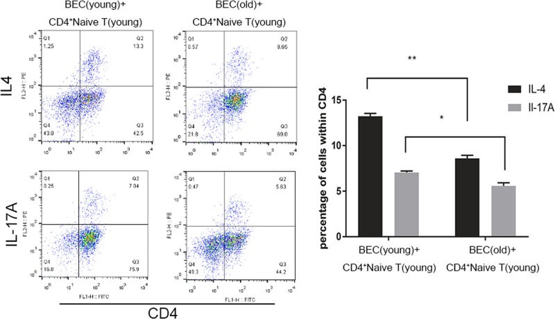
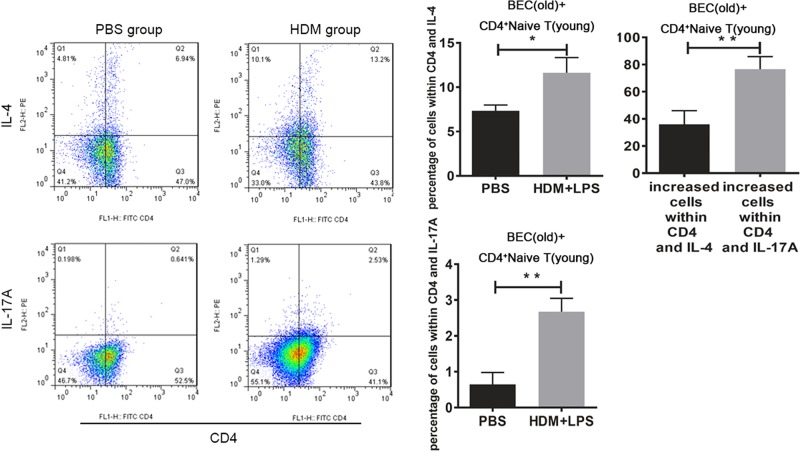
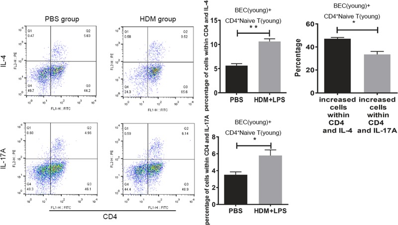
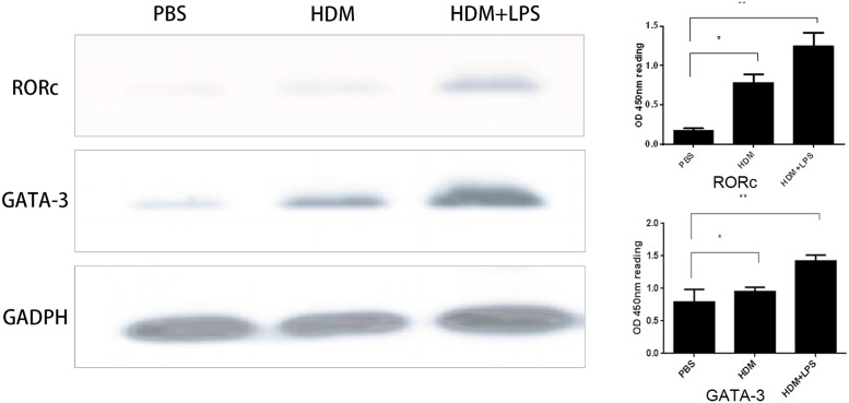
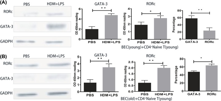
To determine whether or not house dust mite (HDM) and HDM+lipopolysaccharide (LPS) exposure causes a difference in T-cell subsets from young and old mice. The bronchial epithelial cells (BECs) from young and old mice were divided into three groups (PBS (control), HDM, and HDM+LPS). CD4+ naive T cells from the spleen and lymph nodes were collected after 24 h of co-culture with BECs. The number of Th2 and Th17 cells was elevated in the HDM and HDM+LPS groups compared with the control group; these responses were exacerbated when exposed to HDM+LPS. The number of HDM- and HDM+LPS-specific Th2/Th17 cells in young mice was higher than old mice; however, the Th2:Th17 cell ratio was greater in young mice, whereas the Th17:Th2 cell ratio was greater in old mice. The expression of GATA-3 and RORc was increased in the HDM+LPS and HDM groups compared with the PBS group and exhibited most in HDM+LPS group. The expression of HDM+LPS-specific GATA-3 in young mice was higher, while the expression of HDM+LPS-specific RORc in old mice was higher. Murine BECs directly regulated CD4+ naive T-cell differentiation under allergen exposure.
Keywords: Th17 cells; Th2 cells; asthma; epithelial cells.
© 2019 The Author(s).
Conflict of interest statement
The authors declare that there are no competing interests associated with the manuscript.
Figures
Similar articles
-
Dectin-2 promotes house dust mite-induced T helper type 2 and type 17 cell differentiation and allergic airway inflammation in mice.Am J Respir Cell Mol Biol. 2014 Aug;51(2):201-9. doi: 10.1165/rcmb.2013-0522OC. Am J Respir Cell Mol Biol. 2014. PMID: 24588637
-
Enhanced allergic airway disease in old mice is associated with a Th17 response.Clin Exp Allergy. 2014 Oct;44(10):1282-92. doi: 10.1111/cea.12388. Clin Exp Allergy. 2014. PMID: 25109604
-
Aged mice-derived bronchial epithelial cells regulate Th17 cell differentiation in asthma via the MBD2-sICOSL axis.Cell Immunol. 2025 May-Jun;411-412:104954. doi: 10.1016/j.cellimm.2025.104954. Epub 2025 Apr 15. Cell Immunol. 2025. PMID: 40252480
-
STAT3 inhibition prevents lung inflammation, remodeling, and accumulation of Th2 and Th17 cells in a murine asthma model.Allergy. 2016 Dec;71(12):1684-1692. doi: 10.1111/all.12937. Epub 2016 Jun 23. Allergy. 2016. PMID: 27225906
-
Mode of dendritic cell activation: the decisive hand in Th2/Th17 cell differentiation. Implications in asthma severity?Immunobiology. 2015 Feb;220(2):254-61. doi: 10.1016/j.imbio.2014.09.016. Epub 2014 Sep 16. Immunobiology. 2015. PMID: 25245013 Review.
Cited by
-
Role of Sex Hormones at Different Physiobiological Conditions and Therapeutic Potential in MBD2 Mediated Severe Asthma.Oxid Med Cell Longev. 2021 Dec 14;2021:7097797. doi: 10.1155/2021/7097797. eCollection 2021. Oxid Med Cell Longev. 2021. PMID: 35096261 Free PMC article. Review.
-
MBD2 mediates Th17 cell differentiation by regulating MINK1 in Th17-dominant asthma.Front Genet. 2022 Oct 11;13:959059. doi: 10.3389/fgene.2022.959059. eCollection 2022. Front Genet. 2022. PMID: 36303542 Free PMC article.
-
Androgen Plays a Potential Novel Hormonal Therapeutic Role in Th17 Cells Predominant Neutrophilic Severe Asthma by Attenuating BECs Regulated Th17 Cells Differentiation via MBD2 Expression.Oxid Med Cell Longev. 2022 Aug 25;2022:3096528. doi: 10.1155/2022/3096528. eCollection 2022. Oxid Med Cell Longev. 2022. PMID: 36062195 Free PMC article.
-
The role of GLS1-mediated glutaminolysis/2-HG/H3K4me3 and GSH/ROS signals in Th17 responses counteracted by PPARγ agonists.Theranostics. 2021 Mar 4;11(9):4531-4548. doi: 10.7150/thno.54803. eCollection 2021. Theranostics. 2021. PMID: 33754076 Free PMC article.
-
METTL3 mediates SOX5 m6A methylation in bronchial epithelial cells to attenuate Th2 cell differentiation in T2 asthma.Heliyon. 2024 Apr 1;10(7):e28884. doi: 10.1016/j.heliyon.2024.e28884. eCollection 2024 Apr 15. Heliyon. 2024. PMID: 38601672 Free PMC article.
References
-
- Asthma G.I.F. (1995) Global Strategy for Asthma Management and Prevention. NHLBI/WHO Workshop Report, National Institutes of Health National Heart, Lung and Blood institute, Bethesda, MD, NIH publication number 95-3659
Publication types
MeSH terms
Substances
LinkOut - more resources
Full Text Sources
Medical
Research Materials