

miR-125 regulates PI3K/Akt/mTOR signaling pathway in rheumatoid arthritis rats via PARP2
- PMID: 30541899
- PMCID: PMC6328865
- DOI: 10.1042/BSR20180890
miR-125 regulates PI3K/Akt/mTOR signaling pathway in rheumatoid arthritis rats via PARP2
Abstract
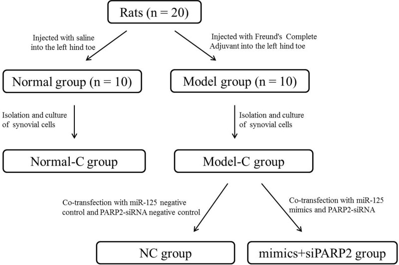
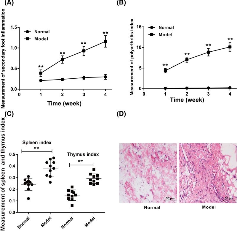
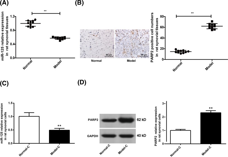
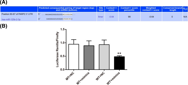
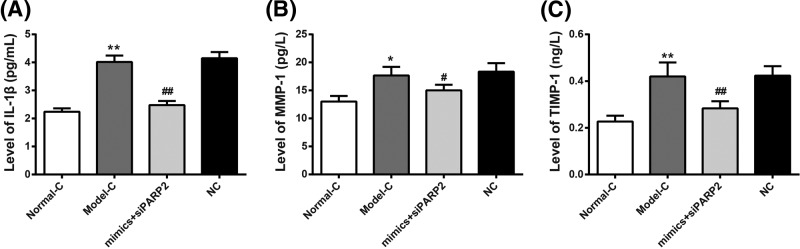
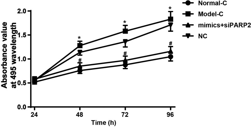
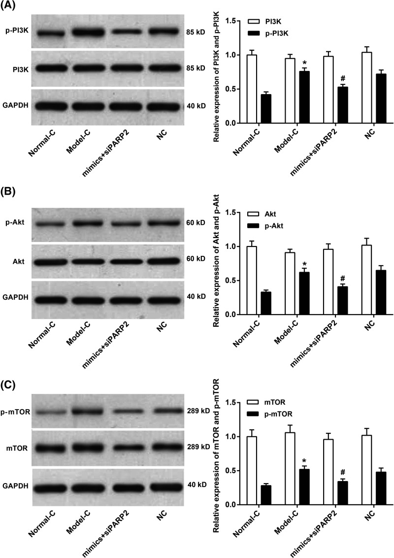
The present study aimed to explore miR-125 effects on rheumatoid arthritis (RA) development to provide a potential target for RA. Briefly, rat RA model was established (Model group) by injection of Freund's Complete Adjuvant into the left hind toe. Normal rats injected with saline in the same location were set as Normal group. All rats' secondary foot swelling degree, polyarthritis index score, spleen and thymus index were measured. Synovial tissues were subjected to Hematoxylin-Eosin (HE) staining and immunohistochemistry. Synovial cells of each group were isolated and named as Normal-C group and Model-C group, respectively. Synovial cells of Model-C group further underwent cotransfection with miR-125 mimics and PARP2-siRNA (mimics+siPARP2 group) or with miR-125 negative control (NC) and PARP2-siRNA NC (NC group). Quantitative reverse transcriptase PCR (qRT-PCR), Western blot, luciferase reporter assay, ELISA, and MTT assay were performed. As a result, compared with Normal group, rats of Model group showed significantly higher secondary foot swelling degree, polyarthritis index score, spleen and thymus index (P<0.01). Down-regulated miR-125 and up-regulated PARP2 was found in synovial tissues of Model group when compared with Normal group (P<0.01). Synovial tissues of Model-C group exhibited severe hyperplasia and inflammatory cell infiltration. Luciferase reporter assay indicated that PARP2 was directly inhibited by miR-125. Compared with NC group, cells of mimics+siPARP2 group had significantly lower IL-1β, MMP-1 and TIMP-1 levels, absorbance value, and p-PI3K, p-Akt and p-mTOR relative expression (P<0.01 or P<0.05). Thus, miR-125 might attenuate RA development by regulating PI3K/Akt/mTOR signaling pathway via directly inhibiting PARP2 expression.
Keywords: PARP2; PI3K/Akt/mTOR signaling pathway; miR-125; rheumatoid arthritis.
© 2018 The Author(s).
Conflict of interest statement
The authors declare that there are no competing interests associated with the manuscript.
Figures
Similar articles
-
microRNA-383 suppresses the PI3K-AKT-MTOR signaling pathway to inhibit development of cervical cancer via down-regulating PARP2.J Cell Biochem. 2018 Jul;119(7):5243-5252. doi: 10.1002/jcb.26585. Epub 2018 Mar 25. J Cell Biochem. 2018. Retraction in: J Cell Biochem. 2021 Jan;122(1):148. doi: 10.1002/jcb.29854. PMID: 29236322 Retracted.
-
[Effect of moxibustion on PI3K/Akt/mTOR signaling pathway in foot-pad synovium in rats with rheumatoid arthritis].Zhongguo Zhen Jiu. 2020 Nov 12;40(11):1211-6. doi: 10.13703/j.0255-2930.20200112-k0003. Zhongguo Zhen Jiu. 2020. PMID: 33788490 Chinese.
-
Effect of moxibustion on autophagy and the inflammatory response of synovial cells in rheumatoid arthritis model rat.J Tradit Chin Med. 2022 Feb;42(1):73-82. doi: 10.19852/j.cnki.jtcm.20210324.002. J Tradit Chin Med. 2022. PMID: 35294125 Free PMC article.
-
Up-regulation of miR-365 promotes the apoptosis and restrains proliferation of synoviocytes through downregulation of IGF1 and the inactivation of the PI3K/AKT/mTOR pathway in mice with rheumatoid arthritis.Int Immunopharmacol. 2020 Feb;79:106067. doi: 10.1016/j.intimp.2019.106067. Epub 2019 Dec 24. Int Immunopharmacol. 2020. PMID: 31881377
-
Moxibustion regulates the polarization of macrophages through the IL-4/STAT6 pathway in rheumatoid arthritis.Cytokine. 2022 Apr;152:155835. doi: 10.1016/j.cyto.2022.155835. Epub 2022 Feb 28. Cytokine. 2022. PMID: 35240467 Review.
Cited by
-
Ethanolic Extract of Propolis Modulates Autophagy-Related microRNAs in Osteoarthritic Chondrocytes.Int J Mol Sci. 2023 Sep 30;24(19):14767. doi: 10.3390/ijms241914767. Int J Mol Sci. 2023. PMID: 37834215 Free PMC article.
-
Unveiling Novel Drug Targets and Emerging Therapies for Rheumatoid Arthritis: A Comprehensive Review.ACS Pharmacol Transl Sci. 2024 May 28;7(6):1664-1693. doi: 10.1021/acsptsci.4c00067. eCollection 2024 Jun 14. ACS Pharmacol Transl Sci. 2024. PMID: 38898941 Free PMC article. Review.
-
Baicalein Induces Apoptosis of Rheumatoid Arthritis Synovial Fibroblasts through Inactivation of the PI3K/Akt/mTOR Pathway.Evid Based Complement Alternat Med. 2022 Sep 7;2022:3643265. doi: 10.1155/2022/3643265. eCollection 2022. Evid Based Complement Alternat Med. 2022. PMID: 36118088 Free PMC article.
-
An Integrative Pharmacology Model for Decoding the Underlying Therapeutic Mechanisms of Ermiao Powder for Rheumatoid Arthritis.Front Pharmacol. 2022 Feb 23;13:801350. doi: 10.3389/fphar.2022.801350. eCollection 2022. Front Pharmacol. 2022. PMID: 35281924 Free PMC article.
-
Orchestrated modulation of rheumatoid arthritis via crosstalking intracellular signaling pathways.Inflammopharmacology. 2021 Aug;29(4):965-974. doi: 10.1007/s10787-021-00800-3. Epub 2021 Mar 19. Inflammopharmacology. 2021. PMID: 33740220 Review.
References
-
- Jiang X. (2015) Gene-environment interactions in rheumatoid arthritis: quantification and characterization of contributing factors. Transplant. Proc. 35, 1123–1124
MeSH terms
Substances
LinkOut - more resources
Full Text Sources
Molecular Biology Databases
Research Materials
Miscellaneous