. 2019 Feb 7;9(2):425-437.
doi: 10.1534/g3.118.200859.

Complex Structural PPT1 Variant Associated with Non-syndromic Canine Retinal Degeneration

Affiliations

Complex Structural PPT1 Variant Associated with Non-syndromic Canine Retinal Degeneration

Leonardo Murgiano et al. G3 (Bethesda). .

Abstract

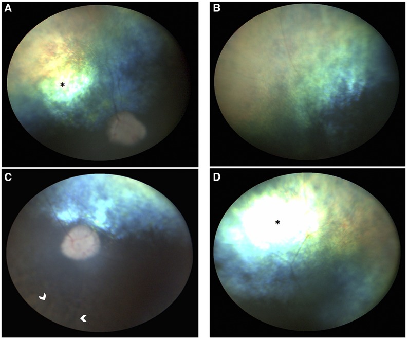
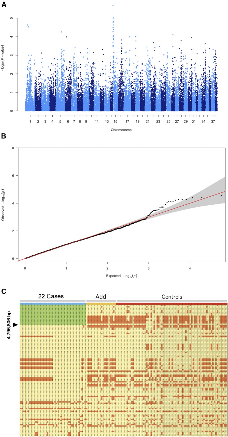
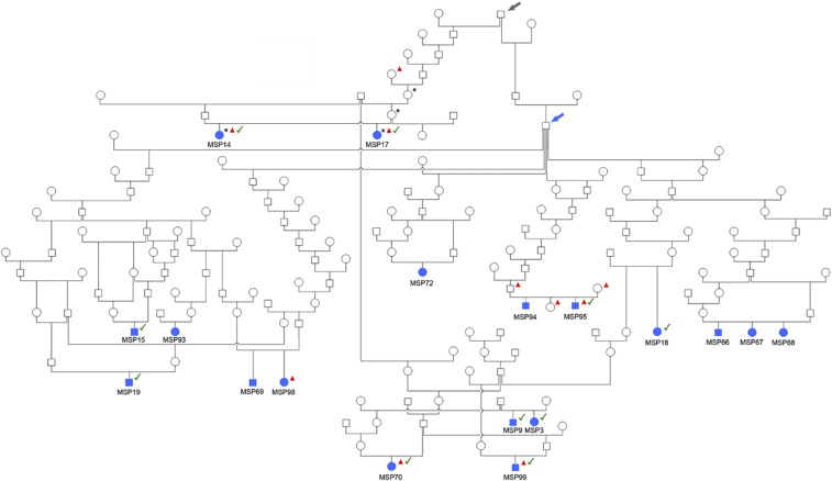
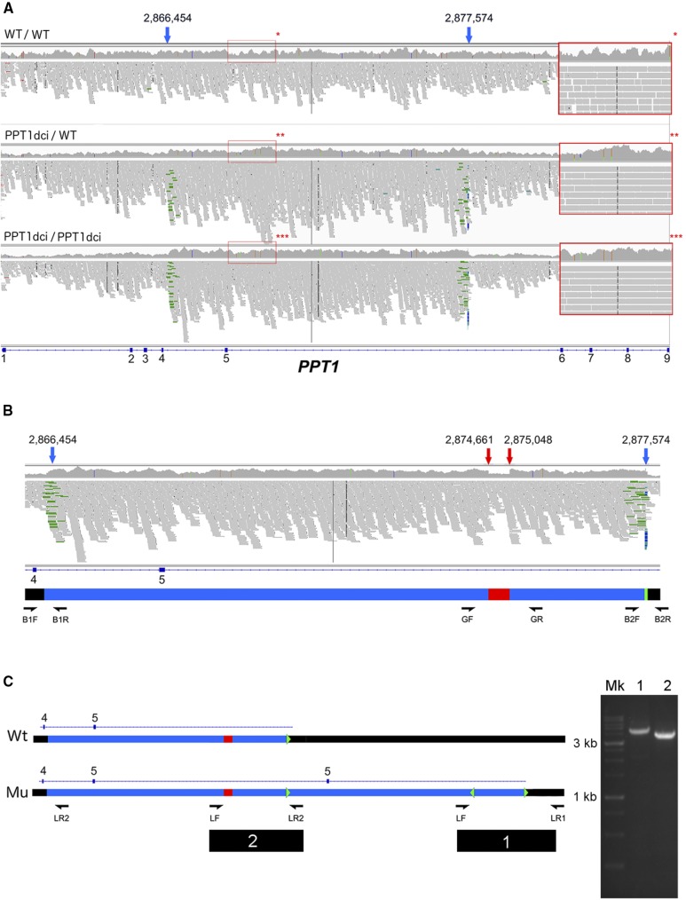
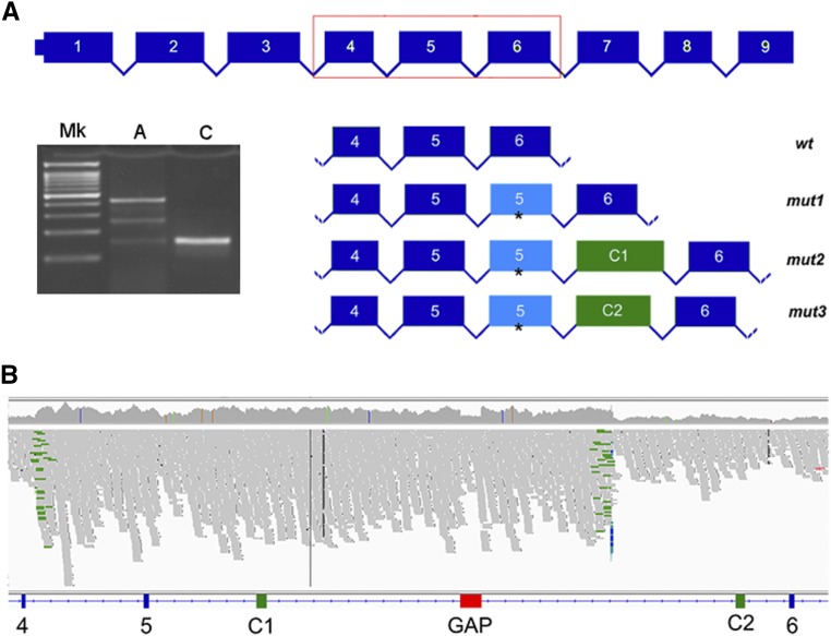
Rod and cone photoreceptors are specialized retinal neurons that have a fundamental role in visual perception, capturing light and transducing it into a neuronal signal. Aberrant functioning of rod and/or cone photoreceptors can ultimately lead to progressive degeneration and eventually blindness. In man, many rod and rod-cone degenerative diseases are classified as forms of retinitis pigmentosa (RP). Dogs also have a comparable disease grouping termed progressive retinal atrophy (PRA). These diseases are generally due to single gene defects and follow Mendelian inheritance.We collected 51 DNA samples from Miniature Schnauzers affected by PRA (average age of diagnosis ∼3.9 ±1 years), as well as from 56 clinically normal controls of the same breed (average age ∼6.6 ±2.8 years). Pedigree analysis suggested monogenic autosomal recessive inheritance of PRA. GWAS and homozygosity mapping defined a critical interval in the first 4,796,806 bp of CFA15. Whole genome sequencing of two affected cases, a carrier and a control identified two candidate variants within the critical interval. One was an intronic SNV in HIVEP3, and the other was a complex structural variant consisting of the duplication of exon 5 of the PPT1 gene along with a conversion and insertion (named PPT1dci ). PPT1dci was confirmed homozygous in a cohort of 22 cases, and 12 more cases were homozygous for the CFA15 haplotype. Additionally, the variant was found homozygous in 6 non-affected dogs of age higher than the average age of onset. The HIVEP3 variant was found heterozygous (n = 4) and homozygous wild-type (n = 1) in cases either homozygous for PPT1dci or for the mapped CFA15 haplotype. We detected the wildtype and three aberrant PPT1 transcripts in isolated white blood cell mRNA extracted from a PRA case homozygous for PPT1dci , and the aberrant transcripts involved inclusion of the duplicated exon 5 and novel exons following the activation of cryptic splice sites. No neurological signs were detected among the dogs homozygous for the PPT1dci variant. Therefore, we propose PPT1dci as causative for a non-syndromic form of PRA (PRA PPT1 ) that shows incomplete penetrance in Miniature Schnauzers, potentially related to the presence of the wild-type transcript. To our knowledge, this is the first case of isolated retinal degeneration associated with a PPT1 variant.

Keywords: PRA; complex variant; dog; palmitoyl protein thioesterase; progressive retinal atrophy; retinal degeneration; whole genome sequencing.


Figures

Figure 1
Figure 1
Fundus photographs of the right (A, B) and left (C, D) eyes of a 4 year old female Miniature Schnauzer homozygous for the PPT1 complex structural variant. Both eyes show marked attenuation and loss of the major and minor retinal arterioles and venules, and the optic disc is pale and undergoing atrophic changes. With the proper angle of illumination, there is pronounced increased reflectivity from the tapetum lucidum (* in A and D), an indication of severe retinal thinning. The non-tapetal region shows patches of retinal pigment epithelial atrophy which appear paler, and these are situated adjacent to hyperpigmented and darker areas (C arrows). Photographs provided by Dr. Julien Charron, Clinique vétérinaire Saint Roch, La Rochelle, France
Figure 2
Figure 2
Mapping. (A) Results of the genome-wide association study (GWAS), obtained analyzing the imputed Affymetrix/Illumina merged data showing the negative log of the raw p-values calculated with the genotypic association test (max p-value 2.20e-06) (B) QQ-plot, showing the observed vs. expected log p-values. The moderate skewing of a marker toward the upper side suggests a weak association with the “affected” condition compared with what would be expected by mere chance. (C) Result of the homozygosity mapping using the 27,061 shared SNPs merged dataset: detail of the markers encompassing the first 14,490,600 Mb of CFA15. In green the candidate region shared exclusively among a sub-group of blue-marked 22 cases. Other cases (“Add”) are marked in yellow-orange, “Controls” in red. The region is shown in Microsoft Excel with the different alleles colored for ease of visualization. We opted to select a candidate region as the combination of the suggestive peak (log –pvalue 05) obtained by the GWAS and the homozygosity mapping.
Figure 3
Figure 3
- Family tree of 18 Miniature Schnauzers cases affected with PRA in which there was sufficient family history to identify a putative common ancestor. Females are shown in circles, males in squares. All the animals indicated with the blue filled symbol are homozygous for the CFA15 haplotype. Dogs indicated with an asterisk (*) were used in WGS. Dogs indicated with a red triangle are cases and controls subjected to targeted sequencing. Dogs with the green tick have been confirmed homozygous for the PPT1 variant through targeted or Sanger sequencing. The blue arrow shows a confirmed common ancestor for many cases, and putative carriers. The gray arrow shows another older, common ancestor in which the carrier status is definitive.
Figure 4
Figure 4
Characterization of the 11Mb-spanning variant. Figure is based on screenshots of the interval visualized with IGV. (A) Duplication of the 5th PPT1 exon visualized with IGV. The 11,120 bp duplication can be observed. From top to bottom, a case, a carrier and a control are shown. Note the inverted pairs, marked in green by IGV, on the variant boundaries present in carrier and case. Compare the coverage of the area with the flanking regions. The blue arrows show the mutation boundaries. In the red squares, a region for each of control (*), carrier (**) and case (***) is shown. Note the heterozygous SNPs contained in the carrier interval. Additionally, compare the coverage of the carrier with the flanking regions, and note the greater coverage of the case in comparison to carrier and control. (B) Critical regions of the variant. The blue arrows show the mutation boundaries (g.2866454_2877574dup), the red arrows the conversion (g.2874661_2875048con2,877,563-2,877,607inv). In detail, the green interval represents the 45 bp interval (2,877,563-2,877,607) duplicated, inverted and inserted in position g.2874661_2875048 in lieu of the bases normally present in such interval. Details for the sequencing primers are shown in File_S2. (C) Representation of the variant – top, the wild type (Wt), bottom the duplication (Mu). Observe the gap region (in red in the reference) missing in the first copy, substituted by an inserted and inversed copy of the 45 bp interval (2,877,563-2,877,607), shown as a green arrowhead. Primer placement for the long-range PCR is shown: on the right, the long PCR products obtained (1 and 2 are shown run on a 1% agarose gel, along with the ladder (Mk). See also File_S2.
Figure 5
Figure 5
- Characterization of the transcripts. (A) PCR carried out with the 4F and 6R primers shown in File_S2. On the gel, is shown the ladder (Mk), RT-PCR of the blood cDNA extracted from one affected individual (A) is shown along with PCR obtained from the control (C). A schematic of (I) the wild-type transcript, wt; (II) a mutant transcript containing a retained exon 5 from the duplication (that is, containing thus two copies of the 5th PPT1 exon), mut 1; (III) a third, longer transcript containing a second copy of the 5th PPT1 exon plus an additional 166 bp fragment, mut2; (IV) a fourth, aberrant transcript containing a second copy of the 5th PPT1 exon plus an additional 141 bp fragment, mut3. The asterisk shows the common premature stop codon in the additional exon 5. (B) Localization on the reference sequence (included an IGV screenshot of the interval) of the C1 and C2 novel exons, in position c.2,870,731-2,870,896 (C1) and 2,879,973-2,880,113 (C2), respectively.

Comment in

  • Formal commentary.
    Aguirre GD, Lohi H, Kaukonen M, Murgiano L. Aguirre GD, et al. PLoS Genet. 2020 Nov 5;16(11):e1009059. doi: 10.1371/journal.pgen.1009059. eCollection 2020 Nov. PLoS Genet. 2020. PMID: 33151924 Free PMC article. No abstract available.

References

    1. Acland G. M., Ray K., Mellersh C. S., Gu W., Langston A. A., et al. , 1998. Linkage analysis and comparative mapping of canine progressive rod-cone degeneration (prcd) establishes potential locus homology with retinitis pigmentosa (RP17) in humans. Proc. Natl. Acad. Sci. USA 95: 3048–3053. 10.1073/pnas.95.6.3048 - DOI - PMC - PubMed
    1. Adzhubei I. A., Schmidt S., Peshkin L., Ramensky V. E., Gerasimova A., et al. , 2010. A method and server for predicting damaging missense mutations. Nat. Methods 7: 248–249. 10.1038/nmeth0410-248 - DOI - PMC - PubMed
    1. Aguirre G., Acland G. M., 2006. Models, Mutants and Man: Searching for Unique Phenotypes and Genes in the Dog Model of Inherited Retinal Degeneration, pp. 291–325 in The Dog and Its Genome, edited by Ostrander E. A., Giger U., Lindblad-Toh K. Cold Spring Harbor Laboratory Press, Cold Spring Harbor, NY.
    1. Aguirre G., O’Brien P., 1986. Morphological and biochemical studies of canine progressive rod-cone degeneration. 3H-fucose autoradiography. Invest. Ophthalmol. Vis. Sci. 27: 635–655. - PubMed
    1. Aguirre G. D., 2017. Concepts and strategies in retinal gene therapy. Invest. Ophthalmol. Vis. Sci. 58: 5399–5411. 10.1167/iovs.17-22978 - DOI - PMC - PubMed

Publication types