. 2018 Dec 12;50(12):1-12.
doi: 10.1038/s12276-018-0167-1.

Increased α2-6 sialylation of endometrial cells contributes to the development of endometriosis

Affiliations

Increased α2-6 sialylation of endometrial cells contributes to the development of endometriosis

Hee-Jin Choi et al. Exp Mol Med. .

Abstract

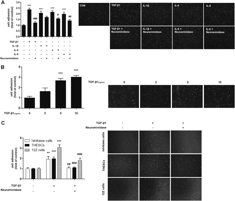
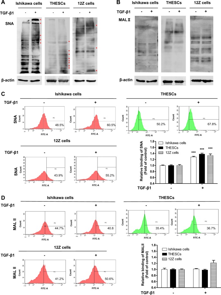
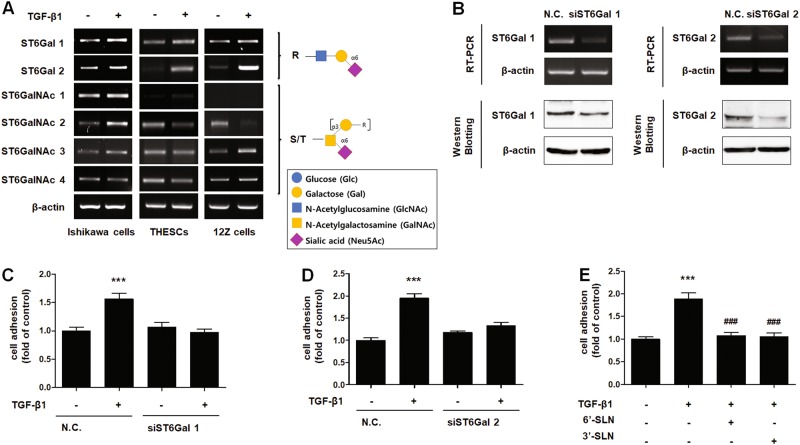
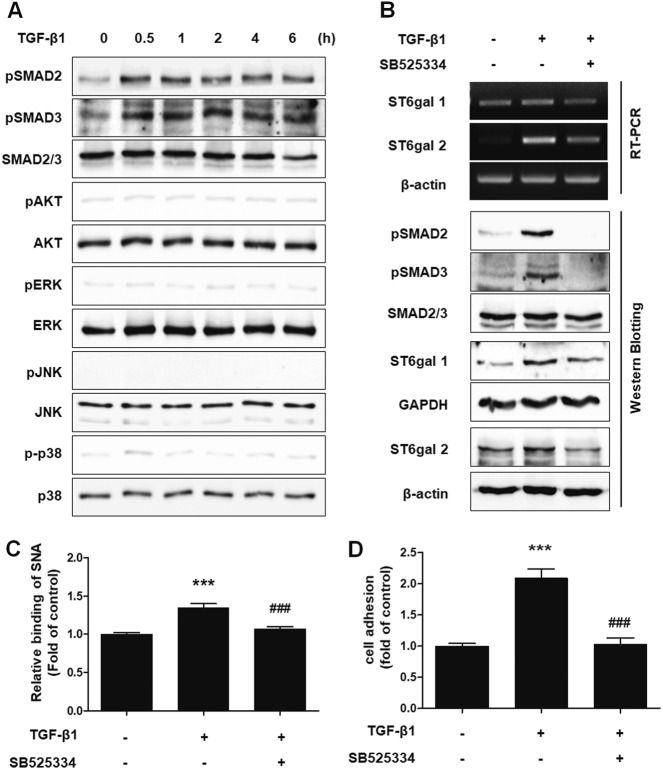
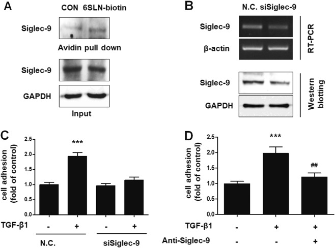
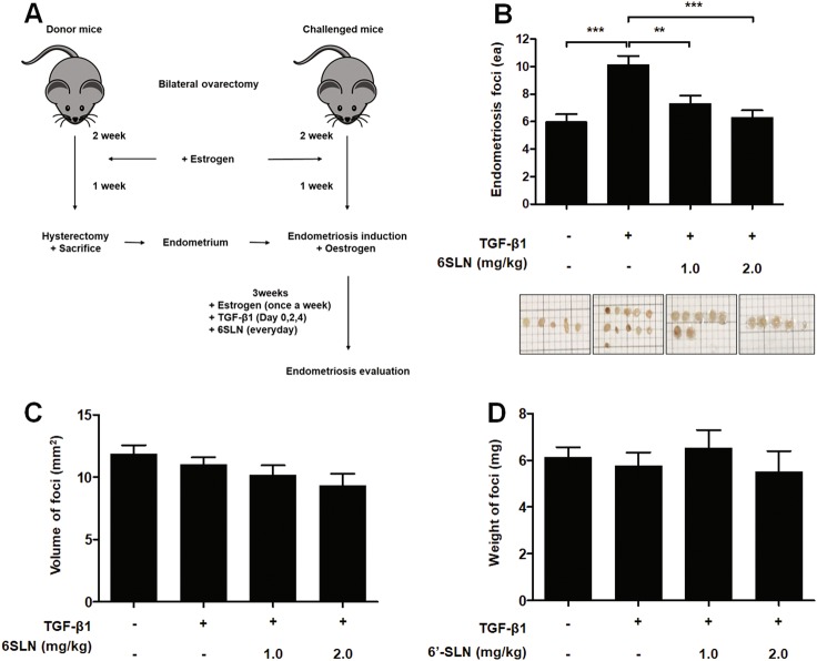
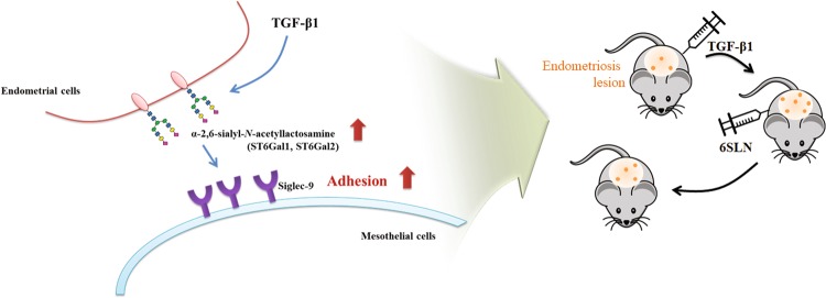
Endometriosis is a disease characterized by implants of endometrial tissue outside the uterine cavity and is strongly associated with infertility. Focal adhesion of endometrial tissue to the peritoneum is an indication of incipient endometriosis. In this study, we examined the effect of various cytokines that are known to be involved in the pathology of endometriosis on endometrial cell adhesion. Among the investigated cytokines, transforming growth factor-β1 (TGF-β1) increased adhesion of endometrial cells to the mesothelium through induction of α2-6 sialylation. The expression levels of β-galactoside α2-6 sialyltransferase (ST6Gal) 1 and ST6Gal2 were increased through activation of TGF-βRI/SMAD2/3 signaling in endometrial cells. In addition, we discovered that terminal sialic acid glycan epitopes of endometrial cells engage with sialic acid-binding immunoglobulin-like lectin-9 expressed on mesothelial cell surfaces. Interestingly, in an in vivo mouse endometriosis model, inhibition of endogenous sialic acid binding by a NeuAcα2-6Galβ1-4GlcNAc injection diminished TGF-β1-induced formation of endometriosis lesions. Based on these results, we suggest that increased sialylation of endometrial cells by TGF-β1 promotes the attachment of endometrium to the peritoneum, encouraging endometriosis outbreaks.


Conflict of interest statement

The authors declare that they have no conflict of interest.

Figures

Fig. 1
Fig. 1. TGF-β1 enhances the adhesion of endometrial cells by increasing sialylation on N-glycoproteins
a–c Fluorescence-labeled endometrial (Ishikawa or THESCs) or endometriotic (12Z) cells were added to a confluent monolayer of Met-5A cells. After 20 min of incubation at 37 °C, the number of adhered Ishikawa, THESCs or 12Z cells was calculated and is expressed as fold-difference relative to the control (mean ± SD). a Ishikawa cells were treated with neuraminidase (4 U/mL) for 1 h and then stimulated with cytokines (10 ng/mL) for 48 h. b Ishikawa cells were stimulated with the indicated concentration of TGF-β1 for 48 h. c Ishikawa, THESCs and 12Z cells were incubated with neuraminidase 1 h before TGF-β1 (10 ng/mL) stimulation. * p < 0.05, ** p < 0.01, and *** p < 0.001 compared with the control group. # p < 0.01 compared with the TGF-β1-treated group
Fig. 2
Fig. 2. α2-6 Sialylation of endometrial cells is increased by TGF-β1 treatment
Ishikawa, THESCs, or 12Z cells were incubated with TGF-β1 for 48 h. The expression of α2-3 or α2-6 sialic acid epitopes in endometrial or endometriotic cells was determined by lectin blot a, b or lectin FACS analysis c, d using biotin-labeled MAL II and SNA, respectively. Data from the lectin FACS analysis are expressed as the mean ± SD of three independent experiments. Red asterisk indicates increased protein expression in the TGF-β1-treated group compared with the control group. * p < 0.05 and *** p < 0.001 compared with the control group
Fig. 3
Fig. 3. TGF-β1-induced α2-6 sialylation is mediated by increased expression of ST6Gal1 and ST6Gal2 in endometrial cells
a Ishikawa, THESCs, and 12Z cells were treated with TGF-β1 for 24 h. Expression of α2-6 sialyltransferases was estimated by RT-PCR. b–d Ishikawa cells were transfected with 200 nM ST6Gal1- or ST6Gal2-targeting siRNA. b Expression of ST6Gal1 or ST6Gal2 in Ishikawa cells was determined by RT-PCR or western blot analysis after 24 h of incubation. c, d siRNA-transfected Ishikawa cells were cultured with TGF-β1 for 48 h, and then, transfected cells were labeled with CMFDA and added onto a Met-5A cell monolayer. The number of Ishikawa cells bound to Met-5A cells was calculated and is expressed as the fold-difference relative to the control (mean ± SD). e Ishikawa cells were incubated with TGF-β1 for 48 h. 3ʹ-SLN or 6ʹ-SLN were applied to treat Met-5A cells 1 h before the adhesion assay. Fluorescence-labeled Ishikawa cells were incubated at 37 °C with a Met-5A cell monolayer, and then, total number of adhered Ishikawa cells was determined. ** p < 0.01 and *** p < 0.001 compared with the control group. # p < 0.05 compared with the TGF-β1-treated group
Fig. 4
Fig. 4. TGF-β1 induces the adhesion of endometrial cells to mesothelial cells through the TGF-βRI/SMAD2 signaling pathway
a Ishikawa cells were cultured with TGF-β1 (10 ng/mL) according to the indicated times. Phosphorylation levels of SMAD2, SMAD3, Akt, ERK, JNK, and p38 were examined by western blot analysis. b–d Ishikawa cells were treated with 10 μm TGF-βRI inhibitor (SB525334) 1 h before TGF-β1 (10 ng/mL) stimulation. b Phosphorylation of SMAD2 and SMAD3 was determined 4 h after TGF-β1 treatment. Expression levels of ST6Gal1 and ST6Gal2 were estimated 24 h after TGF-β1 stimulation. c Ishikawa cells were incubated with TGF-β1 for 48 h. Using biotin-labeled SNA, α2-6 sialic acid epitopes in Ishikawa cells were analyzed via FACS using biotin-labeled SNA. d Tracker™ Green CMFDA-labeled Ishikawa cells were added onto Met-5A cells 48 h after TGF-β1 treatment. After incubation and washing, the number of attached Ishikawa cells was calculated and is expressed as the fold-difference relative to the control (mean ± SD). ** p < 0.01 for comparison between two groups. ##p < 0.01 compared with the TGF-β1-treated group
Fig. 5
Fig. 5. 6ʹ-SLN ligands on the endometrial cells engaged Siglec-9 on Met-5A cells
a To confirm binding between 6ʹ-SLN and Siglec-9, cell lysates from Met-5A cells were subjected to a pull-down assay using biotin-labeled 6ʹ-SLN and incubated with avidin beads. Proteins binding to the avidin beads were resolved and subjected to western blot analysis. b Met-5A cells were incubated with siRNA targeting Siglec-9 for 24 h. Expression of Siglec-9 in Met-5A cells was assessed by RT-PCR and western blot analysis. c siSiglec-9-transfected Met-5A cells were cultured to a confluent monolayer. Tracker™ Green CMFDA-labeled Ishikawa cells were added to the monolayer and incubated for 20 min. Adhered Ishikawa cells were manually counted, and the results are expressed as the fold-difference relative to the control (mean ± SD). d Ishikawa cells were treated with TGF-β1 (10 ng/mL) for 48 h. Met-5A cells were incubated with neutralizing anti-Siglec-9 antibody (2 μg/mL) 24 h before the adhesion assay. Fluorescence-labeled Ishikawa cells were added onto Met-5A cells. After 20 min of incubation and then washing, attached cells were manually counted, and the results are expressed as the fold-difference relative to the control. ** p < 0.01 and *** p < 0.001 compared with the control group. ## p < 0.01 and ### p < 0.001 compared with the TGF-β1-treated group
Fig. 6
Fig. 6. 6ʹ-SLN treatment attenuates TGF-β1-induced endometriosis in C57BL/6 mice
a Schematic diagram of the mouse endometriosis model. Endometriosis was induced at day 0. Donor and challenged mice were subjected to ovariectomy at day − 21 and injected subcutaneously with estrogens (100 mg/kg) at days − 7, 0, + 7, + 14, and + 21. Mice challenged with endometrium were inoculated intraperitoneally with 2.5 μg/kg TGF-β1 (0, + 2, + 4 days) and 6ʹ-SLN (daily). At day + 21 after endometriosis induction, challenged mice were killed, and endometriotic lesions were evaluated. b–d Endometriotic lesions were isolated, and the number, volume and weight of foci were calculated
Fig. 7
Fig. 7
Schematic presentation of TGF-β1-induced adhesion of endometrium to mesothelium through the interaction between α2-6 sialic acid and Siglec-9 in both in vitro and in vivo endometriosis models

Similar articles

Cited by

References

    1. Bulun SE. Endometriosis. N. Engl. J. Med. 2009;360:268–279. doi: 10.1056/NEJMra0804690. - DOI - PubMed
    1. Giudice LC, Kao LC. Endometriosis. Lancet. 2004;364:1789–1799. doi: 10.1016/S0140-6736(04)17403-5. - DOI - PubMed
    1. Young VJ, Brown JK, Saunders PT, Horne AW. The role of the peritoneum in the pathogenesis of endometriosis. Hum. Reprod. Update. 2013;19:558–569. doi: 10.1093/humupd/dmt024. - DOI - PubMed
    1. Miller JE, et al. Implications of immune dysfunction on endometriosis associated infertility. Oncotarget. 2017;8:7138–7147. - PMC - PubMed
    1. Sundqvist J, Andersson KL, Scarselli G, Gemzell-Danielsson K, Lalitkumar PG. Expression of adhesion, attachment and invasion markers in eutopic and ectopic endometrium: a link to the aetiology of endometriosis. Hum. Reprod. 2012;27:2737–2746. doi: 10.1093/humrep/des220. - DOI - PubMed

Publication types

MeSH terms