

In vivo confocal microscopy classification in the diagnosis of meibomian gland dysfunction
- PMID: 30542066
- PMCID: PMC6707180
- DOI: 10.1038/s41433-018-0307-9
In vivo confocal microscopy classification in the diagnosis of meibomian gland dysfunction
Erratum in
-
Correction: In vivo confocal microscopy classification in the diagnosis of meibomian gland dysfunction.Eye (Lond). 2019 May;33(5):860. doi: 10.1038/s41433-019-0373-7. Eye (Lond). 2019. PMID: 30799438 Free PMC article.
Abstract
Aim: Meibomian gland dysfunction (MGD) is one of the most common disorders in ophthalmology. The aim of this study was to evaluate the use of this in vivo confocal microscopy (IVCM)-MGD description to classify patients affected by clinical MGD and measure the correlation with standard clinical criteria and subjective symptoms.
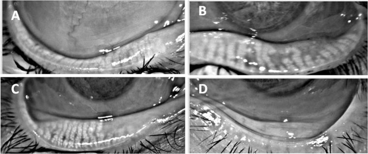
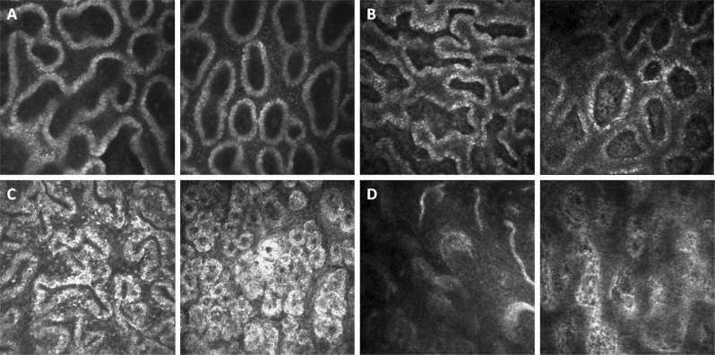
Methods: One hundred eyes of 100 patients suffering from MGD and 15 eyes of normal subjects were included. A comprehensive evaluation with the ocular surface disease index (OSDI), Schirmer test, tear break-up time (TBUT), tear osmolarity, Oxford score, Meibomian gland expression, palpebral IVCM, and meibography was performed. Then each patient was classified using a new IVCM classification: type 0 for normality, type 1 for meibum obstruction, type 2 for inflammation, and type 3 for fibrosis.
Results: The mean age of patients was 52 ± 20 years old, the OSDI was 38 ± 23, the BUT 5 ± 2.6 s, the Schirmer test 13 ± 7 mm, tear osmolarity 300 ± 11 osmol/L, the Oxford score 0.5 ± 0.6, the meibum expression score 1.7 ± 1.02, and the meibography score 1.3 ± 0.9. The IVCM MG classification of the 15 normal subjects was 0. For MGD patients, 29% were in type 1, 40% were type 2, and 31% were type 3. The patients in IVCM MG type 2 had a higher OSDI (p = 0.001) compared with the other types. There was a strong correlation between the IVCM score and the meibography score (r = 0.71 p < 0.0001).
Conclusion: This new IVCM classification provided a practical pathophysiological system for MGD. By giving objective criteria, this IVCM classification may help advance the understanding of patients' symptoms and enhance treatment effectiveness in MGD.
Conflict of interest statement
The authors declare that they have no conflict of interest.
Figures
Comment in
-
Imaging of meibomian glands: from bench to bedside and back.Eye (Lond). 2019 May;33(5):695-697. doi: 10.1038/s41433-019-0351-0. Epub 2019 Feb 6. Eye (Lond). 2019. PMID: 30728489 Free PMC article. No abstract available.
Similar articles
-
[In vivo confocal microscopy evaluation of meibomian glands in meibomian gland dysfunction patients].Zhonghua Yan Ke Za Zhi. 2016 Sep 11;52(9):649-56. doi: 10.3760/cma.j.issn.0412-4081.2016.09.004. Zhonghua Yan Ke Za Zhi. 2016. PMID: 27647244 Chinese.
-
[A new classification for meibomian gland diseases with in vivo confocal microscopy].J Fr Ophtalmol. 2016 Mar;39(3):239-47. doi: 10.1016/j.jfo.2015.07.015. Epub 2016 Feb 16. J Fr Ophtalmol. 2016. PMID: 26896195 French.
-
Functional and Morphological Evaluation of Meibomian Glands in the Assessment of Meibomian Gland Dysfunction Subtype and Severity.Am J Ophthalmol. 2020 Jan;209:160-167. doi: 10.1016/j.ajo.2019.09.005. Epub 2019 Sep 14. Am J Ophthalmol. 2020. PMID: 31526799
-
In vivo Meibomian gland imaging techniques: A review of the literature.J Fr Ophtalmol. 2020 Apr;43(4):e123-e131. doi: 10.1016/j.jfo.2019.11.003. Epub 2020 Jan 9. J Fr Ophtalmol. 2020. PMID: 31928786 Review.
-
[In vivo Meibomian gland imaging techniques: A review of the literature (French translation of the article)].J Fr Ophtalmol. 2020 Jun;43(6):484-493. doi: 10.1016/j.jfo.2019.10.009. Epub 2020 May 11. J Fr Ophtalmol. 2020. PMID: 32409228 Review. French.
Cited by
-
Ductal Hyperkeratinization and Acinar Renewal Abnormality: New Concepts on Pathogenesis of Meibomian Gland Dysfunction.Curr Issues Mol Biol. 2023 Feb 27;45(3):1889-1901. doi: 10.3390/cimb45030122. Curr Issues Mol Biol. 2023. PMID: 36975492 Free PMC article.
-
Efficacy of indirect intense pulsed light irradiation on meibomian gland dysfunction: a randomized controlled study.Int J Ophthalmol. 2024 Nov 18;17(11):2014-2022. doi: 10.18240/ijo.2024.11.06. eCollection 2024. Int J Ophthalmol. 2024. PMID: 39559304 Free PMC article.
-
In Vivo Confocal Microscopy for Automated Detection of Meibomian Gland Dysfunction: A Study Based on Deep Convolutional Neural Networks.J Imaging Inform Med. 2025 Jan 27. doi: 10.1007/s10278-024-01174-y. Online ahead of print. J Imaging Inform Med. 2025. PMID: 39871043
-
Automation of dry eye disease quantitative assessment: A review.Clin Exp Ophthalmol. 2022 Aug;50(6):653-666. doi: 10.1111/ceo.14119. Epub 2022 Jun 27. Clin Exp Ophthalmol. 2022. PMID: 35656580 Free PMC article. Review.
-
Dry Eye Disease Associated with Meibomian Gland Dysfunction: Focus on Tear Film Characteristics and the Therapeutic Landscape.Ophthalmol Ther. 2023 Jun;12(3):1397-1418. doi: 10.1007/s40123-023-00669-1. Epub 2023 Mar 1. Ophthalmol Ther. 2023. PMID: 36856980 Free PMC article. Review.
References
MeSH terms
LinkOut - more resources
Full Text Sources
