

Is Empathy for Pain Unique in Its Neural Correlates? A Meta-Analysis of Neuroimaging Studies of Empathy
- PMID: 30542272
- PMCID: PMC6277791
- DOI: 10.3389/fnbeh.2018.00289
Is Empathy for Pain Unique in Its Neural Correlates? A Meta-Analysis of Neuroimaging Studies of Empathy
Abstract
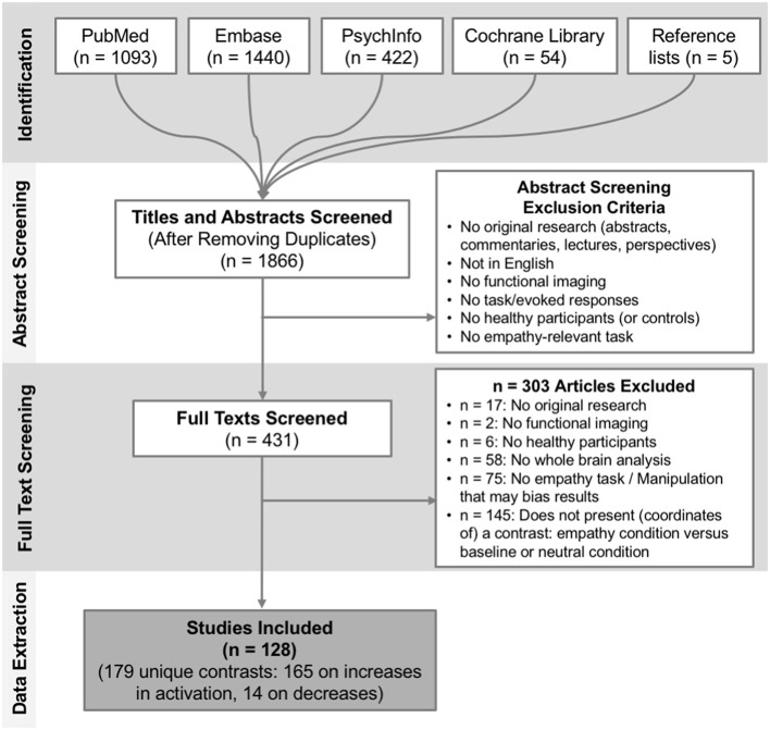
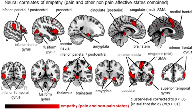
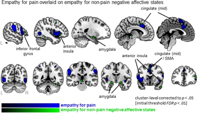
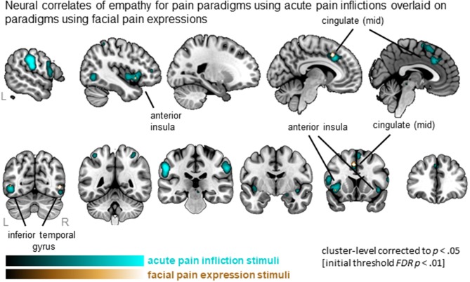
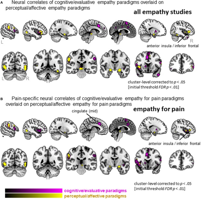
Empathy is an essential component of our social lives, allowing us to understand and share other people's affective and sensory states, including pain. Evidence suggests a core neural network-including anterior insula (AI) and mid-cingulate cortex (MCC)-is involved in empathy for pain. However, a similar network is associated to empathy for non-pain affective states, raising the question whether empathy for pain is unique in its neural correlates. Furthermore, it is yet unclear whether neural correlates converge across different stimuli and paradigms that evoke pain-empathy. We performed a coordinate-based activation likelihood estimation (ALE) meta-analysis to identify neural correlates of empathy, assess commonalities and differences between empathy for pain and for non-pain negative affective states, and differences between pain-empathy evoking stimuli (i.e., facial pain expressions vs. acute pain inflictions) and paradigms (i.e., perceptual/affective vs. cognitive/evaluative paradigms). Following a systematic search, data from 128 functional brain imaging studies presenting whole-brain results of an empathy condition vs. baseline/neutral condition were extracted. Synthesizing neural correlates of empathy confirmed a core network comprising AI, MCC, postcentral gyrus, inferior parietal lobe, thalamus, amygdala, and brainstem. There was considerable overlap in networks for empathy for pain and empathy for non-pain negative affective states. Important differences also arose: empathy for pain uniquely activated bilateral mid-insula and more extensive MCC. Regarding stimuli, painful faces and acute pain inflictions both evoked the core empathy regions, although acute pain inflictions activated additional regions including medial frontal and parietal cortex. Regarding paradigms, both perceptual/affective and cognitive/evaluative paradigms recruited similar neural circuitry, although cognitive/evaluative paradigms activated more left MCC regions while perceptual/affective paradigms activated more right AI. Taken together, our findings reveal that empathy for pain and empathy for non-pain negative affective states share considerable neural correlates, particularly in core empathy regions AI and MCC. Beyond these regions, important differences emerged, limiting generalizability of findings across different affective/sensory states. Within pain-empathy studies, the core regions were recruited robustly irrespective of stimuli or instructions, allowing one to tailor designs according to specific needs to some extent, while ensuring activation of core regions.
Keywords: ALE meta-analysis; brain imaging; empathy; empathy for pain; functional imaging.
Figures
Similar articles
-
Do I feel or do I know? Neuroimaging meta-analyses on the multiple facets of empathy.Cortex. 2020 Aug;129:341-355. doi: 10.1016/j.cortex.2020.04.031. Epub 2020 May 19. Cortex. 2020. PMID: 32562973 Free PMC article.
-
Neural dynamics between anterior insular cortex and right supramarginal gyrus dissociate genuine affect sharing from perceptual saliency of pretended pain.Elife. 2021 Aug 19;10:e69994. doi: 10.7554/eLife.69994. Elife. 2021. PMID: 34409940 Free PMC article.
-
Neural mechanisms of physical and social pain empathy: an activation likelihood estimation meta-analysis of fMRI studies.Cereb Cortex. 2025 Aug 1;35(8):bhaf227. doi: 10.1093/cercor/bhaf227. Cereb Cortex. 2025. PMID: 40833263
-
Meta-analytic evidence for common and distinct neural networks associated with directly experienced pain and empathy for pain.Neuroimage. 2011 Feb 1;54(3):2492-502. doi: 10.1016/j.neuroimage.2010.10.014. Epub 2010 Oct 12. Neuroimage. 2011. PMID: 20946964 Review.
-
Is there a core neural network in empathy? An fMRI based quantitative meta-analysis.Neurosci Biobehav Rev. 2011 Jan;35(3):903-11. doi: 10.1016/j.neubiorev.2010.10.009. Epub 2010 Oct 23. Neurosci Biobehav Rev. 2011. PMID: 20974173 Review.
Cited by
-
Empathic pain evoked by sensory and emotional-communicative cues share common and process-specific neural representations.Elife. 2020 Sep 7;9:e56929. doi: 10.7554/eLife.56929. Elife. 2020. PMID: 32894226 Free PMC article.
-
The influence of social pain experience on empathic neural responses: the moderating role of gender.Exp Brain Res. 2022 Jan;240(1):53-69. doi: 10.1007/s00221-021-06279-2. Epub 2021 Dec 2. Exp Brain Res. 2022. PMID: 34854933
-
Age-related differences in negative cognitive empathy but similarities in positive affective empathy.Brain Struct Funct. 2021 Jul;226(6):1823-1840. doi: 10.1007/s00429-021-02291-y. Epub 2021 May 26. Brain Struct Funct. 2021. PMID: 34037867 Free PMC article.
-
Do I feel or do I know? Neuroimaging meta-analyses on the multiple facets of empathy.Cortex. 2020 Aug;129:341-355. doi: 10.1016/j.cortex.2020.04.031. Epub 2020 May 19. Cortex. 2020. PMID: 32562973 Free PMC article.
-
Inter-subject phase synchronization differentiates neural networks underlying physical pain empathy.Soc Cogn Affect Neurosci. 2020 May 11;15(2):225-233. doi: 10.1093/scan/nsaa025. Soc Cogn Affect Neurosci. 2020. PMID: 32128580 Free PMC article.
References
LinkOut - more resources
Full Text Sources