

Changes in Cortical Thickness in Patients With Early Parkinson's Disease at Different Hoehn and Yahr Stages
- PMID: 30542273
- PMCID: PMC6278611
- DOI: 10.3389/fnhum.2018.00469
Changes in Cortical Thickness in Patients With Early Parkinson's Disease at Different Hoehn and Yahr Stages
Abstract
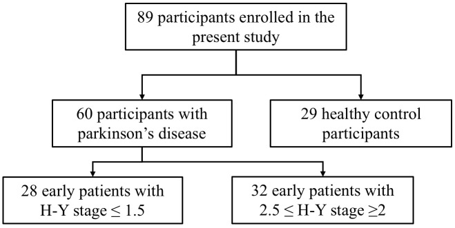
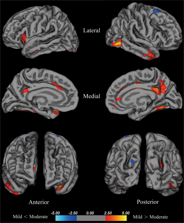
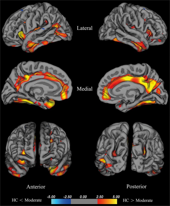
Objectives: This study was designed to explore changes in cortical thickness in patients with early Parkinson's disease (PD) at different Hoehn and Yahr (H-Y) stages and to demonstrate the association of abnormally altered brain regions with part III of the Unified Parkinson's Disease Rating Scale (UPDRS-III). Materials and Methods: Sixty early PD patients and 29 age- and gender-matched healthy controls (HCs) were enrolled in this study. All PD patients underwent comprehensive clinical and neuropsychological evaluations and 3.0 T magnetic resonance scanning. Patients with H-Y stage ≤1.5 were included in the mild group, and all other patients were included in the moderate group. FreeSurfer software was used to calculate cortical thickness. We assessed the relationship between UPDRS-III and regional changes in cortical thinning, including the bilateral fusiform and the temporal lobe. Results: The average cortical thickness of the temporal pole, fusiform gyrus, insula of the left hemisphere and fusiform gyrus, isthmus cingulate cortex, inferior temporal gyrus, middle temporal cortex and posterior cingulate cortex of the right hemisphere exhibited significant decreasing trends in HCs group and PD groups (i.e., the mild group and moderate group). After controlling for the effects of age, gender, and disease duration, the UPDRS-III scores in patients with early PD were correlated with the cortical thickness of the left and right fusiform gyrus and the left temporal pole (p < 0.05). Conclusion: The average cortical thickness of specific brain regions reduced with increasing disease severity in early PD patients at different H-Y stages, and the UPDRS-III scores of early PD patients were correlated with cortical thickness of the bilateral fusiform gyrus and the left temporal pole.
Keywords: FreeSurfer; Hoehn and Yahr stage; Parkinson’s disease; UPDRS part III; magnetic resonance imaging.
Figures
References
LinkOut - more resources
Full Text Sources