Review
. 2018 Nov 13:9:1612.
doi: 10.3389/fphys.2018.01612. eCollection 2018.

Functional Evolution of Subolesin/Akirin

Affiliations
Review

Functional Evolution of Subolesin/Akirin

Sara Artigas-Jerónimo et al. Front Physiol. .

Abstract

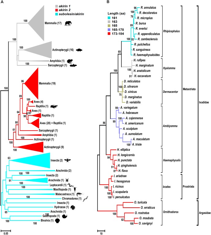
The Subolesin/Akirin constitutes a good model for the study of functional evolution because these proteins have been conserved throughout the metazoan and play a role in the regulation of different biological processes. Here, we investigated the evolutionary history of Subolesin/Akirin with recent results on their structure, protein-protein interactions and function in different species to provide insights into the functional evolution of these regulatory proteins, and their potential as vaccine antigens for the control of ectoparasite infestations and pathogen infection. The results suggest that Subolesin/Akirin evolved conserving not only its sequence and structure, but also its function and role in cell interactome and regulome in response to pathogen infection and other biological processes. This functional conservation provides a platform for further characterization of the function of these regulatory proteins, and how their evolution can meet species-specific demands. Furthermore, the conserved functional evolution of Subolesin/Akirin correlates with the protective capacity shown by these proteins in vaccine formulations for the control of different arthropod and pathogen species. These results encourage further research to characterize the structure and function of these proteins, and to develop new vaccine formulations by combining Subolesin/Akirin with interacting proteins for the control of multiple ectoparasite infestations and pathogen infection.

Keywords: Anaplasma phagocytophilum; immune response; interactome; phylogeny; regulome; tick; vaccine.


Figures

FIGURE 1
FIGURE 1
Phylogenetic analysis of akirin and subolesin nucleotide sequences. (A) A Neighbor Joining (NJ) phylogenetic tree was constructed with 361 nucleotide sequences belonging to 152 families, 73 orders and 15 classes (Mammalia, Actinopterygii, Amphibia, Sarcopterygii, Aves, Reptilia, Arachnida, Malacostraca, Insecta, Leptocardii, Maxillopoda, Chromadorea, Hydrozoa, Gastropoda and Bivalvia) of animals. All branches were collapsed at the class level and the number of orders per cluster is shown inside brackets. GenBank accession numbers and species names are provided in Supplementary Figure S1. Sequences were aligned using MAFFT configured for the maximum accuracy (Katoh and Standley, 2013). The final alignment contained 303 gap-free sites. All ambiguous positions were removed for each sequence pair. The best-fit model of the sequence evolution was selected based on Corrected Akaike Information Criterion (cAIC) and Bayesian Information Criterion (BIC) implemented in Molecular Evolutionary Genetics Analysis (MEGA) version 7. The Kimura 2-parameter model, which showed the lowest values of cAIC and BIC, was chosen for tree reconstruction. The evolutionary history was inferred using the NJ method implemented in MEGA 7 (Kumar et al., 2016). The percentage of replicate trees in which the associated taxa clustered together in the bootstrap test (500 replicates) is shown next to the branches (Felsenstein, 1985). (B) Phylogenetic tree of tick subolesin sequences. A Maximum Parsimony (MP) phylogenetic tree was constructed with 42 nucleotide sequences belonging to 6 and 1 genera of hard (family Ixodidae) and soft (family Argasidae) ticks, respectively. Because the evolution of subolesin in ticks has been less studied when compared to akirins, MP was used to generate a robust hypothesis on the evolution of this molecule in ticks. Sequences were aligned using MAFFT configured for the maximum accuracy (Katoh and Standley, 2013). Then, using the MAFFT alignment as template, a condon aligment was build (HIV database; www.hiv.lanl.gov accessed on 29-12-2017). The final alignment contained 576 total sites of which 329 were gap-free. The evolutionary history was inferred using the MP method (implemented in Molecular Evolutionary Genetics Analysis (MEGA) version 7 (Kumar et al., 2016). The percentage of replicate trees in which the associated taxa clustered together in the bootstrap test (500 replicates) is shown next to the branches (Felsenstein, 1985). The MP tree was obtained using the Subtree-Pruning-Regrafting (SPR) algorithm with search level 1 in which the initial trees were obtained by the random addition of sequences (10 replicates). Sequences were collected from Genbank and transcriptome projects and accession numbers are as follow: Ixodes scapularis (AY652654), I. persulcatus (KM888876), I. ricinus (JX193817), I. ariadnae (KM455971), I. hexagonus (JX193818), Rhipicephalus evertsi (JX193846), R. appendiculatus (DQ159967), R. microplus (EU301808), R. sanguineus (JX193845), R. haemaphysaloides (KP677498), R. annulatus (JX193844), R. decoloratus (JX193843), R. zambeziensis (GFPF01005851), R. bursa (GFZJ01017781), R. pulchellus (GACK01006228), Dermacentor silvarum (JX856138), D. sinicus (KM115649), D. marginatus (KU973622), D. variabilis (AY652657), D. reticulatus (JX193847), Amblyomma variegatum (JX193824), A. hebraeum (EU262598), A. cajennense (JX193823), A. americanum (JX193819), A. maculatum (JX193825), A. aureolatum (GFAC01005925), A. triste (GBBM01002796), A. sculptum (GFAA01000261), Hyalomma anatolicum (KT981976), H. rufipes (JX193849, H. marginatum (DQ159971), H. excavatum (GEFH01000904), Haemaphysalis longicornis (EU289292), Hae. elliptica (JX193850), Hae. qinghaiensis (EU326281, Hae. flava (KJ829652), Hae. punctata (DQ159972), Ornithodoros moubata (JX193852), O. savignyi (JX193851), O. turicata (GDIE01114362), O. erraticus (HM622148) and O. rostratus (GCJJ01005500).)
FIGURE 2
FIGURE 2
Genomic organization of subolesin/akirin orthologs across selected eukaryotic species. The genomic organization of the coding regions of tick (I. scapularis), human (Homo sapiens), mouse (M. musculus), frog (Xenopus laevis), fish (Danio rerio) and mosquito (Anopheles gambiae) subolesin/akirin is shown. The genomic organization of human, mouse, frog and fish akirins was previously reported (Liu et al., 2015). The genomic organization of tick and mosquito subolesin/akirin was collected from VectorBase (https://www.vectorbase.org; Giraldo-Calderón et al., 2015). Latin numerals correspond to the size of exons/introns in base pairs.
FIGURE 3
FIGURE 3
The I. scapularis Subolesin structure and its interactions with DNA and transcription factors. (A) The pairwise sequence alignment of the I. scapularis Subolesin (Sub) and the rat Arkinin2 (Ak2), accession numbers indicated, was generated using the MAFFT alignment program at default settings (Katoh et al., 2017). The NLS 1 and 2 domains (red box), binding sites 1-5 (bold-underlined in Ak2 and cyan for Sub), and the novel DNA binding sites (green and enclosed in a cyan box) are shown. The residues color-coded orange are extensions of the NLS domains. (B) The superposed tertiary structures of Sleeping Beauty (transparent black) and Subolesin (transparent green) are represented with the clamp loop labeled and the five α-helices of Sleeping Beauty (PDB: 5CR4) annotated in roman numerals. The tertiary residue positions of the labeled Subolesin NLS domains and binding sites are, respectively, color-coded as in the pairwise alignment. The Subolesin termini positions are color-labeled (green). (C) The Subolesin-DNA complex, modeled from the Mos1-DNA (PDB: 3HOS) show the residues of the novel DNA-binding site on α-helices IV-V, enclosed by a cyan box that were predicted by I-TASSER (Zhang, 2008). The DNA prime ends are color-labeled for the respective directions (indicated by arrows) of the sense (gray) and antisense (dark gray) strands. The residue positions of the Subolesin clamp loop, NLS domains and binding sites are color-coded as in previous panels A and B. (D) The schematic representation of the upstream DNA (gray helix) interactions with Subolesin NLS2, binding sites 2-5, and the potential clamp loop interaction (via NLS1 and binding site 1) with an unknown co-transcription factor (CTF?) and unknown (?) transcription factor (TF).
FIGURE 4
FIGURE 4
Model for Subolesin/Akirin function in immune response pathways. A simplified annotation of the downstream components of the arthropod IMD and mammalian TNF/TLR pathways (Goto et al., 2008; Beutler and Moresco, 2008; de la Fuente et al., 2008; Naranjo et al., 2013; Shaw et al., 2017). (A) After activation of the arthropod IMD pathway, the TGF-β (TAK1), Tak1-binding protein 2 (TAB2) and the I-KB kinase (IKK) complex are recruited, which leads to phosphorylation of the NF-κB transcription factor, Relish. After phosphorylation, the N-terminal domain of Relish (N-Rel) is cleaved by Caspase-8 homolog Dredd or a similar Caspase and is translocated to the nucleus. Subolesin/Akirin may be post-translationally modified and translocated to the nucleus. In the nucleus, N-Rel interacts with Subolesin/Akirin through unknown proteins to drive the production of anti-microbial peptides and other effector genes. In ticks, N-Rel and Subolesin may be reciprocally regulated. (B) In mammals, the activation of the TNF/TLR signaling pathways also results in the recruitment of the TAB2-TAK1 and IKK complexes, which results in the phosphorylation of the inhibitory regulator of NF-kB, IkB, resulting in the NF-kB translocation to the nucleus. As in arthropods, Akirin2 may be post-translationally modified and translocated to the nucleus. Once in the nucleus, NF-kB interacts with Akirin2 through unknown proteins for the activation of gene expression. In both arthropods and mammals, Subolesin/Akirin are involved in the regulation of genes that are Relish/NF-kB independent.
FIGURE 5
FIGURE 5
Scheme of the evolution and function of Subolesin/Akirin | Functional annotations were done based on published results for Subolesin/Akirin2. References are in the text of the paper. Myogenesis, attributed exclusively to Akirin1, was included and labeled as such. For each taxa, species in which studies were conducted are shown.
FIGURE 6
FIGURE 6
Examples of the role of tick Subolesin in different biological processes | (A) Role of tick Subolesin in A. phagocytophilum infection and blood feeding. The transcriptomics analysis in different I. scapularis tick tissues showed that subolesin (ISCW023283) but not relish (ISCW018935) mRNA levels significantly increased in response to A. phagocytophilum (Ap) infection in both midgut (MG) and salivary glands (SG). In addition, the subolesin gene knockdown phenotype in ticks injected with dsRNA resulted in a significant reduction in the number of female ticks completing feeding, oviposition and fertility. Results were reported by Ayllón et al. (2015). Photo of dissected I. scapularis partially fed adult female ticks courtesy of K. M. Kocan (Oklahoma State University, United States). (B) Role of tick Subolesin in A. phagocytophilum infection, blood feeding and questing speed. The response to A. phagocytophilum and stress increases subolesin levels, which together with heat shock proteins improve tick questing speed and survival. Results were reported by Busby et al. (2012). Photo of questing Ixodes ricinus courtesy of L. Grubhoffer & J. Erhard (Biology Center of the AS CR, Institute of Parasitology, Czechia).
FIGURE 7
FIGURE 7
Subcellular localization of Subolesin/Akirin2. Representative images of immunofluorescence analysis of I. scapularis ISE6 and human HL60 cells incubated with anti-Subolesin and anti-Akirin2 antibodies, respectively. The cells were fixed with 4% paraformaldehyde in PBS for 15 min, permeabilized with 0.5% Triton X-100 in PBS for 5 min and blocked with blocking buffer (3% BSA in PBS) for 1 h. Then, the cells were incubated overnight at 4°C with anti-Subolesin (Antunes et al., 2014) or anti-Akirin2 (Abcam, Cambridge, United Kingdom) antibodies (1/50 dilution in 3% BSA in PBS). After 3 washes with PBS, the slides were incubated with fluorescein isothiocyanate (FITC) conjugated anti-rabbit secondary antibodies (Sigma-Aldrich, St. Louis, MO, United States; green) at 1/160 dilution in 3% BSA in PBS for 1 h at room temperature. Cells were counterstained with ProLong Antifade containing 4′,6-diamidino-2-phenylindole (DAPI) (Molecular Probes, Eugene, OR, United States; blue), and imaged with a Zeiss LSM800 confocal microscope using a 63× oil immersion lens (Carl Zeiss, Oberkochen, Germany). Yellow arrows show examples of protein localization in the nucleus while red arrows illustrate protein localization in the cytoplasm.
FIGURE 8
FIGURE 8
Subolesin regulome in tick cells. The network of proteins and processes associated to transcription in tick cells uninfected and infected with Anaplasma phagocytophilum. The nodes (circles) are either proteins or processes (labeled). The size of each circle is proportional to its centrality index. The networks show (clusters of interacting proteins and processes in colors. The width of each link is proportional to the strength of the interaction. The networks show the topology of the tick interactome and regulome. The networks were built with the annotated proteins represented in either uninfected or infected cells, and a directed network was built for each protein linked to the processes in which it is involved. The weight of each link is proportional to the number of reads of the protein. This weighted degree of each link was used to calculate the centrality indexes, mainly the Betweenness Centrality, which is represented in the panels. Only the proteins annotated as involved in processes associated with transcription (i.e., linked by one or more protein(s) simultaneously annotated as transcripiton or other cellular process). The topology of the networks was obtained with the Lovaine algorithm. In both networks, the topological position of Subolesin is marked with a red arrow. Methods were described in Estrada-Peña et al. (2018).)
FIGURE 9
FIGURE 9
Characterization of the Subolesin/Akirin2 interactome. The information on Subolesin/Akirin-protein physical and functional interactions was compiled from the String protein-protein interactions database v.10.5 (https://string-db.org). The central node of the networks represent Subolesin/Akirin2 while the edges correspond to the predicted functional associations. Only predictions with medium (or better) confidence ( > 0.4) limited to the top 10 interactions with protein-protein interaction (PPI) enrichment p-value ≤ 0.5 were considered. To compare the different species, protein annotations were standardize by identity to I. scapularis/I. ricinus-D. melanogaster-H. sapiens order of priority (see Supplementary Dataset S1 for complete annotations). For illustration purposes, the species included in the analysis correspond to D. melanogaster, I. scapularis, Danio rerio, Mus musculus, Rattus norvegicus, and H. sapiens. Identical proteins in two different species are highlighted in red and blue letters. The functional annotation of the Subolesin/Akirin2 interacting proteins according to the biological processes (level 2) in which they are involved was done using Blast2GO (www.blast2go.com), and represented in pies with different colors for each process and the percentage of proteins on each process. Abbreviations: LO, localization (sepia); RP, rhythmic process (sangria); BIO, biogenesis (blue); SIG, signaling (green); NEG, negative regulation of biological process (black); CP, cellular process (azure); CPR, cell proliferation (white); MCP, multi-organism process (sky); DP, developmental process (gray); LOC, locomotion (violet); MP, metabolic process (red); BR, biological regulation (byzantine); BA, biological adhesion (moss); POS, positive regulation of biological process (yellow); RS, response to stimulus (tea); ISP, immune system process (gold); MOP, multicellular organismal process (orange); REP, reproductive process (smoke). Color code was established according to color thesaurus (https://graf1x.com/list-of-colors-with-color-names/).
FIGURE 10
FIGURE 10
Protective capacity of Subolesin/Akirin in vaccines for the control of arthropod ectoparasite infestations and pathogen infection. The effect of Subolesin/Akirin vaccination is shown on evolutionarily diverse arthropod genera. The effect of the vaccine was recorded on different phases of ectoparasite life cycle in the form of reduction in ectoparasite infestation (number of ectoparasites completing feeding), weight (weight of engorged female ectoparasites), oviposition (number of eggs per female), and fertility (number of larvae per female) in ectoparasites fed on vaccinated hosts when compared to controls. The reduction in pathogen infection was recorded as differences in pathogen levels between ectoparasites fed on vaccinated and control hosts.

Similar articles

Cited by

References

    1. Adipietro K. A., Mainland J. D., Matsunami H. (2012). Functional evolution of mammalian odorant receptors. PLoS Genet. 8:e1002821. 10.1371/journal.pgen.1002821 - DOI - PMC - PubMed
    1. Almazán C., Blas-Machado U., Kocan K. M., Yoshioka J. H., Blouin E. F., Mangold A. J., et al. (2005). Characterization of three Ixodes scapularis cDNAs protective against tick infestations. Vaccine 23 4403–4416. 10.1016/j.vaccine.2005.04.012 - DOI - PubMed
    1. Almazán C., Kocan K. M., Bergman D. K., Garcia-Garcia J. C., Blouin E. F., de la Fuente J. (2003). Identification of protective antigens for the control of Ixodes scapularis infestations using cDNA expression library immunization. Vaccine 21 1492–1501. 10.1016/S0264-410X(02)00683-7 - DOI - PubMed
    1. Almazán C., Lagunes R., Villar M., Canales M., Rosario-Cruz R., Jongejan F., et al. (2010). Identification and characterization of Rhipicephalus (Boophilus) microplus candidate protective antigens for the control of cattle tick infestations. Parasitol. Res. 106 471–479. 10.1007/s00436-009-1689-1 - DOI - PMC - PubMed
    1. Almazán C., Moreno-Cantú O., Moreno-Cid J. A., Galindo R. C., Canales M., Villar M., et al. (2012). Control of tick infestations in cattle vaccinated with bacterial membranes containing surface-exposed tick protective antigens. Vaccine 30 265–272. 10.1016/j.vaccine.2011.10.102 - DOI - PubMed

LinkOut - more resources