. 2018 Nov 13:9:2586.
doi: 10.3389/fimmu.2018.02586. eCollection 2018.

Factors Produced by Macrophages Eliminating Apoptotic Cells Demonstrate Pro-Resolutive Properties and Terminate Ongoing Inflammation

Affiliations

Factors Produced by Macrophages Eliminating Apoptotic Cells Demonstrate Pro-Resolutive Properties and Terminate Ongoing Inflammation

Francis Bonnefoy et al. Front Immunol. .

Abstract

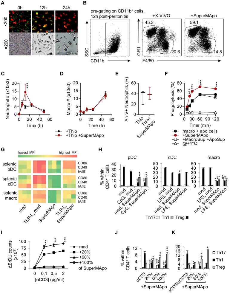
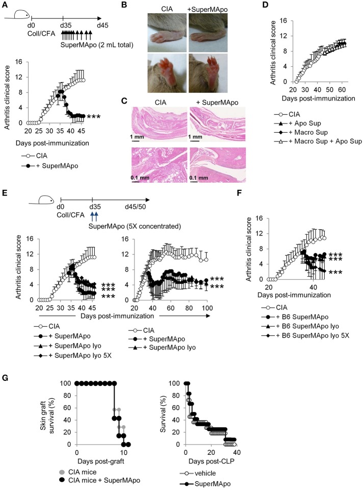
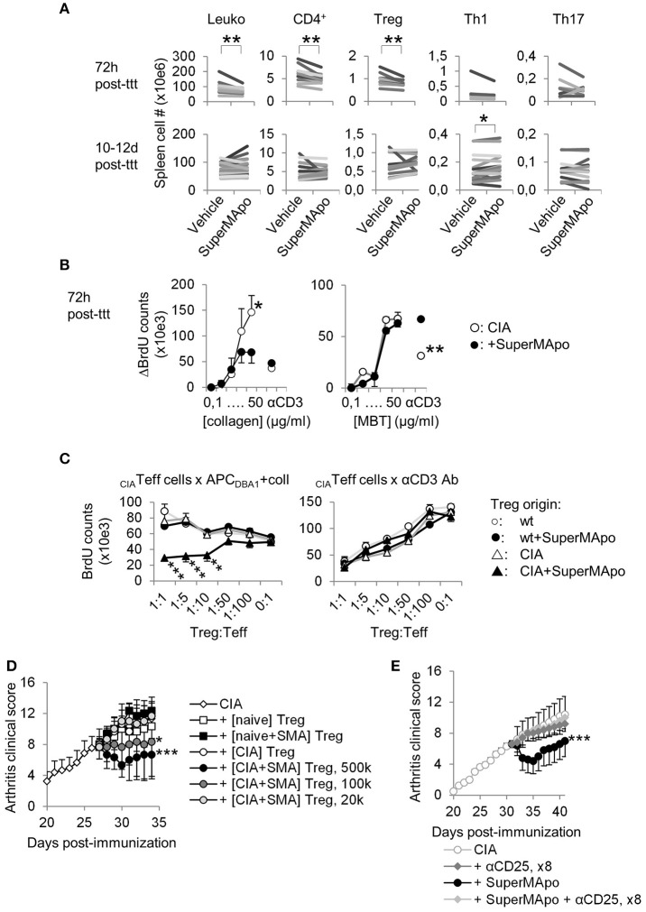
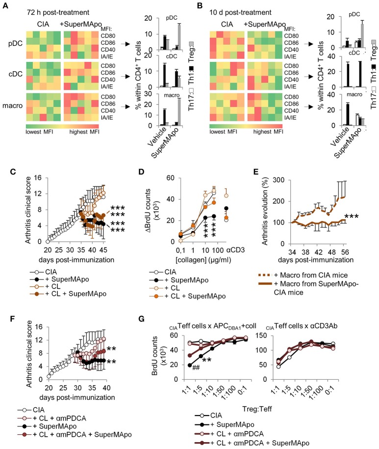
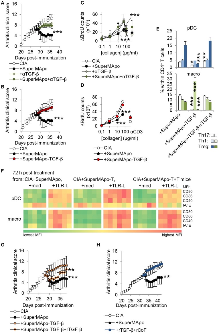
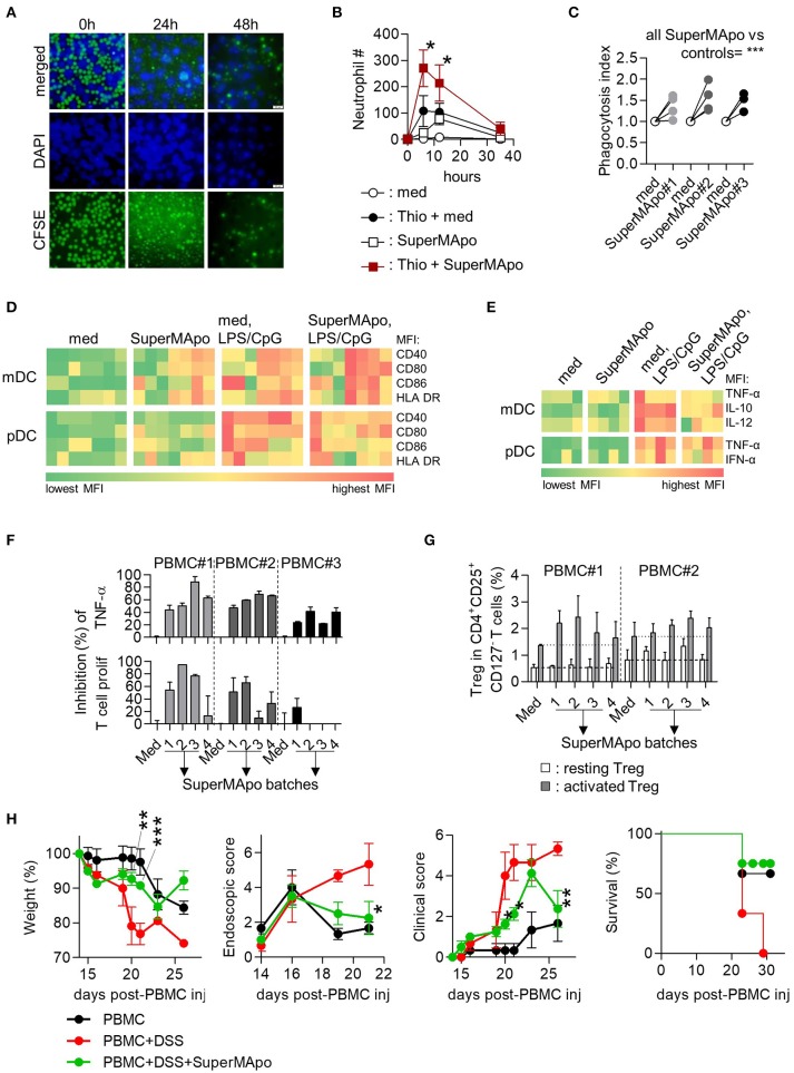
Unresolved inflammation is a common feature in the pathogenesis of chronic inflammatory/autoimmune diseases. The factors produced by macrophages eliminating apoptotic cells during resolution are crucial to terminate inflammation, and for subsequent tissue healing. We demonstrated here that the factors produced by macrophages eliminating apoptotic cells were sufficient to reboot the resolution of inflammation in vivo, and thus definitively terminated ongoing chronic inflammation. These factors were called SuperMApo and revealed pro-resolutive properties and accelerated acute inflammation resolution, as attested by both increased phagocytic capacities of macrophages and enhanced thioglycollate-induced peritonitis resolution. Activated antigen-presenting cells exposed to SuperMApo accelerated their return to homeostasis and demonstrated pro-regulatory T cell properties. In mice with ongoing collagen-induced arthritis, SuperMApo injection resolved and definitively terminated chronic inflammation. The same pro-resolving properties were observed in human settings in addition to xenogeneic colitis and graft-vs.-host disease modulation, highlighting SuperMApo as a new therapeutic opportunity to circumvent inflammatory diseases.

Keywords: apoptotic cells; efferocytosis; macrophages; phagocytosis; resolution of inflammation; tolerance induction.


Figures

Figure 1
Figure 1
Macrophages eliminating apoptotic cells produced factors with pro-resolutive properties. (A) Inverted fluorescence microscopy observation of macrophages stained by PE-F4/80 antibody (red), eliminating CFSE-labeled apoptotic cells (green) at 0, 12, and 24 h of culture. Injection (i.p.) of the supernatant from the previous 48 h culture (SuperMApo) increased neutrophil percentage (B) and number (C), not macrophage number (D) in the peritoneal cavity after peritonitis induction. (E) Neutrophil apoptosis 24 h after peritonitis induction in the presence or not of SuperMApo injection 28 h earlier (n = 3 mice per group; box and whiskers). The gating strategy of peritoneal neutrophils and macrophages is shown in (B), other data are shown as mean ± s.e.m., n = 3. ***P < 0.001, two-way RM ANOVA with Bonferroni post-tests. (F) The percentage of phagocytosis (mean ± s.e.m., n = 3) of apoptotic cells by macrophages was evaluated at various time points in normal culture condition (macro + apo cells) with or without SuperMApo or supernatant from macrophages cultured alone plus the supernatant from apoptotic cells cultured alone (+MacroSup +ApoSup), or at +4°C. ***P < 0.001, two-way RM ANOVA with Bonferroni post-tests. (G) Costimulatory (CD86/CD40) and MHC-II molecule (IA/IE) mean florescence intensity (MFI) expressions evaluated in plasmacytoid DC (pDC), conventional DC (cDC) and macrophages (macro) in spleen cell cultures with or without TLR ligands (TLR-L) or SuperMApo, or medium (med). Each cell in the heat map represents a single well; data are from one experiment representative of three. (H) Ovalbumin (OVA) TCR-specific CD4+CD25 T cell polarization (mean ± s.e.m., n = .3) by pDC, cDC and macrophages cultured as in G in the presence of OVA, was assessed by FACS evaluating IFN-γ (Th1), IL-17 (Th17) and Foxp3 (Treg) intracellular content after 4 days of culture. *P < 0.05, **P < 0.01, ***P < 0.001, 1 way ANOVA with Bonferroni's multiple comparison test. (I) Spleen T cell proliferation in the presence of grading doses of anti-CD3 specific antibody (αCD3) with medium (med) or SuperMApo in different proportions was assessed by BrdU incorporation and counting. ***P < 0.001, med vs. other conditions (mean ± s.e.m., n = 3), two-way RM ANOVA with Tukey's multiple comparison test. CD4 T cell polarization within spleen cells cultured as in I (J), or from naïve CD4+CD25 T cells cultured with anti-CD3/CD28 antibodies (K) in medium (med) or different proportions of SuperMApo was assessed by FACS. *P < 0.05, **P < 0.01, ***P < 0.001, unpaired t-test vs. without SuperMApo (mean ± s.e.m., n = 3). Figure data are issued from representative experiments, repeated at least three times with similar results.
Figure 2
Figure 2
Injection of SuperMApo in ongoing CIA induced a rapid termination of the disease. (A) The clinical arthritic score (mean ± s.e.m., n = 5 per group) after SuperMApo injections was strongly reduced in arthritic mice compared to CIA mice receiving control vehicle injections (CIA). ***P < 0.001, one-way ANOVA with Bonferroni's multiple comparison post-tests. (B) Representative pictures of paw swelling reduction after SuperMApo injection. (C) HES staining of rear ankle joint sections showing vehicle-(CIA) and SuperMApo-treated arthritic mice conditions. (D) The clinical arthritis score (mean ± s.e.m., n = 5 per group) of mice receiving the supernatant from apoptotic cell culture, or from macrophage culture or both the previous together did not demonstrated reduction. (E,F) Arthritic mice treated with lyophilized SuperMApo concentrated (SuperMApo lyo 5X) or not (SuperMApo lyo), demonstrated arthritis clinical score reduction, lasting for more than 60 days, even when issued from a C57Bl/6 (B6) origin. ***P < 0.001, one-way ANOVA with Bonferroni's multiple comparison post-tests, vs. CIA (mean ± s.e.m., n = 5 per group). (G) SuperMApo-treated arthritic mice were able to reject an allogeneic skin graft as control mice did (7 mice per group, data pooled from 2 independent experiments), and naive mice receiving SuperMApo or vehicle demonstrated similar survival rate after cecal ligation and puncture-induced sepsis (11 to 12 mice per group, data pooled from 2 independent experiments). Figure data are issued from representative experiments, repeated at least three times with similar results unless otherwise noted.
Figure 3
Figure 3
Resolution of arthritis implicates the generation of antigen-specific regulatory T cells. (A) Numbers of splenocytes (Leuko), CD4+ T cells and Treg, IFN-γ+ Th1 and IL-17A+ Th17 subsets were decreased early after SuperMApo treatment (72 h) and were observed normalized 10–12 days post-treatment (10–12 days post-ttt), compared to arthritic mice receiving vehicle. Each bar represents means of each group per one experiment with 5 mice per group. **P < 0.01, paired t-test. (B) BrdU counts showing the proliferation of cells from arthritic mice 72 h after receiving SuperMApo or vehicle, in response to increasing doses of collagen or MBT, or to CD3-specific antibody (used as control). Data are shown as mean ± s.e.m. of mouse spleen cells performed in triplicates, 5 mice per group, *P < 0.05, **P < 0.01, 2-way ANOVA with Bonferroni post-tests. (C) BrdU counts showing collagen-specific suppression of cell proliferation by Treg issued from arthritic mice treated with SuperMApo 72 h earlier compared to Treg from other origins. The suppressive activity of all Treg is also shown as control in CD3-specific antibody-stimulated cultures. Data are shown as mean ± s.e.m. of mouse spleen cells performed in triplicates, 5 mice per group, ***P < 0.001, 2-way ANOVA with Bonferroni post-tests. (D) Arthritis clinical score showing that only Treg issued from arthritic mice treated with SuperMApo ([CIA + SMA] Treg) demonstrated the capacity to reduced arthritis after adoptive transfer compared to Treg from other origins (from CIA mice [CIA], naïve mice receiving SuperMApo [naïve+SMA] or not [naïve]), in a cell number-dependent manner. Five mice per group, mean ± s.e.m., *P < 0.05, ***P < 0.001 vs. CIA group, one-way ANOVA with Bonferroni's multiple comparison post-tests. (E) Arthritis clinical score showing that CD25-specific depleting antibody injection inhibited SuperMApo-induced arthritis resolution. Five mice per group, mean ± sem, ***P < 0.001 vs. other groups, one-way ANOVA with Bonferroni's multiple comparison post-tests.
Figure 4
Figure 4
Antigen-presenting cells demonstrate reprogramming in vivo after SuperMApo treatment of CIA mice. (A,B) Costimulatory (CD80/CD86/CD40) and MHC-II molecule (IA/IE) mean florescence intensity (MFI) expressions, evaluated in spleen plasmacytoid DC (pDC), conventional DC (cDC) and macrophages (macro) 72 h and 10 days after SuperMApo or vehicle treatment of arthritic mice. Data from representative experiments showing cell marker expression from individual mouse (5 mice per group). Th17, Th1 and Treg CD4+ T cell polarizations by the same APC are also shown (bars represent mean ± s.e.m. of triplicates of APC isolated from each mouse and cultured with naïve CD4+ T cells). (C) Arthritis clinical score of mice receiving SuperMApo treatment or vehicle and clodronate-loaded (CL) or control liposomes (mean ± s.e.m., 5 mice per group). ***P < 0.001 vs. respective control groups (CIA & CIA + CL), one-way ANOVA with Bonferroni's multiple comparison post-tests. (D) BrdU counts showing the proliferation of cells from arthritic mice 72 h after receiving SuperMApo or vehicle and phagocyte depletion, in response to increasing doses of collagen or to CD3-specific antibody (as control). Data are shown as mean ± s.e.m. of cell triplicates, 5 mice per group, ***P < 0.001, 2-way ANOVA with Bonferroni post-tests. (E) Evolution of arthritis clinical score (in percentage; 100% = arthritis clinical score the day of injection) in mice with ongoing arthritis (mean arthritis clinical score = 7.1 ± 0.6 the day of cell injection) after receiving macrophages from vehicle- or SuperMApo-treated CIA mice. Percentages of clinical score are shown as mean ± s.e.m., 8 mice per group from 2 independent experiments. ***P < 0.001, Wilcoxon signed rank test. (F) Arthritis clinical score of mice receiving SuperMApo treatment or vehicle with or without clodronate-loaded liposomes and anti-mPDCA antibody (CL + αmPDCA) (mean ± s.e.m., 5 mice per group). **P < 0.005 for CIA vs. CIA + SuperMApo & CIA vs. CIA + CL + αmPDCA + SuperMApo, one-way ANOVA with Bonferroni's multiple comparison post-tests. (G) BrdU counts showing collagen-specific suppression of cell proliferation by Treg issued from arthritic mice from F. The suppressive activity of all Treg is also shown as control in CD3-specific antibody-stimulated cultures. Data are shown as mean ± s.e.m. of mouse spleen cells performed in triplicates, 5 mice per group, ##P < 0.005 vs. CL+mPDCA and CIA, **P < 0.005 vs. CIA, 2-way ANOVA with Bonferroni post-tests. Figure data are issued from representative experiments, repeated at least three times with similar results unless otherwise noted.
Figure 5
Figure 5
TGF-β associated with soluble factors within SuperMApo demonstrates pro-resolutive properties. (A,B) Arthitis clinical scores showing the role of TGF-β within SuperMApo on arthritis evolution through TGF-β neutralization (+αTGF-β) or depletion (-TGF-β) from SuperMApo. ***P < 0.001 vs. CIA (5 mice per group, mean ± s.e.m.), one-way ANOVA with Bonferroni's multiple comparison post-tests. (C,D) BrdU counts showing the proliferation of cells from arthritic mice from panels A,B. Data are shown as mean ± s.e.m. of mouse spleen cells performed in triplicates, 5 mice per group, ***P < 0.001, 2-way ANOVA with Bonferroni post-tests. (E) IL-17A+ Th17, IFN-γ+ Th1 and Treg CD4+ T cell polarization by pDC or macrophages isolated from mice from B and/or G (bars represent mean ± s.e.m. of triplicate of APC isolated from each mouse and cultured with naïve CD4+ T cells). *P < 0.05, **P < 0.01, ***P < 0.001, 1way ANOVA with Bonferroni's multiple comparison test. (F) Costimulatory (CD80/CD86/CD40) and MHC-II molecule (IA/IE) mean florescence intensity (MFI) expressions, evaluated in pDC and macrophages (macro) from spleens from mice from B and/or G (-T = SuperMApo depleted for TGF-β; -T+T = -T plus recombinant TGF-β). Data from a representative experiment showing cell marker expression from individual mouse (5 mice per group). (G) Arthritis clinical score showing no arthritis reduction with SuperMApo depleted from TGF-β (+SuperMApo-TGF-β) compared to SuperMApo treatment, and a partial effect of recombinant TGF-β injection on arthritis resolution when injected with SuperMApo depleted from TGF-β (+SuperMApo-TGF-β+rTGF-β). **P < 0.01, ***P < 0.001 vs. control groups (5 mice per group, mean ± s.e.m.), one-way ANOVA with Bonferroni's multiple comparison post-tests. (H) Arthritis clinical score showing that injection of recombinant TGF-β in addition to recombinant cofactors MMP2, ApoE, C1q, Ttr and Thbs1 (+rTGF-β+rCoF), does not favor resolution when compared to SuperMApo injection. **P < 0.01, vs. control and +rTGF-β+rCoF groups (5 mice per group, mean ± s.e.m.), one-way ANOVA with Sidak's multiple comparisons test; representative from 2 independent experiments.
Figure 6
Figure 6
Efferocytosis factors issued from monocyte-derived macrophages demonstrate pro-resolutive properties in vitro and in vivo. (A) Monocyte-derived macrophages were cultured with CFSE-labeled apoptotic PBMC (green) and observed after DAPI staining by inverted fluorescence microscopy at 0, 24, and 48 h of culture. (B) Injection (i.p.) of the supernatant from the previous 48 h culture (SuperMApo) increased neutrophil number (mean ± s.e.m., n = 2-9 mice per group), 12 h after thioglycollate mobilization (Thio + SuperMApo) and control PBMC/neutrophils-reconstituted NSG mice (SuperMApo). *P < 0.05, Kruskal-Wallis test with Dunn's MCT. (C) Monocyte-derived macrophages demonstrated increased capacity to phagocytose apoptotic cells in the presence of SuperMApo compared to medium (med). Data from 4 individual healthy volunteers. ***P < 0.001, paired t test. (D) Costimulatory (CD40/CD80/CD86) and HLA-DR molecule mean florescence intensity (MFI) expressions evaluated in sorted myeloid DC (mDC) and pDC in culture with or without TLR ligands (TLR-L) or SuperMApo, or medium (med). (E) Intracellular cytokine percentages evaluated in sorted myeloid DC (mDC) and pDC as in D. One square represents the mean of duplicate obtain for cells from one healthy volunteer (n = 4 to 7), several SuperMApo batches have been used. (F) SuperMApo inhibition of TNF-α production was evaluated intracellularly by flow cytometry in PBMC monocytes stimulated by PHA as well as T cell proliferation within PHA-stimulated PBMC also evaluated by flow cytometry on CFSE+CD3+ T cells. PBMC were issued from different healthy volunteers (n = 3) and SuperMApo from different batches. Bars represent means ± s.e.m. of duplicate. (G) Foxp3+CD25highCD45RA activated and Foxp3intCD25intCD45RA+ resting Treg in CD4+CD127 T cells were evaluated in PBMC cultured with CD3-specific antibody with or without SuperMApo. Bars represent means ± s.e.m. of triplicate. Dash lines represent Treg levels obtained in medium condition. (H) SuperMApo was injected in PBMC-reconstituted NOG mice receiving DSS (PBMC+DSS+SuperMApo) and then, the weight was evaluated in these mice and controls (PBMC; PBMC+DSS), as well as colonic lesion by video-colonoscopy, the clinical score was determined as well as survival. The data are shown as mean ± s.e.m. for each group, with 3–4 mice per group. *P < 0.05, **P < 0.01, ***P < 0.001, vs. DSS, 2-way RM ANOVA with Bonferroni post-tests.

References

    1. Serhan CN, Savill J. Resolution of inflammation: the beginning programs the end. Nat Immunol. (2005) 6:1191–7. 10.1038/ni1276 - DOI - PubMed
    1. Poon IK, Lucas CD, Rossi AG, Ravichandran KS. Apoptotic cell clearance: basic biology and therapeutic potential. Nat Rev Immunol. (2014) 14:166–80. 10.1038/nri3607 - DOI - PMC - PubMed
    1. Nagata S, Hanayama R, Kawane K. Autoimmunity and the clearance of dead cells. Cell (2010) 140:619–30. 10.1016/j.cell.2010.02.014 - DOI - PubMed
    1. Szondy Z, Garabuczi E, Joos G, Tsay GJ, Sarang Z. Impaired clearance of apoptotic cells in chronic inflammatory diseases: therapeutic implications. Front Immunol. (2014) 5:354. 10.3389/fimmu.2014.00354 - DOI - PMC - PubMed
    1. Perretti M, Leroy X, Bland EJ, Montero-Melendez T. Resolution pharmacology: opportunities for therapeutic innovation in inflammation. Trends Pharmacol Sci. (2015) 36:737–55. 10.1016/j.tips.2015.07.007 - DOI - PubMed

Publication types

LinkOut - more resources