

Contrast-enhanced voiding urosonography with intravesical administration of ultrasound contrast agent for the diagnosis of pediatric vesicoureteral reflux
- PMID: 30542403
- PMCID: PMC6257520
- DOI: 10.3892/etm.2018.6793
Contrast-enhanced voiding urosonography with intravesical administration of ultrasound contrast agent for the diagnosis of pediatric vesicoureteral reflux
Abstract
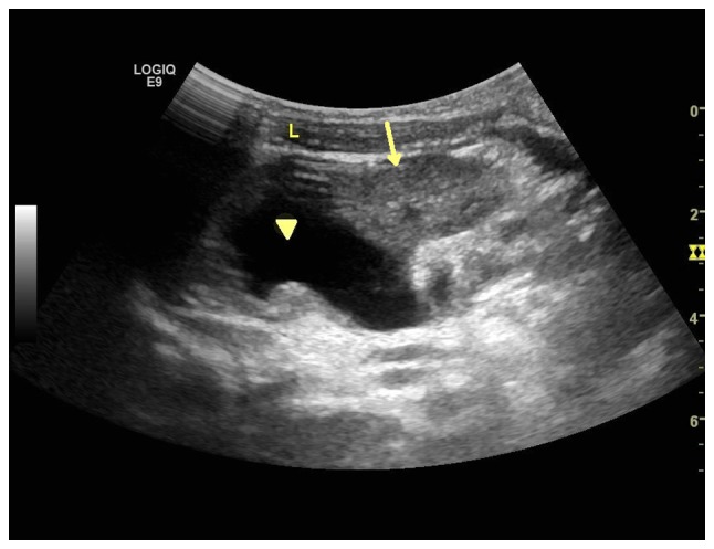
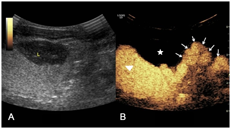
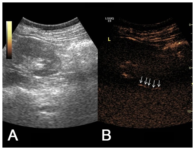
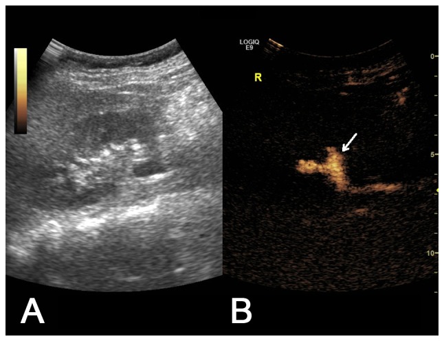
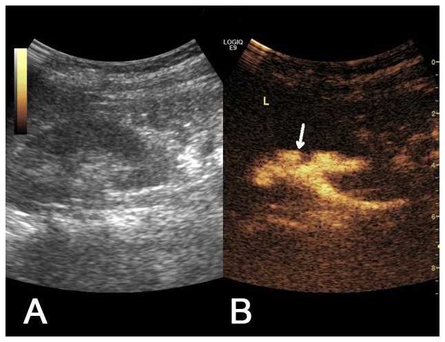
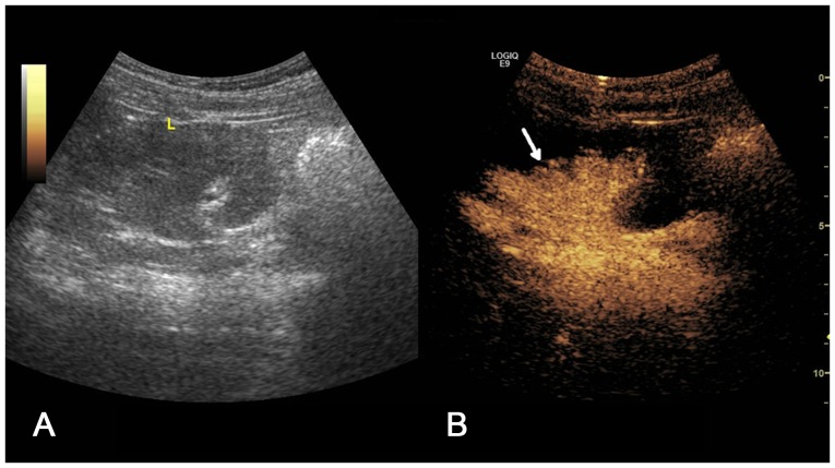
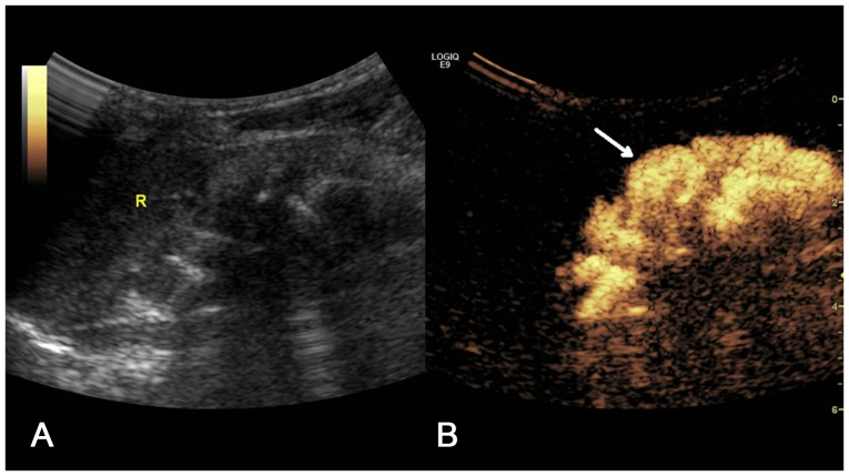
Vesicoureteral reflux (VUR) is one of the most common urinary tract anomalies in children. It has been reported that VUR may be associated with reflux nephropathy. Ultrasound contrast-enhanced voiding urosonography (CeVUS) has become a routine diagnostic method for VUR in a number of European countries; however, it is not widely used in China. The aim of the present study was to analyze the clinical application and evaluate the safety of CeVUS as a diagnostic tool for VUR in children in order to establish a standardized operating procedure for CeVUS in pediatric VUR in China. Between August 2016 and October 2017, 90 children who were susceptible to VUR were admitted into the Pediatric Nephrology Department of Tongji Hospital and underwent CeVUS. The SonoVue second-generation USA contrast agent was administered intravesically via a transurethral bladder catheter at a dose of 1 ml. The occurrence of adverse events was monitored. Urine analysis and culture were performed. A total of 90 children (47 female, 43 male; mean age, 36.6 months) with 178 Pelvi-Ureteral Units (PUUs) underwent CeVUS to screen for VUR. VUR was detected in 44/90 pediatric patients (48.89%) and 65/178 PUUs (36.52%) by CeVUS. The grade distribution of the 65 PUUs with VUS was as follows: Grade I, 3; Grade II, 9; Grade III, 14; Grade IV, 22; and Grade V, 17. The accuracy of CeVUS in the present study were consistent with previous reports. No urethral anomalies were detected and there were no adverse events. CeVUS was demonstrated to be a safe, accurate and reliable imaging technique for detecting VUR in high-risk children, including neonates. Results of the present study indicated that CeVUS can be adopted as the primary screening and follow-up method for pediatric VUR diagnoses in China.
Keywords: children; contrast-enhanced voiding urosonography; urinary tract infection; vesicoureteric reflux.
Figures
Similar articles
-
Contrast-enhanced voiding urosonography, part 1: vesicoureteral reflux evaluation.Pediatr Radiol. 2021 Nov;51(12):2351-2367. doi: 10.1007/s00247-020-04906-8. Epub 2021 Mar 31. Pediatr Radiol. 2021. PMID: 33787945 Review.
-
The learning curve of contrast-enhanced 'microbubble' voiding urosonography-validation study.J Pediatr Urol. 2019 Aug;15(4):385.e1-385.e6. doi: 10.1016/j.jpurol.2019.04.015. Epub 2019 Apr 19. J Pediatr Urol. 2019. PMID: 31133505
-
Contrast media viscosity and its potential effect on the diagnosis of vesicoureteral reflux in children.Eur Radiol. 2025 Mar;35(3):1615-1622. doi: 10.1007/s00330-024-11079-7. Epub 2024 Sep 16. Eur Radiol. 2025. PMID: 39285028
-
Voiding Urosonography with Second-Generation Ultrasound Contrast Agent for Diagnosis of Vesicoureteric Reflux: First Local Pilot Study.Open Access Maced J Med Sci. 2017 Apr 11;5(2):215-221. doi: 10.3889/oamjms.2017.055. eCollection 2017 Apr 15. Open Access Maced J Med Sci. 2017. PMID: 28507631 Free PMC article.
-
The Intrarenal Reflux Diagnosed by Contrast-Enhanced Voiding Urosonography (ceVUS): A Reason for the Reclassification of Vesicoureteral Reflux and New Therapeutic Approach?Biomedicines. 2024 May 5;12(5):1015. doi: 10.3390/biomedicines12051015. Biomedicines. 2024. PMID: 38790977 Free PMC article. Review.
Cited by
-
A narrative review on the applications of intracavitary contrast-enhanced ultrasonography in pediatric lower genitourinary anomalies.Front Pediatr. 2023 May 19;11:984643. doi: 10.3389/fped.2023.984643. eCollection 2023. Front Pediatr. 2023. PMID: 37274817 Free PMC article. Review.
-
Contrast-enhanced voiding urosonography, part 1: vesicoureteral reflux evaluation.Pediatr Radiol. 2021 Nov;51(12):2351-2367. doi: 10.1007/s00247-020-04906-8. Epub 2021 Mar 31. Pediatr Radiol. 2021. PMID: 33787945 Review.
-
Contrast-enhanced voiding urosonography in the assessment of vesical-ureteral reflux: the time has come.Radiol Med. 2021 Jul;126(7):901-909. doi: 10.1007/s11547-021-01360-w. Epub 2021 May 5. Radiol Med. 2021. PMID: 33954899 Review.
-
Contrast-enhanced ultrasound: a comprehensive review of safety in children.Pediatr Radiol. 2021 Nov;51(12):2161-2180. doi: 10.1007/s00247-021-05223-4. Epub 2021 Oct 30. Pediatr Radiol. 2021. PMID: 34716453 Free PMC article. Review.
-
Management of Vesicoureteral Reflux: What Have We Learned Over the Last 20 Years?Front Pediatr. 2021 Mar 31;9:650326. doi: 10.3389/fped.2021.650326. eCollection 2021. Front Pediatr. 2021. PMID: 33869117 Free PMC article. Review.
References
-
- Roić G, Roić AC, Palcić I, Grmoja T, Batos AT. Contrast enhanced voiding urosonography (cevus) in the diagnosis of vesicoureteral reflux. Lijecnicki Vjesnik. 2016;138:39–46. (In Croatian) - PubMed
LinkOut - more resources
Full Text Sources