

Concentration-dependent effects of rapamycin on proliferation, migration and apoptosis of endothelial cells in human venous malformation
- PMID: 30542410
- PMCID: PMC6257489
- DOI: 10.3892/etm.2018.6782
Concentration-dependent effects of rapamycin on proliferation, migration and apoptosis of endothelial cells in human venous malformation
Abstract
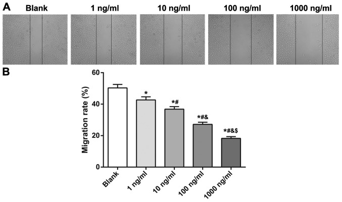
Rapamycin has been reported to be immunosuppressive and anti-proliferative towards vascular endothelial and smooth muscle cells. The purpose of the present study was to investigate the effects of rapamycin on the biological behaviors of endothelial cells that have been separated from the deformed vein in human venous malformation (VM). Cellular morphology was observed using inverted microscopy. An MTT assay was performed to measure the cell viability at different concentrations of rapamycin and different time points. Cell apoptosis and migration were detected using a terminal deoxynucleotidyl-transferase-mediated dUTP nick end labeling assay and a wound-healing assay, respectively. At 48 and 72 h, rapamycin inhibited proliferation of human VM endothelial cells, with the effects becoming more pronounced with increasing concentration. Only rapamycin at a concentration of 1,000 ng/ml had a significant effect at 24 h in repressing proliferation. At 48 h, compared with the blank group, the majority of cells maintained a clear nuclear boundary and a regular shape following treatment with 1 ng/ml rapamycin; 10 and 100 ng/ml rapamycin caused desquamation and rounded shape; and 1,000 ng/ml rapamycin caused even more marked desquamation, rounded shape and necrosis. Rapamycin at concentrations of 1, 10, 100 and 1,000 ng/ml reduced cell viability, increased the number of apoptotic cells and suppressed the migration capacity of human VM endothelial cells, and the effects became more pronounced with increasing concentration, when compared with the blank group. These findings provide evidence that rapamycin induces apoptosis and inhibits proliferation and migration of human VM endothelial cells in a concentration-dependent manner.
Keywords: cell viability; concentration-dependence; endothelial cells; rapamycin; venous malformation.
Figures


Similar articles
-
Inhibitory effect of rapamycin on corneal neovascularization in vitro and in vivo.Invest Ophthalmol Vis Sci. 2005 Feb;46(2):454-60. doi: 10.1167/iovs.04-0753. Invest Ophthalmol Vis Sci. 2005. PMID: 15671269
-
[Effects of Rapamycin and Rapamycin-loaded Poly(lactic-co-glycolic)Acid Nanoparticles on Apoptosis and Expression of bcl-2 and p27(kip1) Proteins of Human Umbilical Arterial Vascular Smooth Muscle Cell].Zhongguo Yi Xue Ke Xue Yuan Xue Bao. 2015 Dec;37(6):633-40. doi: 10.3881/j.issn.1000-503X.2015.06.001. Zhongguo Yi Xue Ke Xue Yuan Xue Bao. 2015. PMID: 26725384 Chinese.
-
Rapamycin-loaded nanoparticles for inhibition of neointimal hyperplasia in experimental vein grafts.Ann Vasc Surg. 2011 May;25(4):538-46. doi: 10.1016/j.avsg.2011.01.003. Ann Vasc Surg. 2011. PMID: 21549923
-
Inhibitory effect of rapamycin on proliferation of human umbilical arterial smooth muscle cells.Immunopharmacol Immunotoxicol. 2019 Aug;41(4):485-489. doi: 10.1080/08923973.2019.1628045. Epub 2019 Jun 23. Immunopharmacol Immunotoxicol. 2019. PMID: 31232124
-
Protective effect of silymarin against rapamycin-induced apoptosis and proliferation inhibition in endothelial progenitor cells.Nat Prod Commun. 2015 Feb;10(2):263-6. Nat Prod Commun. 2015. PMID: 25920256
Cited by
-
Effects of Microvesicles Derived from NK Cells Stimulated with IL-1β on the Phenotype and Functional Activity of Endothelial Cells.Int J Mol Sci. 2021 Dec 20;22(24):13663. doi: 10.3390/ijms222413663. Int J Mol Sci. 2021. PMID: 34948459 Free PMC article.
-
Microtubule Association of EML4-ALK V3 Is Key for the Elongated Cell Morphology and Enhanced Migration Observed in V3 Cells.Cells. 2024 Nov 25;13(23):1954. doi: 10.3390/cells13231954. Cells. 2024. PMID: 39682703 Free PMC article.
-
mTOR-FABP4 signal is activated in brain arteriovenous malformations in humans.J Mol Med (Berl). 2022 Sep;100(9):1287-1297. doi: 10.1007/s00109-022-02237-9. Epub 2022 Jul 25. J Mol Med (Berl). 2022. PMID: 35876909
-
Targeted medical therapies for vascular anomalies.Hematology Am Soc Hematol Educ Program. 2024 Dec 6;2024(1):709-717. doi: 10.1182/hematology.2024000599. Hematology Am Soc Hematol Educ Program. 2024. PMID: 39644074 Free PMC article. Review.
-
Mechanism of lncRNA gadd7 regulating mitofusin 1 expression by recruiting LSD1 to down-regulate H3K9me3 level, and mediating mitophagy in alveolar type II epithelial cell apoptosis in hyperoxia-induced acute lung injury.Cell Biol Toxicol. 2025 Apr 29;41(1):77. doi: 10.1007/s10565-025-10021-x. Cell Biol Toxicol. 2025. PMID: 40301157 Free PMC article.
References
LinkOut - more resources
Full Text Sources